. 2000 Jan;20(1):416-27.
doi: 10.1128/MCB.20.1.416-427.2000.

Characterization of insulin-responsive GLUT4 storage vesicles isolated from 3T3-L1 adipocytes

Affiliations

Characterization of insulin-responsive GLUT4 storage vesicles isolated from 3T3-L1 adipocytes

M Hashiramoto et al. Mol Cell Biol. 2000 Jan.

Abstract

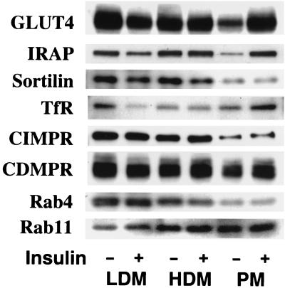
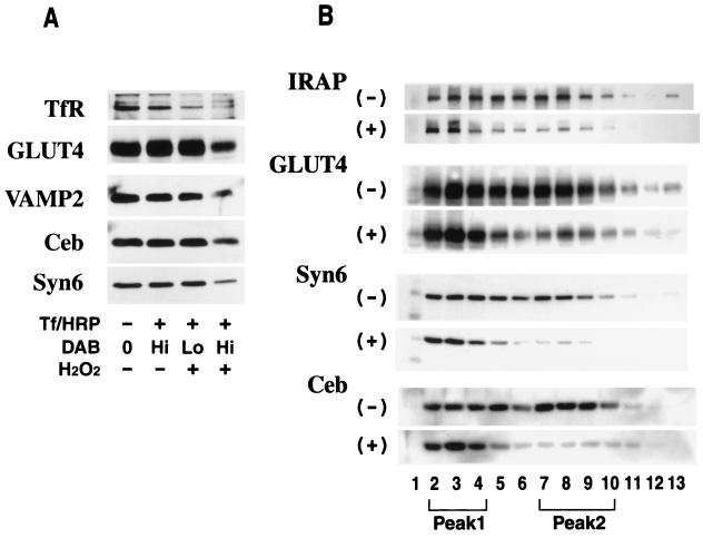
Insulin regulates glucose transport in muscle and adipose tissue by triggering the translocation of a facilitative glucose transporter, GLUT4, from an intracellular compartment to the cell surface. It has previously been suggested that GLUT4 is segregated between endosomes, the trans-Golgi network (TGN), and a postendosomal storage compartment. The aim of the present study was to isolate the GLUT4 storage compartment in order to determine the relationship of this compartment to other organelles, its components, and its presence in different cell types. A crude intracellular membrane fraction was prepared from 3T3-L1 adipocytes and subjected to iodixanol equilibrium sedimentation analysis. Two distinct GLUT4-containing vesicle peaks were resolved by this procedure. The lighter of the two peaks (peak 2) was comprised of two overlapping peaks: peak 2b contained recycling endosomal markers such as the transferrin receptor (TfR), cellubrevin, and Rab4, and peak 2a was enriched in TGN markers (syntaxin 6, the cation-dependent mannose 6-phosphate receptor, sortilin, and sialyltransferase). Peak 1 contained a significant proportion of GLUT4 with a smaller but significant amount of cellubrevin and relatively little TfR. In agreement with these data, internalized transferrin (Tf) accumulated in peak 2 but not peak 1. There was a quantitatively greater loss of GLUT4 from peak 1 than from peak 2 in response to insulin stimulation. These data, combined with the observation that GLUT4 became more sensitive to ablation with Tf-horseradish peroxidase following insulin treatment, suggest that the vesicles enriched in peak 1 are highly insulin responsive. Iodixanol gradient analysis of membranes isolated from other cell types indicated that a substantial proportion of GLUT4 was targeted to peak 1 in skeletal muscle, whereas in CHO cells most of the GLUT4 was targeted to peak 2. These results indicate that in insulin-sensitive cells GLUT4 is targeted to a subpopulation of vesicles that appear, based on their protein composition, to be a derivative of the endosome. We suggest that the biogenesis of this compartment may mediate withdrawal of GLUT4 from the recycling system and provide the basis for the marked insulin responsiveness of GLUT4 that is unique to muscle and adipocytes.


Figures

FIG. 1
FIG. 1
Polypeptide composition of GLUT4-containing vesicles. Vesicle immunoadsorption was carried out by incubating protein G-Sepharose beads coupled to a purified monoclonal GLUT4 antibody (1F8) with pooled velocity sucrose gradient fractions isolated from rat adipocytes for 2 h at 4°C on a rotating wheel in PBS containing 0.1% BSA. After the incubation, beads were washed and the vesicles were eluted from the beads with 0.2 M bicarbonate buffer (pH 11.0). Eluted proteins were pelleted and analyzed by SDS-PAGE followed by silver staining. Proteins with molecular masses of 165, 110, and ∼50 kDa from a large-scale vesicle preparation were excised and subjected to N-terminal sequencing. The amino acid sequences and identities of these proteins are shown at the right. Other proteins were identified by immunoblotting. The upper half is from a 6% resolving gel, and the bottom half is from a 10% gel. CIMP/IGFII-R, CI-MPR and insulin-like growth factor receptor II.
FIG. 2
FIG. 2
The effect of insulin on the subcellular distributions of various proteins in 3T3-L1 adipocytes. 3T3-L1 adipocytes were incubated in the absence (−) or presence (+) of 10−7 M insulin for 20 min at 37°C. Subcellular fractionation was carried out to obtain plasma PM, high density microsome (HDM) and LDM fractions. Immunoblotting was carried out with specific antibodies as indicated. The results shown are from a single preparation of the membrane fractions and are representative of similar determinations.
FIG. 3
FIG. 3
Iodixanol equilibrium gradient sedimentation analysis of 3T3-L1 adipocytes. The LDM fraction prepared from 3T3-L1 adipocytes was subjected to iodixanol density gradient analysis as described in Materials and Methods. Fractions were collected from the bottom of the gradient and used to measure protein (A) or the distribution of various proteins by immunoblotting (B). Note that in panel B, fraction 1 is omitted from the immunoblotting analysis (see Results). The results shown are from a single iodixanol gradient analysis and are representative of six experiments. Syn6, syntaxin 6; Ceb, cellubrevin; OD595, optical density at 595 nm.
FIG. 4
FIG. 4
Iodixanol equilibrium gradient sedimentation analysis of internalized Tf. 3T3-L1 adipocytes were serum starved in DMEM for 16 h and then incubated at 37°C for 2 min with KRP buffer containing 125I-diferric human Tf (∼1.0 μCi/ml) and BSA (1 mg/ml). As a control to estimate nonspecific uptake, duplicate plates were incubated in the presence of excess (20 μM) unlabelled holo Tf. After two quick washes, the cells were incubated at 37°C for 0 (●), 3 (○), and 20 (▴) min to allow Tf internalization. Cells were washed and subjected to subcellular fractionation and iodixanol sedimentation analysis as described in Materials and Methods. Fractions were counted in a gamma counter, and internalized Tf was quantitated by subtracting background values from each fraction. The results shown are representative of two separate experiments.
FIG. 5
FIG. 5
Tf-HRP endosomal ablation in 3T3-L1 adipocytes. 3T3-L1 adipocytes were starved in DMEM for 16 h and incubated at 37°C for 60 min in DME-H containing 25 μg of Tf-HRP conjugate per ml. As indicated above, cells were stimulated with 10−7 M insulin for 20 min prior to the addition of the conjugate and insulin was kept in DME-H throughout the incubation with Tf-HRP. Cells were then chilled on ice and washed in low-pH buffer, and DAB was added at final concentrations of 50 μg/ml (Lo) and 250 μg/ml (Hi) for the blot shown in panel A and 250 μg/ml for the blot shown in panel B, in 20 mM HEPES–70 mM NaCl (pH 7.0). Cells were then incubated in the presence (+) or absence (−) of H2O2 (0.02%, vol/vol). After a 60-min incubation at 4°C in the dark, the reaction was quenched by washing with ice-cold PBS containing 0.5% BSA and three subsequent washes in ice-cold HES buffer. The cells were then subjected to subcellular fractionation to obtain the LDM fraction as described. The LDM was either immunoblotted directly (A) or subjected to iodixanol density gradient analysis before immunoblotting (B). For the blot shown in panel B, the iodixanol gradients were centrifuged for 5 h at 265,000 × gav. The results shown are representative of three separate experiments. Syn6, syntaxin 6; Ceb, cellubrevin.
FIG. 6
FIG. 6
Distribution of epitope-tagged sialyltransferase in 3T3-L1 adipocytes and CHO cells. (A) CHO cells stably transfected with GLUT4 or with the human TfR were transiently transfected with VSV-G-tagged sialyltransferase cDNA. GLUT4-sialyltransferase-expressing cells were double labelled with antibodies specific for each of these proteins and visualized using Texas red-conjugated anti-rabbit (GLUT4) or FITC-conjugated anti-mouse (sialyltransferase) secondary antibodies (image a), and TfR-sialyltransferase-expressing cells were incubated with Texas red-conjugated Tf for 15 min (image b) or 60 min (image c) and fixed and labelled with anti-VSV-G antibodies, followed by FITC-conjugated anti-mouse antibodies. Confocal images were obtained with a Bio-Rad MRC-600 laser confocal imaging system. Bar = 10 μm. (B) Membranes from either CHO cells or 3T3-L1 adipocytes, expressing VSV-G-tagged sialyltransferase, were analyzed by iodixanol gradient sedimentation. Gradient fractions were immunoblotted with an anti-VSV-G antibody to detect sialyltransferase. Numbers represent different fractions.
FIG. 6
FIG. 6
Distribution of epitope-tagged sialyltransferase in 3T3-L1 adipocytes and CHO cells. (A) CHO cells stably transfected with GLUT4 or with the human TfR were transiently transfected with VSV-G-tagged sialyltransferase cDNA. GLUT4-sialyltransferase-expressing cells were double labelled with antibodies specific for each of these proteins and visualized using Texas red-conjugated anti-rabbit (GLUT4) or FITC-conjugated anti-mouse (sialyltransferase) secondary antibodies (image a), and TfR-sialyltransferase-expressing cells were incubated with Texas red-conjugated Tf for 15 min (image b) or 60 min (image c) and fixed and labelled with anti-VSV-G antibodies, followed by FITC-conjugated anti-mouse antibodies. Confocal images were obtained with a Bio-Rad MRC-600 laser confocal imaging system. Bar = 10 μm. (B) Membranes from either CHO cells or 3T3-L1 adipocytes, expressing VSV-G-tagged sialyltransferase, were analyzed by iodixanol gradient sedimentation. Gradient fractions were immunoblotted with an anti-VSV-G antibody to detect sialyltransferase. Numbers represent different fractions.
FIG. 7
FIG. 7
Effect of insulin on the intracellular distribution of GLUT4 in 3T3-L1 adipocytes. (A) 3T3-L1 adipocytes were serum starved for 16 h in DMEM, incubated with or without 10−7 M insulin for 20 min, and then subjected to Tf-HRP ablation as described in the legend for Fig. 5 by using 25 μg of Tf-HRP conjugate per ml and 50 μg of DAB per ml in the presence (+) or absence (−) of 0.02% H2O2. The insulin level was maintained throughout the incubation with the Tf-HRP conjugate. The LDM fraction was obtained as described above and analyzed by SDS-PAGE followed by immunoblotting. Syn 6, syntaxin6; Ceb, cellubrevin. (B) 3T3-L1 adipocytes were serum starved for 16 h in DMEM at 37°C and then left untreated or stimulated with 10−7 M insulin for 20 min. The LDM fraction was obtained and subjected to iodixanol gradient sedimentation as described. The iodixanol fractions were analyzed as described in the legend to Fig. 3 by SDS-PAGE followed by immunoblotting for GLUT4. The results shown are representative of three separate experiments. Numbers represent different fractions.
FIG. 8
FIG. 8
Polypeptide composition of GLUT4-containing vesicles isolated from peak 1 and peak 2. Intracellular membranes were isolated from 3T3-L1 adipocytes and subjected to either velocity sucrose gradient or iodixanol density gradient sedimentation. Fractions enriched in GLUT4 from either the sucrose gradient (SG) or the iodixanol gradient (peak 1 [Pk1] and peak 2 [Pk2]) were pooled and incubated with a GLUT4-specific antibody (1F8) or an irrelevant antibody (CTL) coupled to cellulose fibers. Immunoadsorbed vesicles were eluted with 0.2 M bicarbonate buffer (pH 11.0) and pelleted, and the eluted proteins or the relevant starting materials were subjected to SDS-PAGE and silver staining. The migration of molecular weight standards is indicated at the left hand side (numbers indicate masses).
FIG. 9
FIG. 9
Intracellular distribution of GLUT4 in other cell types. The LDM fraction from rat skeletal muscle (A) and an intracellular membrane fraction from CHO cells stably overexpressing GLUT4 (B) were obtained by differential centrifugation and subjected to iodixanol equilibrium gradient centrifugation. The iodixanol fractions were analyzed as described for Fig. 3 by SDS-PAGE followed by immunoblotting with antibodies specific for GLUT4, syntaxin 6 (Syn6) or cellubrevin (Ceb).

Similar articles

Cited by

References

    1. Birnbaum M J, James D E. The insulin-regulatable glucose transporter GLUT-4. Curr Opin Endocrinol Diabetes. 1995;2:383–391.
    1. Bock J B, Klumperman J, Davanger S, Scheller R H. Syntaxin 6 functions in trans-Golgi network vesicle trafficking. Mol Biol Cell. 1997;8:1261–1271. - PMC - PubMed
    1. Clark S F, Martin S, Carozzi A J, Hill M M, James D E. Intracellular localization of phosphatidylinositide 3-kinase and insulin receptor substrate-1 in adipocytes: potential involvement of a membrane skeleton. J Cell Biol. 1998;140:1211–1225. - PMC - PubMed
    1. Courtoy P J, Quintart J, Baudhuin P. Shift of equilibrium density induced 3,3′-diaminobenzidine cytochemistry: a new procedure for the analysis and purification of peroxidase-containing organelles. J Cell Biol. 1984;98:870–876. - PMC - PubMed
    1. Cushman S W, Wardzala L J. Potential mechanism of insulin action on glucose transport in the isolated rat adipose cell: apparent translocation of intracellular transport systems to the plasma membrane. J Biol Chem. 1980;255:4758–4762. - PubMed

Publication types