. 1999 Dec 15;19(24):10635-46.
doi: 10.1523/JNEUROSCI.19-24-10635.1999.

The general anesthetic propofol slows deactivation and desensitization of GABA(A) receptors

Affiliations

The general anesthetic propofol slows deactivation and desensitization of GABA(A) receptors

D Bai et al. J Neurosci. .

Abstract

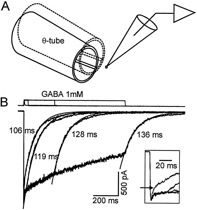
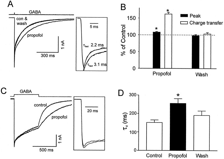
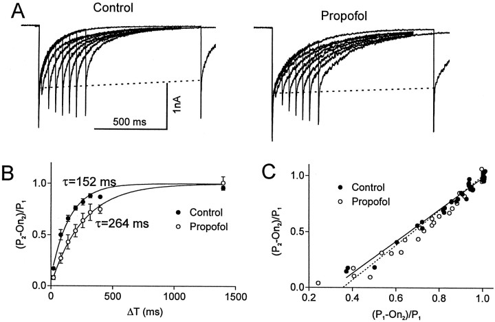
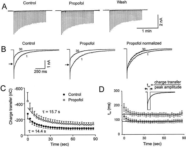
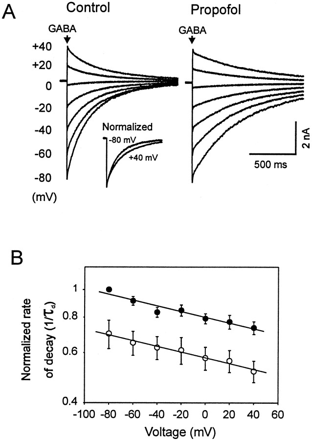
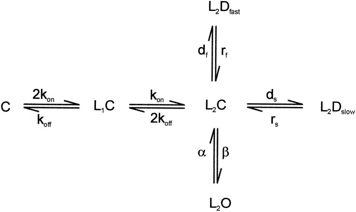
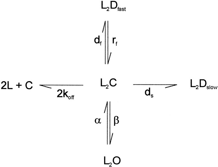
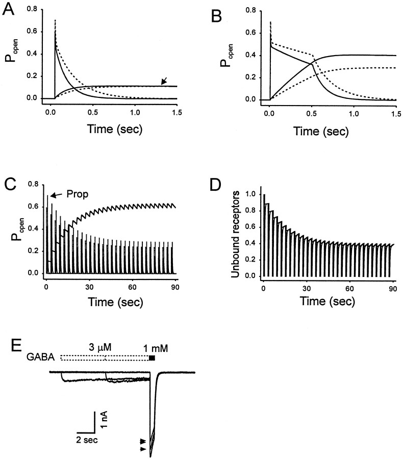
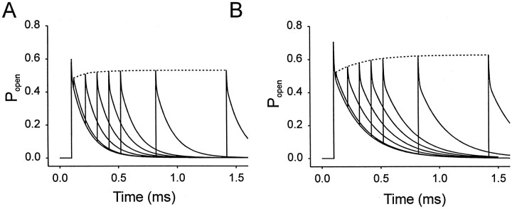
Propofol (2,6-di-isopropylphenol) has multiple actions on GABA(A) receptor function that act in concert to potentiate GABA-evoked currents. To understand how propofol influences inhibitory IPSCs, we examined the effects of propofol on responses to brief applications of saturating concentrations of GABA (1-30 mM). GABA was applied using a fast perfusion system to nucleated patches excised from hippocampal neurons. In this preparation, propofol (10 microM) had no detectable agonist effect but slowed the decay, increased the charge transfer (62%), and enhanced the peak amplitude (8%) of currents induced by brief pulses (3 msec) of GABA. Longer pulses (500 msec) of GABA induced responses that desensitized with fast (tau(f) = 1.5-4.5 msec) and slow (tau(s) = 1-3 sec) components and, after the removal of GABA, deactivated exponentially (tau(d) = 151 msec). Propofol prolonged this deactivation (tau(d) = 255 msec) and reduced the development of both fast and slow desensitization. Recovery from fast desensitization, assessed using pairs of brief pulses of GABA, paralleled the time course of deactivation, indicating that fast desensitization traps GABA on the receptor. With repetitive applications of pulses of GABA (0.33 Hz), the charge transfer per pulse declined exponentially (tau approximately 15 sec) to a steady-state value equal to approximately 40% of the initial response. Despite the increased charge transfer per pulse with propofol, the time course of the decline was unchanged. These experimental data were interpreted using computer simulations and a kinetic model that assumed fast and slow desensitization, as well as channel opening developed in parallel from a pre-open state. Our results suggest that propofol stabilizes the doubly liganded pre-open state without affecting the isomerization rate constants to and from the open state. Also, the rate constants for agonist dissociation and entry into the fast and slow desensitization states were reduced by propofol. The recovery rate constant from fast desensitization was slowed, whereas that from slow desensitization appeared to be unchanged. Taken together, the effects of propofol on GABA(A) receptors enhance channel opening, particularly under conditions that promote desensitization.


Figures

Fig. 1.
Fig. 1.
Experimental setup and GABA-induced currents recorded from nucleated patch excised from cultured hippocampal neuron. A, The drawing illustrates the rapid solution switching system. The theta tubing with control and GABA-containing solutions flowing through the barrels is rapidly moved in front of the nucleated patch. A rapid step-like change in the concentration of GABA (within 2 msec) occurs as the interface of the solutions is moved across the patch. B, The bottom traces represent superimposed currents recorded from the same nucleated patch. GABA (1 mm) was applied for the various time intervals indicated by the top traces. A brief pulse of GABA (1 mm, 3 msec) induced a transient inward current that increased rapidly, then decayed with a fast and a slow time course (as indicated by thearrow in the inset). The slower component declined monoexponentially with a time constant of 106 msec (smooth line). Longer applications of GABA (30, 300, 1000 msec) induced currents with a similar rising phase and peak amplitude. However, in the continued presence of GABA, currents desensitized with two distinct components that we refer to as fast and slow desensitization (see inset). After the removal of GABA, the responses declined to baseline (defined as deactivation). The major component of deactivation was adequately described by a single exponential function, and the smooth lines superimposed on the experimental data represent an exponential function fit to the data. The time constants are shown. A small slow component is evident but is ignored in our analysis. The inset provides a temporal expansion of the onset and initial decay of the currents activated by brief and longer pulses of GABA. The linesabove the recordings indicate the duration of the agonist application. The transient upward deflection illustrates the junctional current measured after the membrane patch was disrupted.
Fig. 2.
Fig. 2.
Propofol prolonged the deactivation and increased the peak amplitude of GABA-induced current. A, Three superimposed responses to brief pulses of GABA (1 mm) were recorded from the same patch in the absence and presence of 10 μm propofol. Each trace represents the average of two to three individual traces. Propofol increased the peak amplitude and charge transfer associated with the responses, an effect that was reversed after the washout of propofol. The top trace shows the open tip junctional current. The inset illustrates the slowing of the fast decay by propofol. The time constant of the fast component of a biexponential function was increased by propofol from 2.2 to 3.1 msec (solid lines). B, The bar graph summarizes the effects of propofol on the peak amplitude and the charge transfer of currents recorded from 17 patches. Values were normalized to those obtained under control conditions. A consistent increase in peak current amplitude (8 ± 2%, n = 17, p < 0.01; filled bar) and in charge transfer (62 ± 5%, n = 17, p < 0.01; open bar) was observed. C, Superimposed traces of currents activated by longer (500 msec) pulses of GABA (1 mm) in the absence and presence of propofol (10 μm) are shown. Note that the predominant effect of propofol is to prolong the deactivation. Similar to the results obtained for brief pulses of GABA, the peak current amplitude was increased (shown in inset). A temporal expansion of the currents is shown in the inset. Propofol slows the initial decay of the response. D, Thebar graph illustrates that the time constant of deactivation (τd) was reversibly increased by propofol.
Fig. 3.
Fig. 3.
Recovery from fast desensitization was slowed by propofol. A, Superimposed currents evoked by paired pulses of GABA (3 mm, 3 msec) are illustrated. Desensitization produced by the conditioning pulse was investigated by applying a second test pulse. The test pulse produced negligible additional current when the time interval between the pulses was 20 msec. As the interval between the pulses increased, the amplitude of the test response gradually increased to a level that was similar but not quite equal to that of the conditioning pulse. The reduction in the amplitude of the test response was primarily caused by loss of the fast component that occurred within the first 10 msec. The dashed lines indicate that the slow component of the decay is relatively stable. The time course of recovery from desensitization was slowed by propofol (10 μm). B, The ratio of the amplitude of test pulse and the conditioning pulse [(P2 − On2)/P1)] is plotted versus the time interval between the two pulses (Δt). To control for rundown of the response during the recordings, the amplitude of the conditioning response was normalized to the initial value of P1. The data pointsrepresent the average values for currents recorded from three difference patches in the absence (filled circle) and presence of propofol (open circles). The time course of the recovery was best fit by a single exponential function (τcontrol = 152 msec, τpropofol = 264 msec). C, The relationship between the fraction of receptor population that has recovered from desensitization (P2 − On2)/P1 and the extent of deactivation (P1 − On2)/P1 is shown. Note that the plots can be superimposed in the absence and presence of propofol, suggesting that deactivation parallels desensitization and propofol does not influence this relationship.
Fig. 4.
Fig. 4.
Propofol reduces slow desensitization. A, Current traces illustrate the response to repetitive applications of brief (3 msec) pulses of GABA (30 pulses administered at a rate of 1 per 3 sec). Under control conditions, the peak amplitude of the current gradually declined to a steady-state level. Propofol reversibly increased the duration of each response and also increased the amplitude of each of the 30 responses to a similar extent. B, A temporal expansion of currents evoked by the 1st and 30th application of GABA is shown. Responses are superimposed, and thearrows indicate the peak amplitude of the 30th response. Note that in the absence or presence of propofol, the decay of the 30th response was accelerated compared with the initial response. This effect is further illustrated in the panel to theright where the peak amplitude is normalized to the maximum response. C, The charge transfer associated with each response during the repetitive applications of GABA is shown. Data points are the average values obtained from seven different patches. After the first two pulses, in the absence of propofol (filled circle), the charge transfer declined monoexponentially with a time constant of 14.4 sec to a steady-state value 39% of the initial response. In the presence of propofol (open circle), the time constant was 15.7 sec, and the steady-state value was 38% of the initial value. The first two data points were not included in the exponential fit. Propofol significantly increased the charge transfer of all 30 pulses to a similar extent (two-way ANOVA, p < 0.01). D, The decline in ton (charge transfer/amplitude) for the 30 pulses activated by GABA is shown. Propofol increased ton of GABA-evoked currents (two-way ANOVA, p < 0.01). The straight line plotted through the last 28 data points represents a linear regression with the slope restricted to zero. The intercept of this line for ton was 90 and 142 msec for currents recorded in the absence and presence of propofol, respectively. Theinset illustrates the calculation of ton.
Fig. 5.
Fig. 5.
Slowing of deactivation by propofol is voltage-independent. A, Currents evoked by GABA (3 mm, 3 msec) at various holding potentials are superimposed. The rate of decay (1/τd) was increased at hyperpolarizing potentials. The inset illustrates the superimposed currents obtained at holding potentials of +40 mV (inverted) and −80 mV normalized to the peak amplitude for responses. The decay was increased by propofol at all holding potentials. B, Data obtained from five different patches were averaged and plotted versus 1/τd (log scale). A linear relationship was observed between 1/τd and holding potential (voltage) under control conditions (filled circle) and in the presence of propofol (open circles). Propofol produced a similar decrease in the rate of current decay at all holding potentials as indicated by the slope of the lines (n = 5, two-way ANOVA, p < 0.01).
Fig. FS1.
Fig. FS1.
Fig. FS2.
Fig. FS2.
Fig. 6.
Fig. 6.
Simulations of the effects of propofol on GABA-induced current and predesensitization of GABA currents. A, Simulations of GABAAR-mediated activity after the application of brief pulses of GABA (3 mm, 3 msec). Popen represents fraction of channels in open state. The solid declining line depicts the rapid decrease in open probability. Superimposed is a plot of the probability of slow desensitization (L2Dslow) in the absence (solid line) or presence (dashed line) of propofol. The slower buildup of the L2Dslow state under control conditions is indicated by the solid inclining line(arrow). Propofol causes an increase in the open probability and a small decrease in slow desensitization as indicated by thedotted lines. B, Simulations of the longer (500 msec) pulses of GABA is shown. Again, superimposed is a plot of L2Dslow. Note the increase in open probability and slower buildup of L2Dslow in the presence of propofol (dashed lines). Propofol (10 μm) increased the probability of channel opening and reduced slow desensitization. C, Simulation of the application of 30 brief pulses of GABA at 30 pulses administered at a rate of 1 per 3 sec in the absence and presence of propofol. The buildup of the L2Dslow state was extensive in the absence and presence of propofol. D, Simulation of the same experiment as in C only now monitoring the level of unbound receptors (C). E, Experimental data illustrate that the preapplication of 3 μm GABA decreased the amplitude of current evoked by a saturating concentration of GABA (1 mm), but this effect is far from equilibrium even with longer applications. Increasing the duration of the preapplication from 5 sec (arrow) to 10 sec (double arrows) doubled the effect from an 8% decrease to a 17% decrease. However, our model parameters predict that at equilibrium, 3 μm GABA will reduce the test response by 16 and 24% in the absence and presence of propofol, respectively. Thus, a 10 sec predesensitization period will underestimate the affinity of the slow desensitized state for GABA because the IC50 for predesensitization is 3 μm at equilibrium.
Fig. 7.
Fig. 7.
Simulation of the recovery from desensitization. Using our proposed model, we simulated the currents activated by paired pulses of GABA. GABA (3 mm for 3 msec) was applied at intervals of 20, 120, 220, 320, 420, 720, 1320, and 1920 msec. Thedotted line represents the fit of a monoexponential equation to the peak amplitude of the second pulse. B, Simulated currents for responses activated in the presence of propofol (10 μm).

References

    1. Bai D, MacDonald JF, Orser BA. Midazolam and propofol modulation of tonic GABAergic current and transient IPSCs in cultured hippocampal neurons. Soc Neurosci Abstr. 1998;24:1832.
    1. Berger T, Schwarz C, Kraushaar U, Monyer H. Dentate gyrus basket cell GABAA receptors are blocked by Zn2+ via changes of their desensitization kinetics: an in situ patch-clamp and single-cell PCR study. J Neurosci. 1998;18:2437–2448. - PMC - PubMed
    1. Brickley SG, Cull-Candy SG, Farrant M. Development of a tonic form of synaptic inhibition in rat cerebellar granule cells resulting from persistent activation of GABAA receptors. J Physiol (Lond) 1996;497:753–759. - PMC - PubMed
    1. Brickley SG, Cull-Candy SG, Farrant M. Single-channel properties of synaptic and extrasynaptic GABAA receptors suggest differential targeting of receptor subtypes. J Neurosci. 1999;19:2960–2973. - PMC - PubMed
    1. Celentano JJ, Wong RK. Multiphasic desensitization of the GABAA receptor in outside-out patches. Biophys J. 1994;66:1039–1050. - PMC - PubMed

Publication types

LinkOut - more resources