. 1999 Dec 15;19(24):10767-77.
doi: 10.1523/JNEUROSCI.19-24-10767.1999.

Compromised glutamate transport in human glioma cells: reduction-mislocalization of sodium-dependent glutamate transporters and enhanced activity of cystine-glutamate exchange

Affiliations

Compromised glutamate transport in human glioma cells: reduction-mislocalization of sodium-dependent glutamate transporters and enhanced activity of cystine-glutamate exchange

Z C Ye et al. J Neurosci. .

Abstract

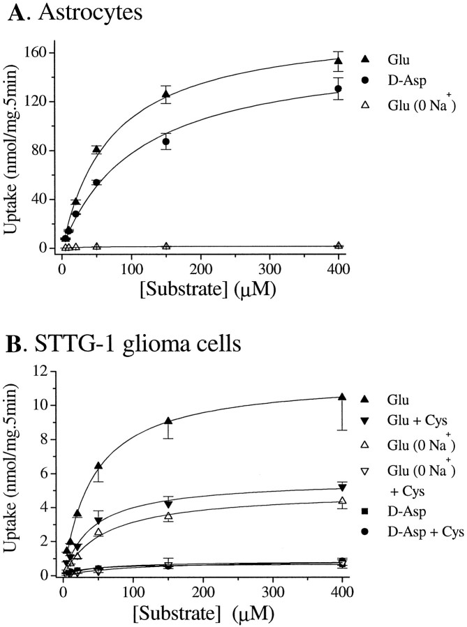
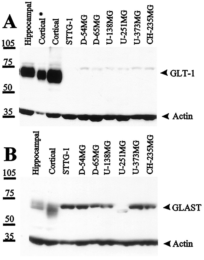
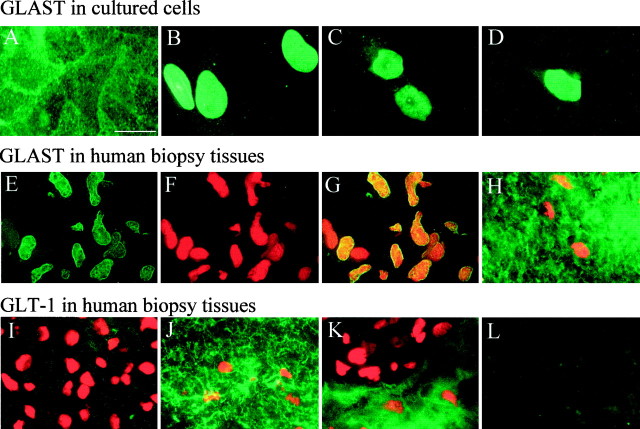
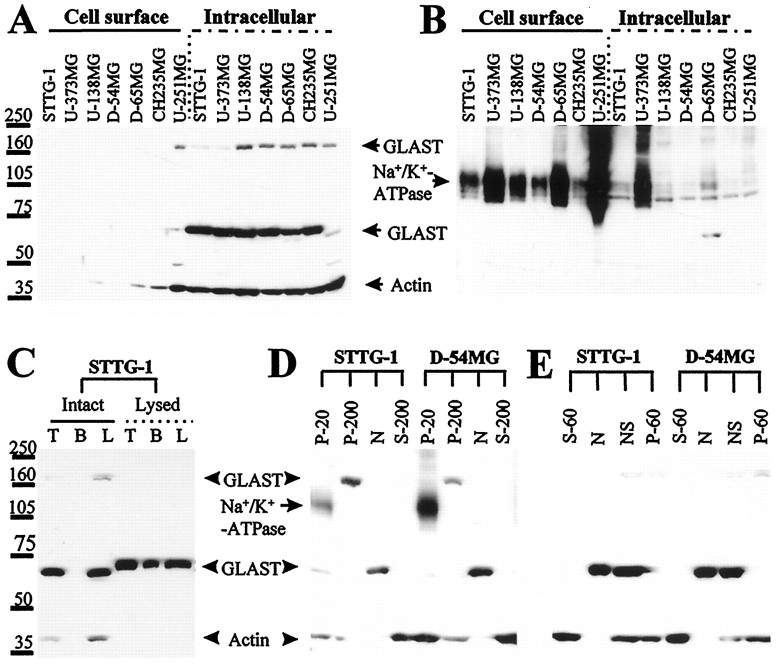
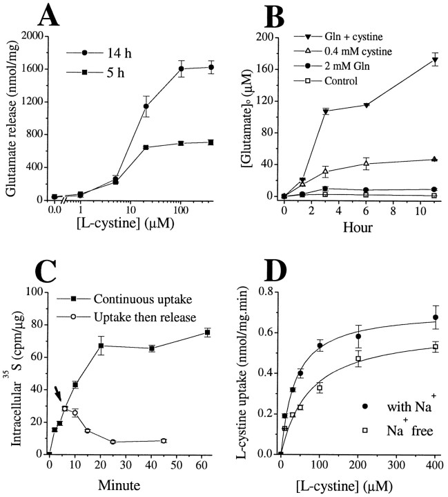
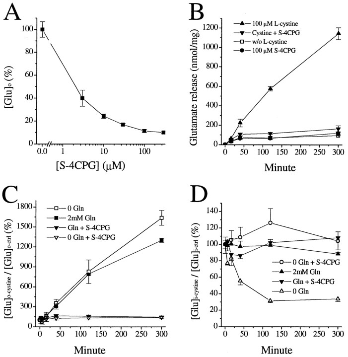
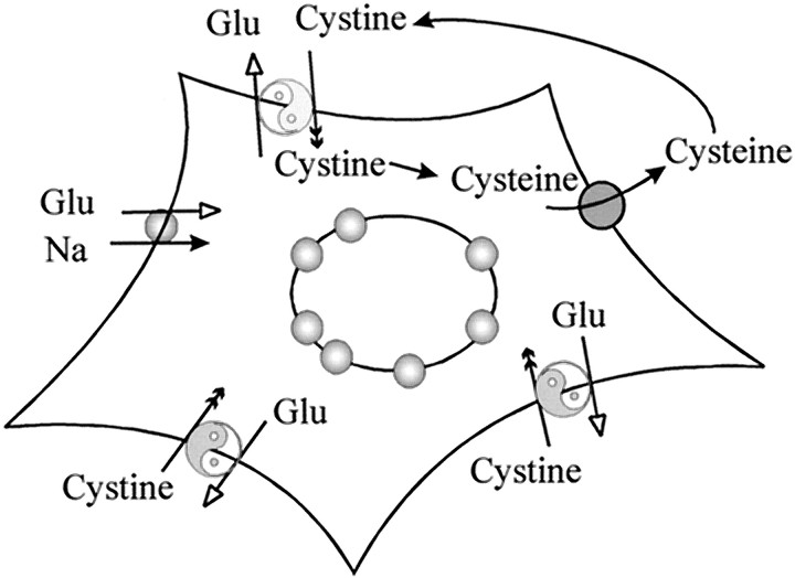
Elevated levels of extracellular glutamate ([Glu](o)) can induce seizures and cause excitotoxic neuronal cell death. This is normally prevented by astrocytic glutamate uptake. Neoplastic transformation of human astrocytes causes malignant gliomas, which are often associated with seizures and neuronal necrosis. Here, we show that Na(+)-dependent glutamate uptake in glioma cell lines derived from human tumors (STTG-1, D-54MG, D-65MG, U-373MG, U-251MG, U-138MG, and CH-235MG) is up to 100-fold lower than in astrocytes. Immunohistochemistry and subcellular fractionation show very low expression levels of the astrocytic glutamate transporter GLT-1 but normal expression levels of another glial glutamate transporter, GLAST. However, in glioma cells, essentially all GLAST protein was found in cell nuclei rather than the plasma membrane. Similarly, brain tissues from glioblastoma patients also display reduction of GLT-1 and mislocalization of GLAST. In glioma cell lines, over 50% of glutamate transport was Na(+)-independent and mediated by a cystine-glutamate exchanger (system x(c)(-)). Extracellular L-cystine dose-dependently induced glutamate release from glioma cells. Glutamate release was enhanced by extracellular glutamine and inhibited by (S)-4-carboxyphenylglycine, which blocked cystine-glutamate exchange. These data suggest that the unusual release of glutamate from glioma cells is caused by reduction-mislocalization of Na(+)-dependent glutamate transporters in conjunction with upregulation of cystine-glutamate exchange. The resulting glutamate release from glioma cells may contribute to tumor-associated necrosis and possibly to seizures in peritumoral brain tissue.


Figures

Fig. 1.
Fig. 1.
Na+-dependent and Na+-independent glutamate transport in astrocytes and glioma cells. A, Astrocytes transport glutamate andd-aspartate at similar rates, and this transport is primarily dependent on the presence of Na+.B, STTG-1 glioma cells transport glutamate at much lower rates than astrocytes and are only partially depend on Na+. l-Cystine (100 μm) reduced glutamate transport by ∼50% in the presence of Na+ but almost completely blocked Na+-independent glutamate uptake. STTG-1 cells transport d-aspartate much less efficiently than glutamate, and this transport is insensitive to l-cystine. Data are means ± SE; n = 4–6.
Fig. 2.
Fig. 2.
Differential expression of glutamate transporters GLT-1 and GLAST in cultured rat hippocampal and cortical astrocytes and seven human glioma cell lines; 30 μg (Cortical*, 10 μg) of protein of each whole-cell lysate were used and probed for the expression of GLT-1 and GLAST. Blots were also probed with anti-actin as loading control. Rat astrocytes (12 d in vitro) expressed abundant GLT-1, whereas human glioma cells virtually lacked GLT-1, except for a small amount of protein appearing as faint a band that was ∼10 kDa bigger than rat GLT-1. Most glioma cell lines expressed comparable amounts of GLAST as rat astrocytes, except U-251MG expressed less amount of GLAST that appeared ∼5 kDa smaller.
Fig. 3.
Fig. 3.
Immunohistochemical localization of glutamate transporters in cultured cells and human brain tissues.A, GLAST staining of cultured rat hippocampal astrocytes shows abundant staining in all aspects of the cell with more intense labeling at cell–cell junctions. B–D, Representative examples of GLAST expression in STTG-1 (B), D54-MG (C), and D65-MG (D) cells show intensive GLAST staining in the cell nuclei with little staining on cell processes. E–L, Immunohistochemical staining of biopsy sections. E–G show a representative GBM section stained for GLAST (E), propidium iodide (F), and superimposition of these to show colocalization in cell nuclei (G).H shows double staining of GLAST and propidium iodide in uninvolved brain tissue from the same patient (note that the density of nuclei is much lower than in GBM tissues). I,K, GBM tumor tissues double-stained for GLT-1 and propidium iodide. Areas with high nuclear densities exhibited no GLT-1 immunoreactivity, whereas comparison tissue from the same patient shows strong immunoreactivity for GLT-1 (J). Occasionally, tumor tissue showed areas that appeared to be tumor margins in which the area with low density of nuclei stained prominently for GLT-1 (presumably normal brain), but areas with high densities of nuclei (presumably tumor) lack GLT-1 staining (K). L, Secondary antibody and reagent control. Section adjacent to (J) stained with FITC-conjugated goat anti-rabbit only. Scale bar: A–L, 20 μm.
Fig. 4.
Fig. 4.
Localization of GLAST in human glioma cell lines.A, Cell surface proteins were separated from intracellular proteins through biotinylation, followed by separation with a biotin–avidin interaction. No cell surface expression of GLAST was detected in these cells, whereas GLAST remained at the intracellular fraction. B, Blot of A was stripped and reprobed for the cell surface protein Na+/K+-ATPase. It appeared that the majority of Na+/K+-ATPase was located in the biotinylated cell surface fraction. C, GLAST in osmotically lysed STTG-1 cells but not in intact ones was accessible for biotinylation. Biotinylation increases the apparent molecular weight by ∼5 kDa. T, Total lysate;B, biotinylated fraction; L, total lysate minus biotinylated proteins. D, Subcellular fractionation of glioma cells; the blot was probed with GLAST plus actin, subsequently stripped, and probed with Na+/K+-ATPase. Then, the two staining patterns were superimposed for comparison. GLAST monomers were found in the nuclear fraction (N), and the multimers were found in the P-200 fraction. The P-20 fraction that contained the cell surface marker Na+/K+-ATPase had a small amount of GLAST, which was likely caused by the presence of endoplasmic reticulum in the P-20 fraction. E, Subcellular fractionation of glioma cells using digitonin. Only the N and NS part that were pelleted by centrifugation at 500 × gcontained significant amounts of GLAST. Fraction N and NS were further separated by centrifugation against a 30% sucrose solution. Pellet N contained purified nuclei, and the supernatant NS consisted of nuclei that were associated with actin and potentially some other membrane structures.
Fig. 5.
Fig. 5.
Cystine–glutamate exchange mediates glutamate release and cystine uptake in human glioma cells (STTG-1 cells).A, l-Cystine stimulated glutamate release in a dose- and time-dependent manner, EC50 of ∼15 μm. B, l-Cystine (0.4 mm) gradually increased extracellular glutamate concentrations, and this effect was enhanced by the presence of glutamine (2.0 mm). C, Intracellular35S reading reached a plateau in the continuous presence of 1 μCi/ml 35S-l-cystine and 100 μm unlabeled l-cystine. After 6 min incubation, some cells were switched to 100 μm unlabeledl-cystine alone (arrow), and the intracellular 35S reading gradually declined.D, l-Cystine uptake did not depend on the presence of Na+. Data are means ± SE;n = 4–6.
Fig. 6.
Fig. 6.
Mutual competitive inhibition of glutamate andl-cystine uptake in STTG-1 glioma cells. A, Lineweaver–Burk plot of l-cystine uptake in the presence of glutamate. B, Glutamate competitively inhibitedl-cystine uptake with a Ki of ∼330 μm. C, D,l-Cystine competitively inhibited Na+-independent glutamate transport with a Ki of ∼15 μm. Data are means ± SE;n = 4.
Fig. 7.
Fig. 7.
Inhibition of cystine-induced glutamate release byS-4CPG. A, Dose-dependent reduction of extracellular glutamate levels ([Glu]o) sampled 5 hr after incubating glioma cells in glutamate-depleted culture media, in the presence of various concentration of S-4CPG. B,S-4CPG specifically inhibited cystine-induced [Glu]o elevation but was without effect in the absence ofl-cystine. C,S-4CPG exerted a similar degree of inhibition onl-cystine-induced glutamate release, regardless of the presence of extracellular glutamine. D,S-4CPG inhibited the cystine-induced decline of intracellular glutamate content ([Glu]i), as did 2 mm glutamine. Data are means ± SE;n = 4.
Fig. 8.
Fig. 8.
Glutamate handling by glioma cells. Reduction–mislocalization of Na+-dependent glutamate transporters makes glioma cells incapable of sufficiently removing glutamate from the extracellular space; cystine–glutamate exchangers mediate glutamate efflux and cystine uptake in the presence of extracellular l-cystine. The transported cystine can be reduced to cysteine and released to the extracellular space, where cysteine is likely to be oxidized to cystine and again serves as substrate for cystine–glutamate exchange.

References

    1. Arriza JL, Eliasof S, Kavanaugh MP, Amara SG. Excitatory amino acid transporter 5, a retinal glutamate transporter coupled to a chloride conductance. Proc Natl Acad Sci USA. 1997;94:4155–4160. - PMC - PubMed
    1. Attwell D, Barbour B, Szatkowski M. Nonvesicular release of neurotransmitter. Neuron. 1993;11:401–407. - PubMed
    1. Bannai S, Ishii T. Transport of cystine and cysteine and cell growth in cultured human diploid fibroblasts: effect of glutamate and homocysteate. J Cell Physiol. 1982;112:265–272. - PubMed
    1. Bannai S, Ishii T. A novel function of glutamine in cell culture: utilization of glutamine for the uptake of cystine in human fibroblasts. J Cell Physiol. 1988;137:360–366. - PubMed
    1. Bannai S, Kitamura E. Transport interaction of l-cystine and l-glutamate in human diploid fibroblasts in culture. J Biol Chem. 1980;255:2372–2376. - PubMed

Publication types

MeSH terms