. 1999 Dec 15;19(24):10778-88.
doi: 10.1523/JNEUROSCI.19-24-10778.1999.

The chondroitin sulfate proteoglycans neurocan and phosphacan are expressed by reactive astrocytes in the chronic CNS glial scar

Affiliations

The chondroitin sulfate proteoglycans neurocan and phosphacan are expressed by reactive astrocytes in the chronic CNS glial scar

R J McKeon et al. J Neurosci. .

Abstract

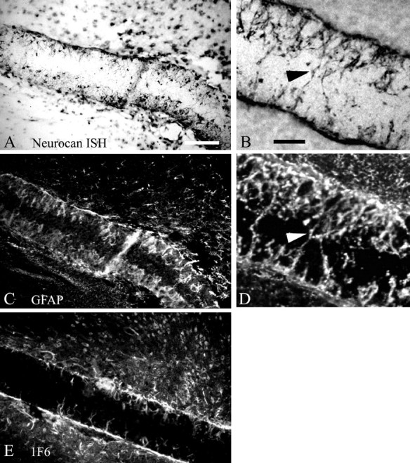
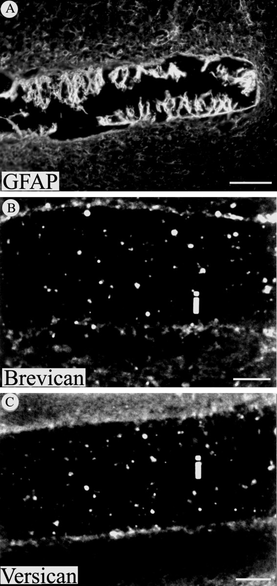
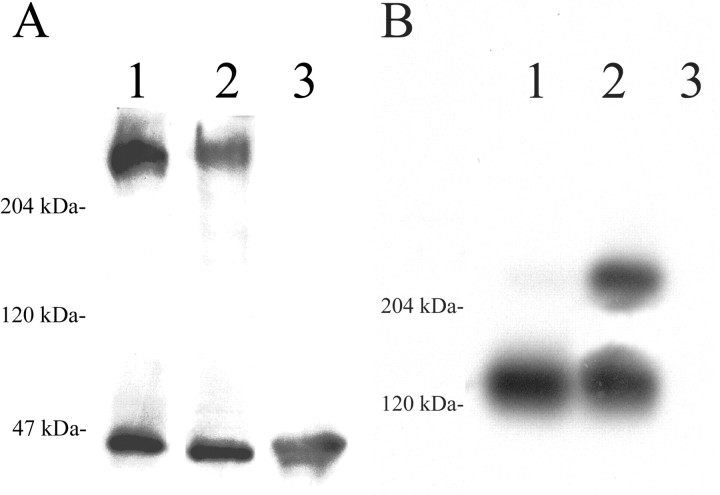
Chondroitin sulfate proteoglycans (CS-PGs) expressed by reactive astrocytes may contribute to the axon growth-inhibitory environment of the injured CNS. The specific potentially inhibitory CS-PGs present in areas of reactive gliosis, however, have yet to be thoroughly examined. In this study, we used immunohistochemistry, combined immunohistochemistry-in situ hybridization, immunoblot analysis, and reverse transcription-PCR to examine the expression of specific CS-PGs by reactive astrocytes in an in vivo model of reactive gliosis: that is, the glial scar, after cortical injury. Neurocan and phosphacan can be localized to reactive astrocytes 30 d after CNS injury, whereas brevican and versican are not expressed in the chronic glial scar. Neurocan is also expressed by astrocytes in primary cell culture. Relative to the amount present in cultured astrocytes or uninjured cortex, neurocan expression increases significantly in the glial scar resulting from cortical injury, including the re-expression of the neonatal isoform of neurocan. In contrast, phosphacan protein levels are decreased in the glial scar compared with the uninjured brain. Because these CS-PGs are capable of inhibiting neurite outgrowth in vitro, our data suggest that phosphacan and neurocan in areas of reactive gliosis may contribute to axonal regenerative failure after CNS injury.


Figures

Fig. 1.
Fig. 1.
Schematic representation of phosphacan and neurocan to indicate location of epitopes and RNA probes used for immunohistochemistry and in situ hybridization.A, The three phosphacan protein isoforms are depicted asopen rectangles. The RPTP1 antibody recognizes a site including the fibronectin type III repeat (F) that is common to the RPTPβ short form (top), RPTPβ long form (middle), and secreted phosphacan (bottom). The 3F8 antibody recognizes a portion of the extracellular domain present in the RPTPβ long form and in secreted phosphacan but not in the RPTPβ short form. CA, Carbonic anhydrase domain; PTP, protein tyrosine phosphatase domains 1 and 2; T, transmembrane domain.Filled circles indicate consensus glycosaminoglycan attachment sites. The unique 3′ untranslated region (3′UTR) of the splice variant encoding phosphacan (phosphacan mRNA, jagged line) was selected for generating specific RT-PCR primers and riboprobes. B, The 1F6 antibody recognizes an N-terminal epitope of neurocan. This epitope is contained in both the full-length 245 kDa neurocan protein (neonatal form) and in a 130 kDa proteolytic fragment that persists in adult animals. A unique 5′ region of the neurocan mRNA (jagged line) was used to generate specific RT-PCR primers and riboprobes. See Table 1 for specific sequences.
Fig. 2.
Fig. 2.
Phosphacan is expressed by reactive astrocytes in gliotic tissue. A, In situ hybridization with a digoxigenin-labeled antisense riboprobe indicates that phosphacan is synthesized by reactive astrocytes surrounding and penetrating the filter implant. The higher magnificationinsets indicate the coincidence of phosphacan mRNA (top) and GFAP (bottom) expression on the same section through the filter implant. The arrowsindicate a phosphacan mRNA and GFAP containing reactive astrocyte cellular process. B, The anti-phosphacan antibody RPTP1 indicates that this CS-PG is expressed in the filter implant.C, To demonstrate the cell type specificity of phosphacan expression in the chronic glial scar, a second anti-phosphacan antibody, 3F8, and anti-GFAP were used for double immunohistochemistry and detected by confocal microscopy. Localization of phosphacan to reactive astrocytes is indicated by the extensive overlap of phosphacan and GFAP staining on cells surrounding the implant (arrowhead) and on processes that project into the implant (top left in all panels inC). Scale bar: A, B, 50 μm; C, 20 μm.
Fig. 3.
Fig. 3.
Localization of neurocan after filter implantation. A, B, A digoxigenin-labeled antisense riboprobe was used to localize neurocan mRNA within filters implanted in different animals. C, D, GFAP immunohistochemistry was performed on the same section as shown inA or B, respectively, to demonstrate coincidence of neurocan mRNA and GFAP expression, indicating that neurocan is expressed by reactive astrocytes. Neurocan protein is also detectable in the filter implant by immunohistochemistry using the 1F6 antibody (E). Arrowheads inB and D indicate reactive astrocytic processes within the implant. Scale bars, 100 μm.
Fig. 4.
Fig. 4.
The CS-PG brevican and versican are not detected in the glial scar 30 d after injury. A, GFAP immunoreactivity is evident around the implanted filter and especially on astrocytic processes that extend into the implant. In contrast, immunohistochemistry for brevican (B) and versican (C) was negative at this same time point on sections from injured cortex containing filter implants (i). Scale bars: A, 100 μm;B, C, 50 μm.
Fig. 5.
Fig. 5.
RT-PCR analysis of gene expression in the glial scar. cDNA prepared from implanted nitrocellulose filters (F), uninjured adult rat cortex (C), and primary astrocyte cultures (A) was subjected to PCR amplification with gene-specific oligonucleotide primer pairs (see Table 1). Similar cDNA amounts were used in this analysis as indicated by the approximately equal amount of GAPDH amplification. Phosphacan mRNA is expressed in the filter implant at levels comparable with uninjured cortex. Neurocan mRNA levels are substantially elevated in the filter implant. The absence of neurofilament amplification in the filter implant supports the contention that this tissue does not include neuronal elements.GAPDH, Glyceraldehyde phosphate dehydrogenase (25 PCR cycles); GFAP, glial fibrillary acidic protein (25 PCR cycles); NCAN, neurocan (27 PCR cycles);PCAN, phosphacan (27 PCR cycles); NF-M, medium neurofilament subunit (29 cycles).
Fig. 6.
Fig. 6.
Immunoblot analysis of CS-PG expression in the glial scar. A, Levels of phosphacan protein in extracts prepared from gliotic tissue retrieved from filter implants (lane 2) are decreased to ∼67% of the levels in age-matched, uninjured cerebral cortex (lane 1), as detected with the 3F8 antibody. In this case, the phosphacan/MAPK values (in densitometric units) were 385,647/228,085 (ratio of 1.6881) and 228,141/202,899 (ratio of 1.1244) for uninjured cortex and filter implant, respectively. B, The 130 kDa proteolytic fragment of neurocan predominates in protein extracts from the cerebral cortex of uninjured age-matched control animals (lane 1), as detected using the 1F6 antibody to neurocan. Alternatively, in gliotic tissue retrieved from filter implants the full-length 245 kDa neonatal form of the neurocan protein is significantly upregulated (lane 2). In this case, the neurocan 245 kDa/neurocan 130 kDa values (see Materials and Methods) were 8454/378662 (ratio of 0.0223) and 284810/355734 (ratio of 0.8006) for uninjured cortex and filter implant, respectively. Neither phosphacan nor neurocan is detected in adult skeletal muscle (A and B,lane 3). ForA and B, 30 μg of protein were loaded in each lane.

Similar articles

Cited by

References

    1. Altschul SF, Madden TL, Schaffer AA, Zhang J, Zhang Z, Miller W, Lipman DJ. Gapped BLAST and PSI-BLAST: a new generation of protein database search programs. Nucleic Acids Res. 1997;25:3389–3402. - PMC - PubMed
    1. Ascher RA, Fidler PS, Rogers JH, Fawcett JW. TGF-β stimulates neurocan synthesis in cultured rat astrocytes. Soc Neurosci Abstr. 1998;24:56.
    1. Bicknese AR, Sheppard AM, O'Leary DM, Pearlman AL. Thalamocortical axons extend along a chondroitin sulfate proteoglycan-enriched pathway coincident with the neocortical subplate and distinct from the efferent path. J Neurosci. 1994;14:3500–3510. - PMC - PubMed
    1. Bovolenta P, Wandosell F, Nieto-Sampedro M. Characterization of a neurite outgrowth inhibitor expressed after CNS injury. Eur J Neurosci. 1993;5:454–465. - PubMed
    1. Bovolenta P, Fernaud-Espinosa I, Méndez-Otero R, Nieto-Sampedro M. Neurite outgrowth inhibitor of gliotic brain tissue. Mode of action and cellular localization, studied with specific monoclonal antibodies. Eur J Neurosci. 1997;9:977–989. - PubMed

Publication types

MeSH terms