. 1999 Dec 15;19(24):10813-28.
doi: 10.1523/JNEUROSCI.19-24-10813.1999.

Gap junctional coupling and patterns of connexin expression among neonatal rat lumbar spinal motor neurons

Affiliations

Gap junctional coupling and patterns of connexin expression among neonatal rat lumbar spinal motor neurons

Q Chang et al. J Neurosci. .

Abstract

Interneuronal gap junctional coupling is a hallmark of neural development whose functional significance is poorly understood. We have characterized the extent of electrical coupling and dye coupling and patterns of gap junction protein expression in lumbar spinal motor neurons of neonatal rats. Intracellular recordings showed that neonatal motor neurons are transiently electrically coupled and that electrical coupling is reversibly abolished by halothane, a gap junction blocker. Iontophoretic injection of Neurobiotin, a low molecular weight compound that passes across most gap junctions, into single motor neurons resulted in clusters of many labeled motor neurons at postnatal day 0 (P0)-P2, and single labeled motor neurons after P7. The compact distribution of dye-labeled motor neurons suggested that, after birth, gap junctional coupling is spatially restricted. RT-PCR, in situ hybridization, and immunostaining showed that motor neurons express five connexins, Cx36, Cx37, Cx40, Cx43, and Cx45, a repertoire distinct from that expressed by other neurons or glia. Although all five connexins are widely expressed among motor neurons in embryonic and neonatal life, Cx36, Cx37, and Cx43 continue to be expressed in many adult motor neurons, and expression of Cx45, and in particular Cx40, decreases after birth. The disappearance of electrical and dye coupling despite the persistent expression of several gap junction proteins suggests that gap junctional communication among motor neurons may be modulated by mechanisms that affect gap junction assembly, permeability, or open state.


Figures
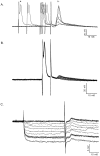
Fig. 1.
Fig. 1.
Characterization of electrical coupling among developing motor neurons. Motor neurons were identified by intracellular impalement in hemisected spinal cord preparations from P0–P8 rats by the presence of an antidromic action potential after ventral root stimulation. Shown are coupling potentials characterized in P0–P2 motor neurons identified by antidromic ventral root stimulation. A, Intracellular current injection was used to elicit an action potential (a.) in the impaled motor neuron during antidromic ventral root stimulation. By adjusting the timing of the intracellular current pulse (a.) relative to antidromic ventral root stimulation, the antidromic action potential (b.) elicited by ventral root stimulation failed, revealing an initial segment spike (c.). As the interval between the intracellular current pulse and antidromic stimulation was decreased further, the initial segment spike also failed. This failure occurs by collision of the antidromic spike by the intracellularly evoked action potential, which transiently inactivates voltage-gated sodium channels. The remaining depolarizing potential (d.) is a putative coupling potential (5.9 mV amplitude, 1.5 msec rise time; 3.9 msec latency from onset of antidromic stimulation; see Table 1 for summary). Calibration: 20 mV, 10 msec.B, Coupling potential amplitude was graded as the intensity of antidromic stimulation was graded. Partial action potentials, which became apparent as collision tests were performed, occurred in an all-or-nothing fashion as antidromic stimulation was graded. Calibration: 20 mV, 10 msec. C, Intracellular hyperpolarization did not affect the amplitude of the coupling potential, in this case detected by straddling threshold for action potential generation. An electrical potential would be insensitive to changes in membrane potential. Coupling potential amplitude = 1.3 mV; amplitude after hyperpolarization to −100 mV, 1.3 mV. Those cells that had coupling potentials as characterized by one or more of these criteria are included in Table 1. Calibration: 5 mV, 10 msec.
Fig. 2.
Fig. 2.
Coupling potentials are abolished by halothane, a gap junction blocker. To determine whether coupling potentials could be reversibly abolished by a gap junction blocker, preparations were superfused with halothane-saturated Ringer's solution after characterization of coupling potentials as shown in Figure 1.A, A collision test was initially used to determine whether a coupling potential was present. Coupling potential, 5.6 mV; average of 10 sweeps, P0 spinal cord. B, After several minutes of exposure to halothane-saturated Ringer's, coupling potentials were abolished. Shown are eight successive sweeps illustrating the decrease in coupling potential amplitude until no potential could be detected. No change in resting membrane potential was observed during perfusion with halothane-saturated Ringer's.C, Washout of halothane with normal Ringer's led to a complete recovery of the coupling potential amplitude, 5.6 mV; average of 20 sweeps 15 min after replacement of halothane with normal Ringer's. Calibration: 10 mV, 10 msec.
Fig. 3.
Fig. 3.
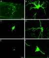
Developing motor neurons are extensively dye coupled around the time of birth. The number of dye-labeled cells was determined 2–4 hr after injection of a single characterized motor neuron with Neurobiotin followed by histology. A, Single plane projection of confocal stack of images from a P2 spinal cord, showing ventral and lateral location of dendrites of injected motor neuron and 10 additional Neurobiotin-labeled cells. Ventral edge of section is shown at top of panel. Scale bar, 25 μm.B, Single plane projection of confocal stack of images of injected motor neuron cell body shown in A (adjacent section), with its proximal dendrites and surrounded by five additional Neurobiotin-labeled cells. A total of 19 labeled cells were in this cluster, spanning five adjacent sections of 20 μm each. Scale bar, 10 μm. C, Single plane projection of confocal stack of images from a P4 spinal cord, showing cell body and proximal dendrites of injected motor neuron. Scale bar, 10 μm. D, Single plane projection of confocal stack of images of an adjacent section, showing one additional Neurobiotin-labeled cell body. Scale bar, 10 μm. E, Single plane projection of confocal stack of images from a P7 spinal cord, showing single cell body and some of the dendritic arbor of the injected motor neuron. Ventral edge of the section is at top of panel. Scale bar, 25 μm.F, Single plane projection of confocal stack of images at higher magnification, confirming that only one Neurobiotin-labeled cell body is present. Scale bar, 10 μm.
Fig. 4.
Fig. 4.
Extent of dye coupling among motor neurons during the first week after birth. The number of cells in each dye-labeled motor neuron cluster is shown plotted against postnatal age in days. Between P0–P2 and P7–P8, there is a sharp decrease in the number of Neurobiotin-labeled motor neurons per cluster.
Fig. 5.
Fig. 5.
Distribution of dye-labeled neurons suggests limited spatial extent of gap junctional coupling after birth. The distribution of dye-labeled motor neurons is shown for each cluster from P0–P2 (A) and P3–P4 (B) spinal cord segments L3, L4, and L5. Each cluster is represented by a different symbol. In P0–P2 spinal cords, each dye-labeled cluster of motor neurons occupied a mean volume of 139 × 130 × 96 μm in the rostral-caudal, dorsal-ventral, and medial-lateral dimensions of the ventral horn, respectively (Table1). The dimensions of individual motor pools at these ages are more than twice as large. Similarly, at P3–P4, each dye-labeled cluster of motor neurons occupied a mean volume of 70 × 70 × 50 μm in the rostral-caudal, dorsal-ventral, and medial-lateral dimensions of the ventral horn, respectively (Table 1). Given that there is relatively little overlap among motor pools in the rat ventral spinal cord, the distribution of dye-labeled motor neurons strongly suggests that, after birth at least, dye coupling is present among motor neurons that innervate the same skeletal muscle. Scale bar, 100 μm.
Fig. 6.
Fig. 6.
RT-PCR analysis of connexins expressed by embryonic motor neurons and in neonatal spinal cord. RT-PCR analysis was performed on motor neuron RNA. PCR products were amplified using primers for each of the 13 known rodent connexins. Bands were typically observed in the motor neuron cDNA lanes, but not RNA lanes, using primers specific for Cx36, Cx37, Cx40, Cx43, and Cx45. Primers specific for Cx36 amplified a 979 bp band in motor neuron and eye cDNA. Primers specific for Cx37, Cx40, Cx43, and Cx45 amplified 422, 308, 292, and 1217 bp bands, respectively, in motor neuron and heart cDNA. In contrast, primers against the other known rodent connexins amplified the predicted size band from tissues known to express that particular connexin (for example, skin, heart, liver, eye, and testis) but failed to amplify the same size band in motor neurons. In each case, PCR products were eluted from gels, cloned, and sequenced to verify their identity. Horizontal lines at leftindicate markers (from top to bottom, 1.2, 0.6, and 0.3 kB).
Fig. 7.
Fig. 7.
Northern and Western blot analyses of connexin-specific reagents. A, Shown are Northern blots of poly(A+) RNA extracted from E15 rat embryos (lanes a, b, e,h, and k), spinal cords of wild-type P1 mice (c, f, and i), or spinal cords of mutant Cx37−/− (d), Cx40−/− (g), and Cx43−/− (j) P1 mice. A rat Cx36 cRNA probe detected a single band of 2.9 kb in E15 rat embryos (a). A rat Cx37 cRNA probe detected a single 1.5 kb band in E15 rat embryos (b) and wild-type P1 mouse spinal cords (c). No band was detected in spinal cords from P1 Cx37−/− mouse (d). A rat Cx40 cRNA probe detected a single 3.4 kb band in E15 rat embryos (e) and wild-type P1 mouse spinal cords (f). No band was detected in spinal cords from P1 Cx40−/− mouse (g). A rat Cx43 cRNA probe detected a single 3 kb band in E15 rat embryos (h) and wild-type P1 mouse spinal cords (i). No band was detected in spinal cords from P1 Cx43−/− mouse (j). A rat Cx45 cRNA probe detected a single 2.2 kb band in E15 rat embryos (k). B, Shown are Western blots of membrane preparations from E15 rat embryos (lanes a,d, and g), spinal cords from wild-type P1 mice (b, e), spinal cords from mutant P1 Cx40−/− (c) and Cx43−/− (f) mice, or HeLa cells transfected with Cx45 cDNA (h) and untransfected HeLa cells (i). The anti-Cx40 antibody recognized a single ∼40 kDa band in E15 rat embryos (a) and wild-type P1 mouse spinal cord (b). No band was detected in P1 Cx40−/− mouse spinal cord (c). The anti-Cx43 antibody recognized a single band of ∼43 kDa in E15 rat embryos (d) and wild-type P1 mouse spinal cords (e). No band was detected in P1 Cx43−/− mouse spinal cords (f). The anti-Cx45 antibody recognized a single band of ∼45 kDa in E15 rat embryos (g) and Cx45-transfected HeLa cells (h). No band was detected in untransfected HeLa cells (i).
Fig. 8.
Fig. 8.
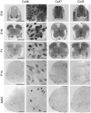
Cx36, Cx37, and Cx43 are expressed by developing and adult motor neurons. Shown are photographs of E15–adult lumbar spinal cord after in situ hybridization for Cx36 (left, left middle), Cx37 (right middle), and Cx43 (right) specific transcripts. Low- and high-power photomicrographs are shown for Cx36 hybridization signal; in left middle column, black arrows indicate examples of the relatively rare motor neurons that appeared negative for Cx36 mRNA. Similar observations were made with Cx37 and Cx43 probes. High-power images are representative fields from ventrolateral spinal cord where motor neurons are located. The temporal and spatial expression patterns of Cx36, Cx37, and Cx43 were similar, in that these connexins were expressed throughout the spinal cord at E15 and in the vast majority of motor neurons from E18 through P14 (see Table 2 for quantification). Cx43, and to a lesser extent Cx37, was expressed in dorsal root ganglia (E15, top row, lateral to spinal cord; data at other ages not shown). Cx36, Cx37, and Cx43 were also expressed in the dorsal horn. Surprisingly, expression was maintained in a substantial proportion of motor neurons in adult animals, despite the lack of functional gap junctional coupling after P3–P4 (also see Table 2). Scale bars: 500 μm (low power Cx36, Cx37, and Cx43); 50 μm (high power Cx36).
Fig. 9.
Fig. 9.
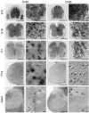
Motor neuron Cx45 and Cx40 expression decrease after birth. Shown are low- and high-power photographs of E15–adult lumbar spinal cord after in situ hybridization for Cx45-specific (left, left middle) and Cx40-specific (right middle,right) transcripts. High-power images are representative fields in ventrolateral spinal cord where motor neurons are located. Black arrows indicate the motor neurons that appeared negative for a particular connexin mRNA. At E15, Cx45 and Cx40 were expressed relatively uniformly throughout the spinal cord, and from E15 to E18, in most if not all motor neurons. Cx45 mRNA was also detected in dorsal root ganglia (left, E15 panel) and in scattered cells in the dorsal horn. After birth, however, the proportion of motor neurons expressing Cx45 mRNA decreased, with only ∼45% of motor neurons remaining positive in P14 and adult rat spinal cord. Cx40 was expressed throughout the spinal cord at E15–P1, including in ventral motor neurons and dorsal root ganglia (right middle, portions shown in E15 panel). Expression decreased after this time, with only ∼10% of motor neurons remaining positive at P14 or in adults. No differences were observed in the proportion of motor neurons positive for Cx45 or Cx40 transcripts in medial, dorsolateral, or ventrolateral motor columns at any of the ages examined. Scale bars: 500 μm (low power Cx45 and Cx40); 50 μm (high power Cx45 and Cx40).
Fig. 10.
Fig. 10.
Connexin protein expression in developing and adult motor neurons. Confocal microscopic examination of immunostained sections revealed punctate membrane and diffuse cytoplasmic staining for Cx40, Cx43, and Cx45. Shown in each panel is a single plane projection of a confocal stack of images. Top row, At E15, punctate Cx40 immunoreactivity was observed surrounding most Islet-1-positive cells (left panel) in the ventral spinal cord. The inset in second panel from left shows the overlay of Islet-1 and Cx40 immunostaining of this field. More diffuse staining within the cytoplasm was also observed. Scale bars, 100 μm. At P1, few motor neurons with punctate membrane staining and diffuse cytoplasmic staining are observed. In adult spinal cord, few motor neurons are immunopositive for Cx40. Shown is one example of an adult motor neuron with characteristic punctate membrane as well as diffuse cytoplasmic staining; this was one of two positive motor neurons in the section.Middle row, At E15, punctate membrane Cx43 immunoreactivity and more diffuse cytoplasmic staining were observed surrounding most motor neurons. Cx43 immunostaining was sensitive to fixation, and this precluded double-labeling with Islet-1. Scale is same as above. At P1, most motor neurons have both punctate membrane staining and diffuse cytoplasmic staining. In adult spinal cord, dense punctate staining was observed in the neuropil surrounding motor neurons; this reflects Cx43 expression in glia (cf. Theriault et al., 1997). Punctate membrane as well as diffuse cytoplasmic staining are also observed in most motor neurons. Scale bar for P1 and adult panels: 50 μm. Bottom row, At E15, punctate membrane as well as diffuse cytoplasmic Cx45 immunoreactivity was observed surrounding most Islet-1-positive cells (left panel) in the ventral spinal cord. Scale bar, 200 μm. The inset insecond panel from left shows Cx45 staining in motor neurons at higher magnification. Scale bar, 50 μm. At P1, most motor neurons have both punctate membrane staining and diffuse cytoplasmic staining. In adult spinal cord, about half of the motor neurons are immunopositive for Cx45. The temporal and spatial expression patterns of Cx40, Cx43, and Cx45 were similar in the medial, dorsolateral, and ventrolateral motor columns and similar to those observed for mRNA.

Similar articles

Cited by

References

    1. Baker R, Llinas R. Electrotonic coupling between neurones in the rat mesencephalic nucleus. J Physiol (Lond) 1971;212:45–63. - PMC - PubMed
    1. Balice-Gordon RJ, Lichtman JW. Long-term synapse loss induced by focal blockade of postsynaptic receptors. Nature. 1994;372:519–524. - PubMed
    1. Balice-Gordon RJ, Thompson WJ. Synaptic rearrangements and alterations in motor unit properties in neonatal rat extensor digitorum longus muscle. J Physiol (Lond) 1988;398:191–210. - PMC - PubMed
    1. Bergoffen J, Scherer SS, Wang S, Scott M, Bone LJ, Paul DL, Chen K, Lensch MW, Chance PF, Fischbeck KH. Connexin mutations in X-linked charcot-marie-tooth disease. Science. 1993;262:2039–2042. - PubMed
    1. Beyer EC, Paul DL, Goodenough DA. Connexin43: a protein from rat heart homologous to a gap junction protein from liver. J Cell Biol. 1987;105:2621–2629. - PMC - PubMed

Publication types

MeSH terms

LinkOut - more resources