. 1999 Dec 15;19(24):10877-85.
doi: 10.1523/JNEUROSCI.19-24-10877.1999.

Graded and areal expression patterns of regulatory genes and cadherins in embryonic neocortex independent of thalamocortical input

Affiliations

Graded and areal expression patterns of regulatory genes and cadherins in embryonic neocortex independent of thalamocortical input

Y Nakagawa et al. J Neurosci. .

Abstract

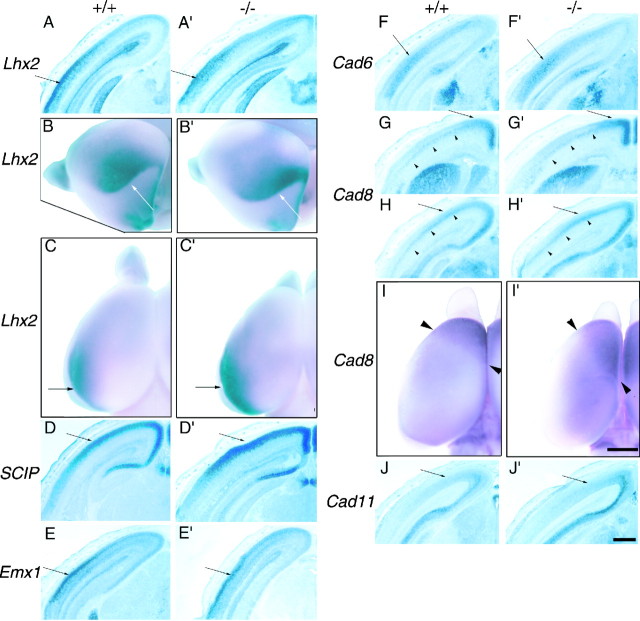
The differentiation of areas of the mammalian neocortex has been hypothesized to be controlled by intrinsic genetic programs and extrinsic influences such as those mediated by thalamocortical afferents (TCAs). To address the interplay between these intrinsic and extrinsic mechanisms in the process of arealization, we have analyzed the requirement of TCAs in establishing or maintaining graded or areal patterns of gene expression in the developing mouse neocortex. We describe the differential expression of Lhx2, SCIP, and Emx1, representatives of three different classes of transcription factors, and the type II classical cadherins Cad6, Cad8, and Cad11, which are expressed in graded or areal patterns, as well as layer-specific patterns, in the cortical plate. The differential expression of Lhx2, SCIP, Emx1, and Cad8 in the cortical plate is not evident until after TCAs reach the cortex, whereas Cad6 and Cad11 show subtle graded patterns of expression before the arrival of TCAs, which later become stronger. We find that these genes exhibit normal-appearing graded or areal expression patterns in Mash-1 mutant mice that fail to develop a TCA projection. These findings show that TCAs are not required for the establishment or maintenance of the graded and areal expression patterns of these genes and strongly suggest that their regulation is intrinsic to the developing neocortex.


Figures

Fig. 1.
Fig. 1.
Graded expression of regulatory genes in the E10.5–E15.5 mouse neocortex. Coronal (A–C, E–K) and sagittal (D) sections of mouse forebrain show the expression of Lhx2(A–D), SCIP(E–G), and Emx1(H–K). A, Lhx2shows a graded pattern in the dorsal telencephalon at E12.5 with higher expression medially than laterally (arrowheads).B, At E14.5, Lhx2 expression is high in the VZ and SVZ and graded with a high-medial-to-low-lateral pattern but is very low in the CP with no obviously graded patterns along the tangential axes. C, D, At E15.5,Lhx2 expression exhibits a dramatic increase in the upper CP of the caudolateral neocortex (arrowheads), strongly graded in high-lateral-to-low-medial (B) and high-caudal-to-low-rostral (C) patterns.E, At E12.5, SCIP expression is detected in the PP of the rostral cortex (arrowheads). SCIP is expressed in a more ventral region, which appears to be the lateral ganglionic eminence (E, arrows). F, G, At E14.5, SCIP expression is limited to the IZ and is graded in a high-lateral-to-low-medial pattern (F;arrowheads) that is still present at E15.5 (G). At E15.5, it is also detected in the upper CP (G; arrowheads), but the caudolateral part of the neocortex, where Lhx2 is highly expressed, shows much weaker expression (G; arrow).H, Emx1 is in a slightly graded pattern as early as E10.5, with higher expression more medially than laterally (arrowheads). I, The same graded expression is detected at E12.5 (arrowheads).J, At E14.5, Emx1 is expressed in the VZ/SVZ and IZ of the neocortex in a slightly graded, high-medial-to-low-lateral pattern, whereas the level of expression in the CP is very low. K, At E15.5, Emx1 is most highly expressed in the upper CP of the caudolateral neocortex (arrowheads). B and J are from adjacent sections, and F is caudal to them.C and K are from adjacent sections, andG is rostral to them. A is slightly caudal to I. In this and all subsequent figures, dorsal is to the top and midline is to the rightin coronal sections, and dorsal is to the top and caudal is to the right in sagittal sections, except forH, which is a coronal section showing both sides of the telencephalon. Scale bar, 100 μm.
Fig. 2.
Fig. 2.
Graded expression of cadherin genes in the E14.5 and E15.5 mouse neocortex. Coronal sections of mouse forebrain show the expression of Cad6 (A–C) andCad8 (D–F), and sagittal sections show that of Cad11 (G, H).A, At E14.5, expression of Cad6 is already graded in a high-lateral-to-low-medial pattern both in the CP (arrowheads) and in the VZ/SVZ (arrows).B, C, At E15.5, this graded expression becomes more pronounced in the CP (B, C; arrowheads), but the expression declines in the VZ/SVZ, especially at rostral levels (B). The medial part of the neocortex expresses a very low level of Cad6 (B, C;arrows). D, Cad8expression is detected only in the intermediate zone at E14.5 and is not graded (arrowheads). E, F, But at E15.5, it is graded in a high-medial-to-low-lateral pattern in the upper CP (arrowheads for high-medial expression). Rostrocaudal differences in expression are not clear for eitherCad6 or Cad8 (data not shown).G, Cad11 expression in the CP is slightly graded in a high-caudal-to-low-rostral pattern at E14.5 (arrowheads). H, At E15.5, this graded expression becomes more evident (arrowheads).A and D as well as C andF are from adjacent sections. E is slightly caudal to B. Scale bar, 100 μm.
Fig. 3.
Fig. 3.
Differential expression of regulatory genes and cadherins in the P2 mouse neocortex. A–L, Coronal sections of mouse forebrain show graded expression and areal patterns (A–G) and layer specificity (H–L) of the expression ofLhx2 (A, H), SCIP(B, I), Emx1 (C, J), Cad6 (D, K),Cad8 (E, F, L; E is rostral to F), and Cad11(G). H′–L′, These panels show 4,6-diamidino-2-phenylindole counterstaining ofH–L, respectively. The boxes inA–D and F show the approximate locations of panels of a higher magnification, H–L. The graded patterns of gene expression that are observed at E15.5 are maintained at P2. A, H, Lhx2 is highly expressed in layers 2/3, 5, and 6 of the caudolateral neocortex, corresponding to the putative auditory area (A; box,H), whereas its expression level is much lower in the more medial, putative visual area (A;singlearrow); expression increases again more medially (A; doublearrows). B, I, SCIPexpression is mainly detected in layers 2/3 and layer 5 (I) and is lowest in the auditory area (B; singlearrow) and much higher in the visual area (B; box)—a graded expression pattern opposite to that of Lhx2. The area indicated by doublearrows (B), where Lhx2 expression is slightly higher than that in the putative visual area, exhibits a lower expression level ofSCIP compared with that in the visual area. C, J, Emx1 is expressed in a tangential pattern similar to that of Lhx2 (C;box for the auditory area with higher expression,singlearrow for the visual area with lower expression, and doublearrows for the area with slightly higher expression) and is higher in layers 2/3, 4, and 6 than in layer 5 (J). D, K, Cad6 expression is graded in a high-lateral-to-low-medial pattern (D) with the highest level in layer 5 and the lower aspect in layers 2/3 (K); the putative auditory area shows a high level of expression (D; box), whereas the visual area shows much lower expression (D;doublearrows) with a different layer specificity (mainly in layer 4). Expression between these areas is at lower levels in all layers (D; singlearrow). E, F, L, Cad8 is more highly expressed medially than laterally in layer 5 (E, F; arrowheads) and, in addition, is expressed in the upper layers of the putative motor (E;arrow) and visual (F; box,L) areas. G, Cad11 is expressed higher in the putative visual (doublearrows) than in the auditory (singlearrow) area, without much layer preference. A–D, F, and G are from adjacent sections. Scale bars:A–G, 1 mm; H–L, H′–L′, 100 μm. iz, Intermediate zone; mz,marginal zone; sp, subplate.
Fig. 4.
Fig. 4.
Graded and/or areal distributions of regulatory genes and cadherins are not altered in the E18.5 Mash-1mutant neocortex. Coronal sections (A, D–H, J, A′, D′–H′, J′) and whole mounts (B, C, I, B′, C′, I′) of wild-type (A–J) and Mash-1 null (A′–J′) mutant forebrains show the comparison ofLhx2 (A–C, A′–C′), SCIP(D, D′), Emx1 (E, E′),Cad6 (F, F′), Cad8(G–I, G′–I′), and Cad11 (J, J′) expression between the two genotypes. TheMash-1 mutant does not show any significant differences from wild type in graded, areal, or laminar cortical expression patterns of the genes examined. The whole-mount panelsshow the left hemispheres; B and B′ are the caudolateral view with the olfactory bulbs on theleft, and C, I, C′, and I′are dorsal views with the midline to the right.A–C, A′–C′, Lhx2 is highly expressed in the caudolateral neocortex in both genotypes (arrows).D–F, J, D′–F′, J′, Tangentially graded patterns ofSCIP, Emx1, Cad6, andCad11 are similar between the wild type and the mutant (arrows show areas with higher expression). G, H, G′, H′, Cad8 expression in layer 5 is graded in a high-medial-to-low-lateral pattern in both genotypes (arrowheads), and expression in the upper layers of the frontal (G, G′; arrows) and occipital (H, H′; arrows) cortex is also unchanged in the mutant. I, I′, The whole-mount pictures show theCad8 expression in the frontal cortex, with thearrowheads showing the caudal boundary of the expression domains. In the Mash-1 mutant, the medial portion of this expression domain extends further caudally than in the wild type. Scale bars: sections, 500 μm; whole mounts, 1 mm.

Similar articles

Cited by

References

    1. Arimatsu Y, Miyamoto M, Nihonmatsu I, Hirata K, Uratani Y, Hatanaka Y, Takiguchi-Hayashi K. Early regional specification for a molecular neuronal phenotype in the rat neocortex. Proc Natl Acad Sci USA. 1992;89:8879–8883. - PMC - PubMed
    1. Barbe MF, Levitt P. The early commitment of fetal neurons to the limbic cortex. J Neurosci. 1991;11:519–533. - PMC - PubMed
    1. Bicknese AR, Sheppard AM, O'Leary DDM, Pearlman AL. Thalamocortical axons extend along a chondroitin sulfate proteoglycan-enriched pathway coincident with the neocortical subplate and distinct from the efferent path. J Neurosci. 1994;14:3500–3510. - PMC - PubMed
    1. Briata P, Di Blas E, Gulisano M, Mallamaci A, Iannone R, Boncinelli E, Corte G. EMX1 homeoprotein is expressed in cell nuclei of the developing cerebral cortex and in the axons of the olfactory sensory neurons. Mech Dev. 1996;57:169–180. - PubMed
    1. Caviness VS., Jr Neocortical histogenesis in normal and reeler mice: a developmental study based upon [3H]thymidine autoradiography. Brain Res. 1982;256:293–302. - PubMed

Publication types

MeSH terms

LinkOut - more resources