. 1999 Dec 15;19(24):10898-907.
doi: 10.1523/JNEUROSCI.19-24-10898.1999.

Cerebral microvascular obstruction by fibrin is associated with upregulation of PAI-1 acutely after onset of focal embolic ischemia in rats

Affiliations

Cerebral microvascular obstruction by fibrin is associated with upregulation of PAI-1 acutely after onset of focal embolic ischemia in rats

Z G Zhang et al. J Neurosci. .

Abstract

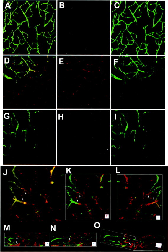
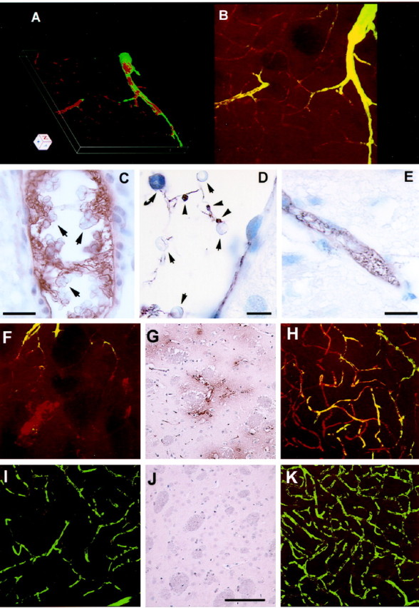
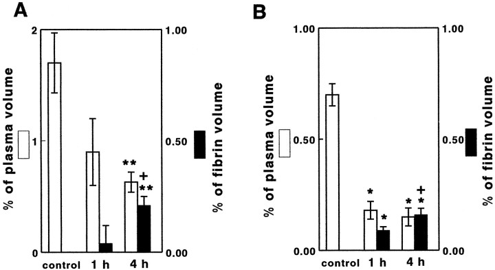
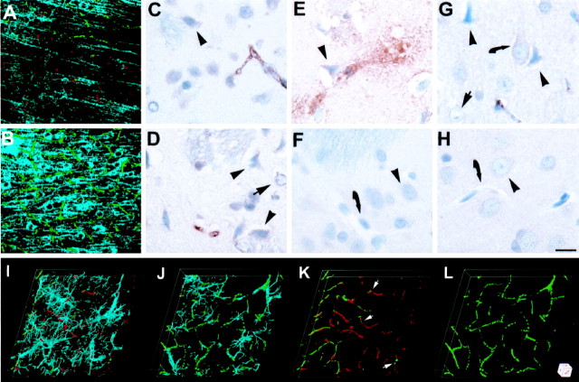
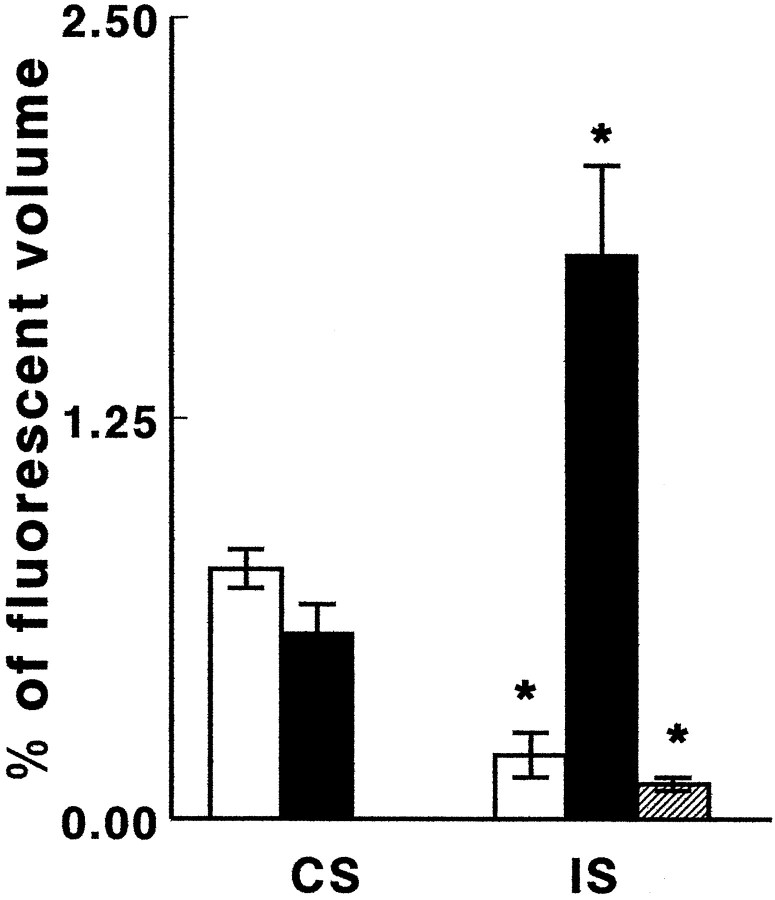
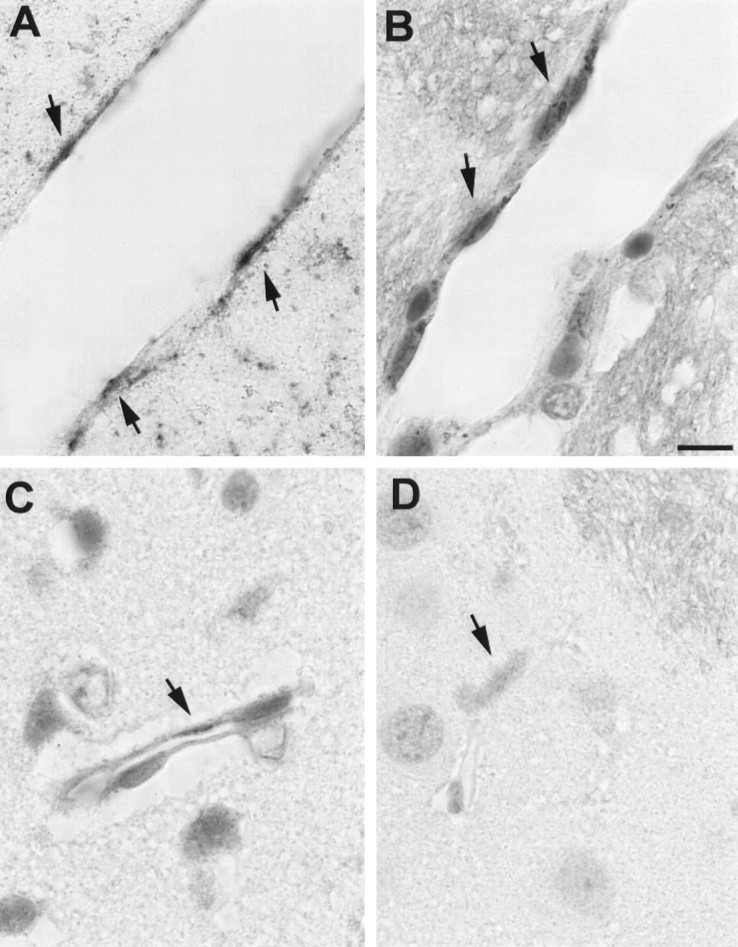
The mechanisms underlying cerebral microvascular perfusion deficit resulting from occlusion of the middle cerebral artery (MCA) require elucidation. We, therefore, tested the hypothesis that intravascular fibrin deposition in situ directly obstructs cerebral microcirculation and that local changes in type 1 plasminogen activator inhibitor (PAI-1) gene expression contribute to intravascular fibrin deposition after embolic MCA occlusion. Using laser-scanning confocal microscopy (LSCM) in combination with immunofluorescent staining, we simultaneously measured in three dimensions the distribution of microvascular plasma perfusion deficit and fibrin(ogen) immunoreactivity in a rat model of focal cerebral embolic ischemia (n = 12). In addition, using in situ hybridization and immunostaining, we analyzed expression of PAI-1 in ischemic brain (n = 13). A significant (p < 0.05) reduction of cerebral microvascular plasma perfusion accompanied a significant (p < 0.05) increase of intravascular and extravascular fibrin deposition in the ischemic lesion. Microvascular plasma perfusion deficit and fibrin deposition expanded concomitantly from the subcortex to the cortex during 1 and 4 hr of embolic MCA occlusion. Three-dimensional analysis revealed that intravascular fibrin deposition directly blocks microvascular plasma perfusion. Vascular plugs contained erythrocytes, polymorphonuclear leukocytes, and platelets enmeshed in fibrin. In situ hybridization demonstrated induction of PAI-1 mRNA in vascular endothelial cells in the ischemic region at 1 hr of ischemia. PAI-1 mRNA significantly increased at 4 hr of ischemia. Immunohistochemical staining showed the same pattern of increased PAI-1 antigen in the endothelial cells. These data demonstrate, for the first time, that progressive intravascular fibrin deposition directly blocks cerebral microvascular plasma perfusion in the ischemic region during acute focal cerebral embolic ischemia, and upregulation of the PAI-1 gene in the ischemic lesion may foster fibrin deposition through suppression of fibrinolysis.


Figures

Fig. 1.
Fig. 1.
Fibrin deposition and cerebral microvascular plasma perfusion from LSCM. Two-dimensional images (x-yprojections, 260.6 × 260.6 μm2) through the stack of 20 optical sections (1 μm/section) of plasma perfusion in capillary networks and fibrin(ogen) immunoreactivity in the ipsilateral caudate putamen from a sham-operated control rat (A–C) and from a rat subjected to 1 hr of embolic MCA occlusion (D–I). Plasma perfusion indicated by intraluminal FITC–dextran showsgreen color, and fibrin immunoreactivity exhibitsred color. A, D, and G are merged images from red (B, E, andH) and green (C, F, and I). The plasma-demarcated capillary networks show a broad array of twist, turns, and junctions in the caudate putamen from a sham-operated rat (A, C). The absence of plasma perfusion (green) in the ipsilateral caudate putamen and increase of fibrin(ogen) immunoreactivity (red) are obvious at 1 hr of embolic MCA occlusion (D–F). Fibrin(ogen) immunoreactivity (red) was not detected when primary antibody was omitted on an adjacent section (G–I). High magnification of three-dimensional reconstructions (J–O, 130.3 × 130.3 × 40 μm3) of plasma perfusion in capillary networks and fibrin(ogen) immunoreactivity from the region in D.J is a merged image before three-dimensional rendering, and K–O are images rendered in three-dimensional space through a stack of 40 optical sections (1 μm/section).K is at x = 0, andy = 0; L is at x= 0, and y = 180; M is atx = 270, and y = 0;N is at x = 270, andy = 90; O is atx = 290, and y = 30. Fibrin deposition (red) directly obstructs plasma perfusion (green), as indicated by arrow andarrowhead.
Fig. 2.
Fig. 2.
Intravascular fibrin deposition, erythrocytes, PMN leukocytes, and platelets from rats subjected to 1 hr of embolic MCA occlusion. A is a three-dimensional reconstructed image (260.6 × 260.6 × 20 μm3) through a stack of 20 optical sections (1 μm/section) of plasma perfusion and fibrin(ogen) immunoreactivity from B, which is a merged image. Intravascular fibrin deposition in a relative large vessel causes the vessel to narrow and decreases plasma perfusion in capillaries (A, B). Erythrocytes (arrows), PMN leukocytes (curved arrow), and platelets (arrowheads) were connected with fibrin within venules (C, D) and capillaries (E) in the ipsilateral caudate putamen from extensively perfused brain tissue. F, H, I, andK are images (x-y projections, 260.6 × 260.6 μm2) through the stack of 20 optical sections (1 μm/section) of plasma perfusion in capillary networks (green) and fibrin(ogen) immunoreactivity (red) in the ipsilateral caudate putamen (F) and in the ipsilateral cortex (H) from a rat subjected to 4 hr of embolic MCA occlusion. Fibrin(ogen) immunoreactivity (red) was present in extravascular space with little plasma perfusion (F, green), and fibrin(ogen) immunoreactivity was not present in the homologous tissue of the contralateral hemisphere (I). Parenchymal fibrin deposition was also observed in the ipsilateral caudate putamen (G) but not the homologous area of the contralateral hemisphere (J) from extensively perfused brain tissue at 4 hr of MCA occlusion. Mixture (H, yellow) of microvascular plasma perfusion (H, green) with intravascular fibrin deposition (H, red) was detected in the ipsilateral parietal cortex (H) compared with the contralateral homologous tissue (K, green only). Scale bars:D, 10 μm; C, E, 20 μm; G, J, 100 μm.
Fig. 3.
Fig. 3.
Bar graphs show volumes of cerebral microvascular plasma (open bars) and fibrin(ogen) immunoreactivity (filled bars) in the cortex (A) and the subcortex (B) at 1 and 4 hr after embolic MCA occlusion. Control = homologous tissue in the contralateral hemisphere. *p < 0.05, significantly different from the control group; **p< 0.01, significantly different from the control group; and +p < 0.01, significantly different from 1 hr group.
Fig. 4.
Fig. 4.
Fibrin deposition and ischemic cell damage.A and B are images (x-yprojections, 260.6 × 260.6 μm2) through the stack of 20 optical sections (1 μm/section) of plasma perfusion in capillary networks (green), fibrin(ogen) immunoreactivity (red), and MAP-2 immunoreactivity (blue) from a rat subjected to 1 hr of MCA occlusion. Increase in fibrin(ogen) immunoreactivity (A, red) and loss of plasma perfusion (A, green) and MAP-2 immunoreactivity (A, blue) on the ipsilateral hemisphere are evident (A) compared with the contralateral hemisphere (B). Dark neurons (C, arrowhead), shrunken neurons (D, arrowheads), and swollen astrocytes (D, arrow) were present in the striatum with intravascular fibrin deposition from extensively perfused brains at 1 (C) and 4 (D) hr of embolic MCA occlusion. Shrunken neurons (arrowhead) with vacuoles were present in the striatum with extravascular fibrin deposition (E) compared with intact neurons (arrowhead) in the contralateral striatum with patent vessels (curved arrow) at 4 hr of embolic MCA occlusion (F). Shrunken neurons (arrowheads), intact neurons (curved arrow), and swollen astrocytes (arrow) were present in the cortex with intravascular fibrin deposition (G) compared with intact neurons (arrowhead) in the contralateral cortex with patent vessels (curved arrow) at 4 hr of ischemia (H). I–L are three-dimensional reconstructions of microvascular plasma perfusion (green), fibrin(ogen) immunoreactivity (red), and GFAP immunoreactivity (blue) in the caudate putamen from a rat subjected to 1 hr of embolic stroke. Enlargement of GFAP-immunoreactive cell bodies and processes (blue) surrounded vessels (green) in the ischemic region (I) compared with the homologous tissue in the contralateral hemisphere (J). Microvascular plasma perfusion (K, green, arrows) was directly blocked by fibrin deposition (K, red, arrows) when GFAP immunoreactivity was removed (K, L). The image size is 260.1 × 260.1 × 20 μm3 for I–K. Scale bar:C–H, 10 μm.
Fig. 5.
Fig. 5.
Bar graph shows volumes of perfused cerebral microvascular plasma (open bars), fibrin(ogen) immunoreactivity (hatched bar), and GFAP immunoreactivity (filled bars) at 1 hr of embolic MCA occlusion. CS, Contralateral striatum;IS, ipsilateral striatum.
Fig. 6.
Fig. 6.
Endothelial cells express PAI-1. PAI-1 mRNA (A, arrows) and PAI-1 antigen (B, C, arrows) were present in the cytoplasm of endothelial cells in venules (A, B) and capillaries (C) in the ipsilateral striatum compared with PAI-1-immunonegative vessels in the contralateral hemisphere (D, arrow) at 4 hr of embolic MCA occlusion. Immunoreactivity of PAI-1 was visualized by diaminobenzidine.

Similar articles

Cited by

References

    1. Adams H. Preliminary safety report of an ongoing dose-escalation trial Abciximab in acute ischemic stroke. Stroke. 1999;30:244.
    1. Bereczki D, Wei L, Otsuka T, Acuff V, Pettigrew K, Patlak C, Fenstermacher J. Hypoxia increases velocity of blood flow through parenchymal microvascular systems in rat brain. J Cereb Blood Flow Metab. 1992;13:475–486. - PubMed
    1. Braaten JV, Handt S, Jerome WG, Kirkpatrick J, Lewis JC, Hantgan RR. Regulation of fibrinolysis by platelet-released plasminogen activator inhibitor 1: light scattering and ultrastructural examination of lysis of a model platelet-fibrin thrombus. Blood. 1993;81:1290–1299. - PubMed
    1. Buchweitz-Milton E, Weiss HR. Perfused microvascular morphometry during middle cerebral artery occlusion. Am J Physiol. 1988;255:H623–H628. - PubMed
    1. Collen D, Lijnen HR. Basic and clinical aspects of fibrinolysis and thrombolysis. Blood. 1991;78:3114–3124. - PubMed

Publication types

MeSH terms

LinkOut - more resources