Comparative Study
. 1999 Dec 15;19(24):10908-22.
doi: 10.1523/JNEUROSCI.19-24-10908.1999.

Corticostriatal projections from rat barrel cortex have an anisotropic organization that correlates with vibrissal whisking behavior

Affiliations
Comparative Study

Corticostriatal projections from rat barrel cortex have an anisotropic organization that correlates with vibrissal whisking behavior

K D Alloway et al. J Neurosci. .

Abstract

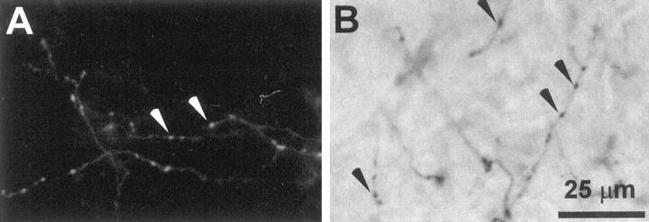
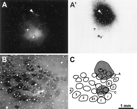
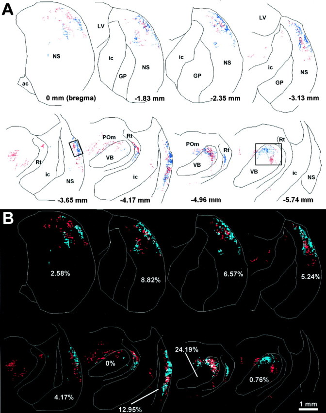
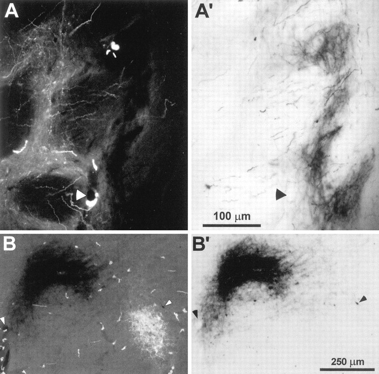
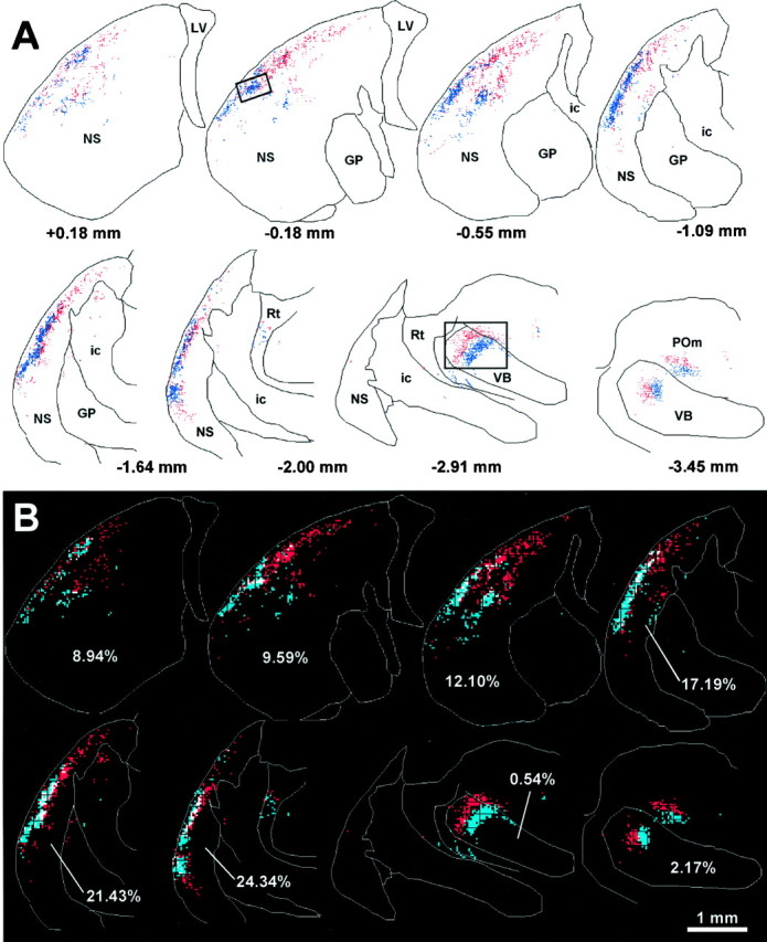
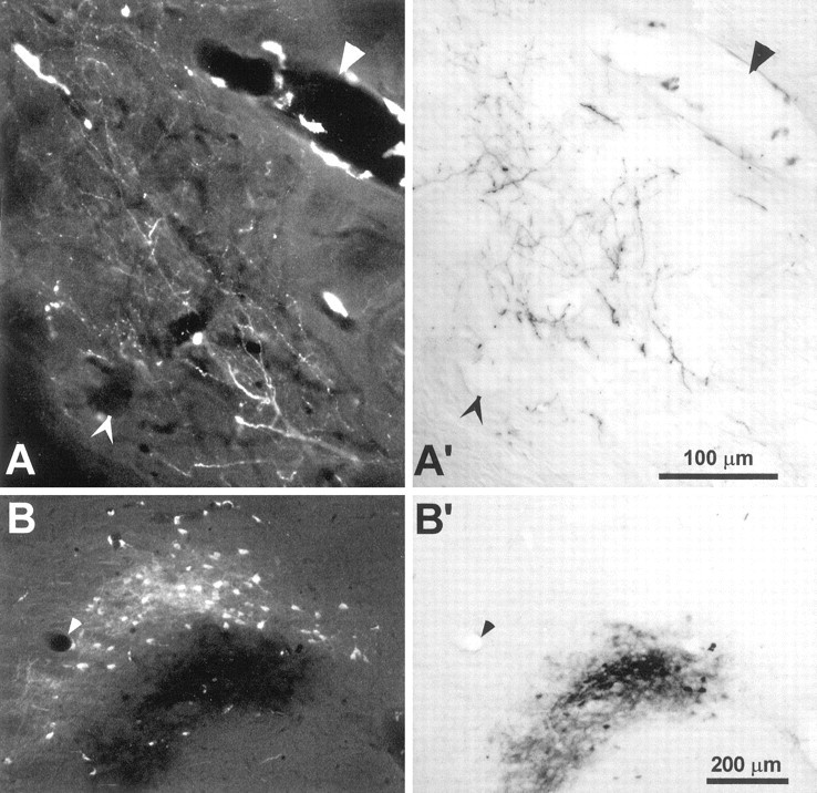
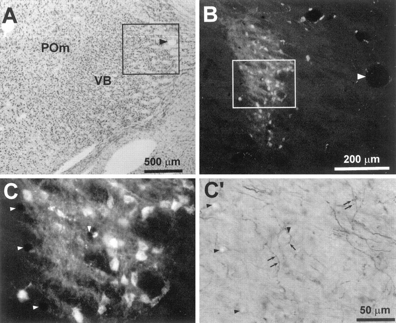
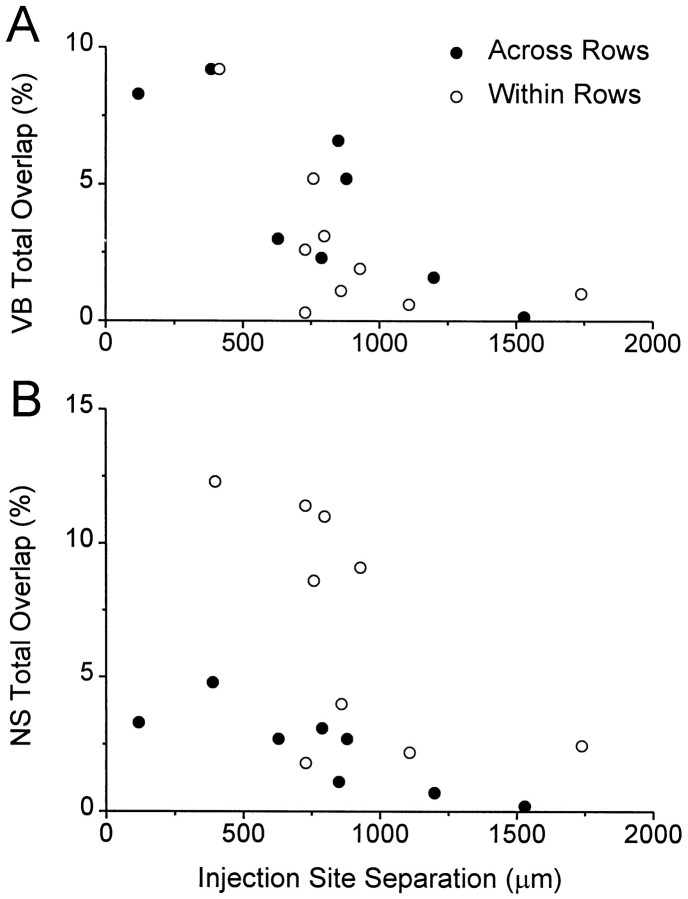
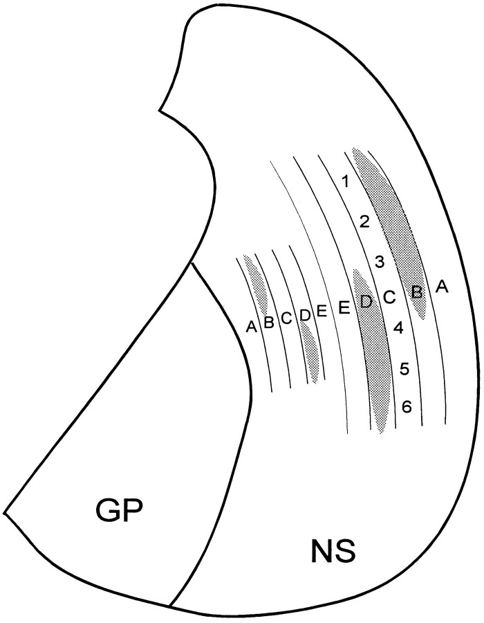
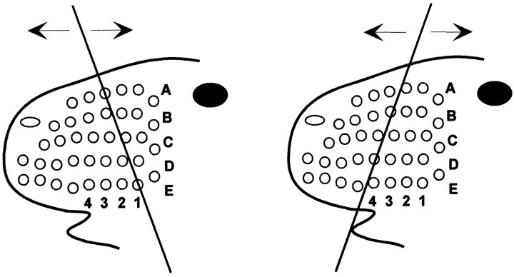
To elucidate the detailed organization of corticostriatal projections from rodent somatosensory cortex, the anterograde tracers biotinylated dextran amine (BDA) and fluoro-ruby (FR) were injected into separate parts of the whisker "barrel" representation. In one group of rats, the two tracers were injected into different barrel columns residing in the same row; in the other group of rats, the tracers were deposited into barrel columns residing in different rows. Reconstructions of labeled axonal varicosities in the neostriatum and ventrobasal thalamus were analyzed quantitatively to compare the extent of overlapping projections to these subcortical structures. For both groups of animals, corticostriatal projections terminated in densely packed clusters that occupied curved lamellar-shaped regions along the dorsolateral edge of the neostriatum. When the tracers were injected into different whisker barrel rows, the distribution of BDA- and FR-labeled terminals in the neostriatum followed a crude somatotopic organization in which the amount of overlap was approximately the same as in the ventrobasal thalamus. When both tracers were injected into the same whisker barrel row, however, the amount of corticostriatal overlap was significantly higher than the amount of overlap observed in the ventrobasal thalamus. These results indicate that corticostriatal projections from whisker barrel cortex have an anisotropic organization that correlates with the pattern of vibrissal movements during whisking behavior.


Figures

Fig. 1.
Fig. 1.
Photomicrographs of isolated branches of corticostriatal terminals labeled by injections of FR or BDA into SI barrel cortex. A, FR-labeled corticostriatal terminals in the neostriatum of rat D45. B, BDA-labeled axons in the neostriatal neuropil of rat D48; some axons appear blurred because they lie outside the focal plane. Arrowheads indicate some of the beaded varicosities appearing along the length of the labeled axons. The scale bar applies to both panels.
Fig. 2.
Fig. 2.
Topography of FR and BDA injections into different rows of SI barrel cortex of rat D11. A, Photomicrograph of a tangential section through layer V of SI cortex reveals the extent of the FR injection. A′. Another view of the same section in A indicates that BDA was injected into a region of SI that did not overlap with the FR injection site.B, A tangential section through layer IV of SI cortex was labeled for CO to indicate the location of individual whisker barrels in the right hemisphere of rat D11. C, Outline drawing of the CO-labeled barrels shown in B.Hatching indicates the relative location of the FR and BDA injections sites shown in A and A′, respectively. This reconstruction indicates that BDA diffused into barrels A2, A3, A4, B2, and B3, whereas FR diffused into barrels C1, C2, D1, and D2. All panels are illustrated at identical scales;arrowheads or filled circles indicate the same set of blood vessels in each panel.
Fig. 3.
Fig. 3.
Topography of labeled terminal varicosities in the neostriatum and thalamus after injections of FR and BDA into different barrel rows of rat D11. A, Reconstructions of selected coronal sections show the relative pattern of corticostriatal projections from the FR and BDA injection sites. In these and subsequent reconstructions, each red or blue dot marks the location of a beaded varicosity labeled with FR or BDA, respectively. Photomicrographs of the region shown in the rectangular insets appear in Figure 4. ac, Anterior commissure; ic, internal capsule; GP, globus pallidus; LV, lateral ventricle;NS, neostriatum; POm, medial posterior nucleus; Rt, nucleus reticularis; VB, ventrobasal complex. B, Overlap analysis of FR- and BDA-labeled varicosities in the neostriatum and thalamus. Each reconstructed plot in A was subdivided into a grid of 35 μm2 bins, and the number of FR- and BDA-labeled varicosities in each bin was counted. Bins containing at least two FR- or BDA-labeled varicosities were colored red orblue, respectively; those containing at least two of each type of labeled varicosity were colored white. Percentages indicate the proportion of white-colored bins in the neostriatum or ventrobasal complex of each section.
Fig. 4.
Fig. 4.
Photomicrographs of FR and BDA labeling in the neostriatum and ventrobasal complex of rat D11. A,FR-labeled corticostriatal terminals enclosed by theinset in Figure 3A (3.65 mm posterior to bregma). A′, Another view of the same section inA reveals BDA-labeled terminals in a neighboring region of neuropil. B, FR and BDA labeling in the nucleus VPM as indicated by the inset in Figure 3A(5.74 mm posterior to bregma). B′, Another view of the same section in B reveals BDA-labeled corticothalamic terminals. Magnification of A and B are the same as A′ and B′, respectively;arrowheads indicate the same blood vessels in each pair of panels.
Fig. 5.
Fig. 5.
Topography of FR and BDA injections into the same rows of SI barrel cortex in rat D30. A, Photomicrograph of a tangential section through layer V of SI cortex reveals the FR injection site. A′, Another view of the same section inA indicates that the BDA injection site did not overlap with the FR injection site. B, A tangential section through layer IV of SI cortex was labeled for CO to indicate the location of individual whisker barrels in the left hemisphere of rat D30. The center of the FR injection site appears in the septal region between two whisker barrels. C, Outline drawing of the CO-labeled barrels shown in B. Hatching indicates the relative location of the FR and BDA injections sites shown inA and A′, respectively. This reconstruction indicated that the injected BDA diffused into barrels D4 and D5, whereas the injected FR diffused into barrels C2 and D2. All panels are illustrated at identical scales; arrowheadsor filled circles indicate the same set of blood vessels in each panel.
Fig. 6.
Fig. 6.
Topography of corticostriatal and corticothalamic projections after injections of FR and BDA into the same whisker barrel rows of rat D30. Reconstructed plots and overlap analysis of selected coronal sections are depicted as in Figure 3. Photomicrographs of the region enclosed by the insets appear in Figure 7.
Fig. 7.
Fig. 7.
Photomicrographs of FR and BDA labeling in the neostriatum and ventrobasal complex of rat D30. A,FR-labeled corticostriatal terminals enclosed by theinset in Figure 6A (0.18 mm posterior to bregma). A′, Another view of the same section in A reveals BDA-labeled terminals in an overlapping part of the neostriatal neuropil. B, FR and BDA labeling in the nucleus VPM (2.91 posterior to bregma).B′, Another view of the same section in Breveals BDA-labeled corticothalamic terminals. Magnification ofA and B are the same as A′and B′, respectively; arrowheads indicate the same blood vessels in each pair of panels. Near some blood vessels in A and B are FR-labeled perivascular cells whose processes were easily discriminated from labeled corticostriatal or corticothalamic terminals.
Fig. 8.
Fig. 8.
Photomicrographs indicating overlapping corticothalamic projections in the ventrobasal complex of rat D30.A, Thionin-stained coronal section (5.02 mm posterior to bregma) indicating the cytoarchitecture of the ventrobasal complex and nucleus POm. The inset encloses the region appearing inB; arrowhead indicates a prominent blood vessel. B, FR-labeled cell bodies and corticothalamic terminals in an adjacent coronal section (5.08 mm posterior to bregma).Arrowhead indicates the same blood vessel indicated inA. C, C′, Microscopic views of BDA- and FR-labeled processes appearing in theinset of B. Arrowsindicate some of the BDA-labeled varicosities that overlapped with FR-labeled processes; arrowheads indicate the same blood vessels appearing in both panels.
Fig. 9.
Fig. 9.
Scatter diagrams illustrating changes in the amount of overlap as a function of the distance separating the injection sites in layer V. A, The amount of overlap in the ventrobasal complex was similar for both the “within row” and “across row” groups. Most of the individual variation was caused by differences in the cortical distances separating the injection sites.B, The amount of overlap in the neostriatum also varied inversely with the distance separating the cortical injection sites, but was noticeably greater among projections originating from barrel columns in the same row of SI cortex.
Fig. 10.
Fig. 10.
Diagram of a coronal section through the neostriatum illustrating the topography of projections from the SI whisker barrel cortex. The lateral neostriatum contains a prominent representation of rows A–E; the medial neostriatum contains a smaller, mirror-image representation of rowsE–A. The relative location of whiskers within each row is represented by numbers 1 (posterior whisker) through6 (anterior) in row C. The gray regions illustrate the divergence of corticostriatal projections by showing areas that would be occupied by labeled terminals after tracer injections in barrel columns B2 and D5.GP, Globus pallidus; NS, neostriatum.
Fig. 11.
Fig. 11.
Schematic diagram illustrating the rostrocaudal back-and-forth vibrissal movements during whisking behavior with respect to an external object at different orientations. Although the relative pattern of whisker stimulation across rows may vary significantly with changes in head orientation or stimulus angle, the temporal sequence of stimulation within a whisker row remains unaffected.

References

    1. Alloway KD, Mutic JJ, Hoover JE. Divergent corticostriatal projections from a single cortical column in the somatosensory cortex of rats. Brain Res. 1998;785:341–346. - PubMed
    1. Apicella P, Scarnati E, Ljungberg T, Schultz W. Neuronal activity in monkey striatum related to the expectation of predictable environmental events. J Neurophysiol. 1992;68:945–960. - PubMed
    1. Armstrong-James M, Millar J. Carbon fibre microelectrodes. J Neurosci Methods. 1978;1:279–287. - PubMed
    1. Bernardo KL, McCasland JS, Woolsey TA. Local axonal trajectories in mouse barrel cortex. Exp Brain Res. 1990a;82:247–253. - PubMed
    1. Bernardo KL, McCasland JS, Woolsey TA, Strominger RN. Local intra- and interlaminar connections in mouse barrel cortex. J Comp Neurol. 1990b;291:231–255. - PubMed

Publication types

LinkOut - more resources