. 1999 Dec 15;19(24):10931-9.
doi: 10.1523/JNEUROSCI.19-24-10931.1999.

Saccadic dysmetria and adaptation after lesions of the cerebellar cortex

Affiliations

Saccadic dysmetria and adaptation after lesions of the cerebellar cortex

S Barash et al. J Neurosci. .

Abstract

We studied the effects of small lesions of the oculomotor vermis of the cerebellar cortex on the ability of monkeys to execute and adapt saccadic eye movements. For saccades in one horizontal direction, the lesions led to an initial gross hypometria and a permanent abolition of the capacity for rapid adaptation. Mean saccade amplitude recovered from the initial hypometria, although variability remained high. A series of hundreds of repetitive saccades in the same direction resulted in gradual decrement of amplitude. Saccades in other directions were less strongly affected by the lesions. We suggest the following. (1) The cerebellar cortex is constantly recalibrating the saccadic system, thus compensating for rapid biomechanical changes such as might be caused by muscle fatigue. (2) A mechanism capable of slow recovery from dysmetria is revealed despite the permanent absence of rapid adaptation.


Figures
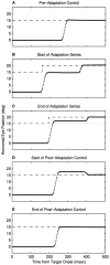
Fig. 1.
Fig. 1.
Examples of trials illustrating saccadic adaptation and extinction. Each panel shows the horizontal eye position and target position during trials from a block designed to study saccadic adaptation. Rightward saccades. A full record of saccade sizes in this block is shown in Figure 2B.
Fig. 2.
Fig. 2.
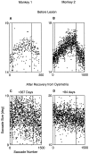
Saccadic adaptation is abolished by the cerebellar lesion. Panels show full records of saccade sizes for block of trials recorded at the days indicated above the panels. All movements are to the right. The two vertical lines in each panel indicate the beginning and end of adaptation trials. The scale of A and Bis stretched twice relative to C andD.
Fig. 3.
Fig. 3.
Lesions of the oculomotor vermis induce gross dysmetria, but with time the dysmetria recovers. Saccades from prelesion period (A, D), early postlesion (day + 3) (B, E), and late postlesion (3 months, 1 year) (C, F). Monkey 1 (A–C), leftward saccades; monkey 2 (D–F), rightwards saccades. Ten saccades per panel. In trials early after the lesion, two and sometimes three saccades are needed to fixate the target (B,E). Whereas the mean saccade size recovers (C, F), the variability in both fixation and saccade size persists (B, C,E, F).
Fig. 4.
Fig. 4.
Histograms of saccade size documentating postlesion hypometria and recovery. A–C, Monkey 1;D–F, monkey 2. A and Drepresent prelesion period; B and E show early postlesion period; C and F show late postlesion period. Saccades in same directions as Figure 1. All appropriate control saccades recorded in this study are included.Thick vertical lines represent means of distributions. Values on each panel show mean ± SD of gain (in percents).
Fig. 5.
Fig. 5.
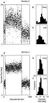
In the absence of saccadic adaptation, prolonged effort induces saccadic errors. A, Record of saccade size in the format of Figure 4; however, instead of adaptation trials, the monkey makes 200 control saccades. Saccades become smaller during the block. B, Histogram of first 100 trials;C, histogram of last 100 trials (1501–1600).D–F, Same format for monkey 3 who has an intact cerebellum. There is no analogous decrease in saccade size.
Fig. 6.
Fig. 6.
Histological sections through the lesion of monkey 1. Sections are 60-μm-thick, Nissl stained. The calibration mark shown in section 1 is valid for all sections. Section numbers in the block are specified. Every 12th section is shown; that is, the distance between displayed sections is 0.72 mm.
Fig. 7.
Fig. 7.
Histological sections through the lesion of monkey 2. Sections are 60-μm-thick, Nissl stained. The scale bar shown insection 2 is valid for all sections. Section numbers in the block are specified. Every 12th section is shown; that is, the distance between displayed sections is 0.72 mm.

References

    1. Albano JE, King WM. Rapid adaptation of saccadic amplitude in humans and monkeys. Invest Ophthalmol Vis Sci. 1989;30:1883–1893. - PubMed
    1. Aschoff JC, Cohen B. Changes in saccadic eye movements produced by cerebellar cortical lesions. Exp Neurol. 1971;32:123–133. - PubMed
    1. Barash S, Melikyan A, Sivakov A, Tauber M. Shift of visual fixation dependent on background illumination. J Neurophysiol. 1998;79:2766–2781. - PubMed
    1. Botzel K, Rottach K, Buettner U. Normal and pathological saccadic dysmetria. Brain. 1993;116:337–353. - PubMed
    1. Buettner U, Fuhry L. Eye movements. Curr Opin Neurol. 1995;8:77–82. - PubMed

Publication types