. 1999 Dec 15;19(24):11049-60.
doi: 10.1523/JNEUROSCI.19-24-11049.1999.

Dopamine terminals in the rat prefrontal cortex synapse on pyramidal cells that project to the nucleus accumbens

Affiliations

Dopamine terminals in the rat prefrontal cortex synapse on pyramidal cells that project to the nucleus accumbens

D B Carr et al. J Neurosci. .

Abstract

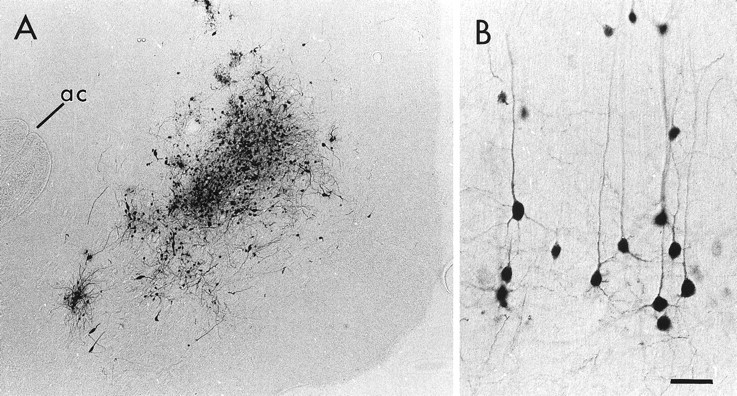
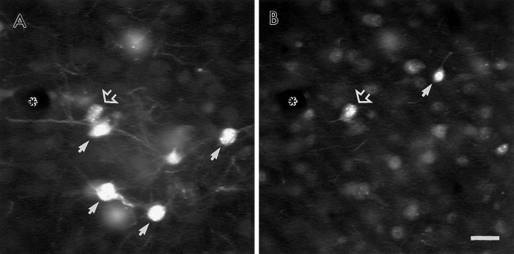
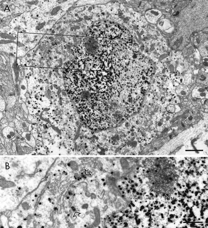
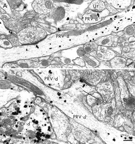
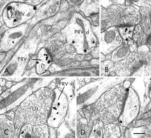
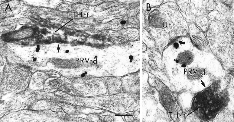
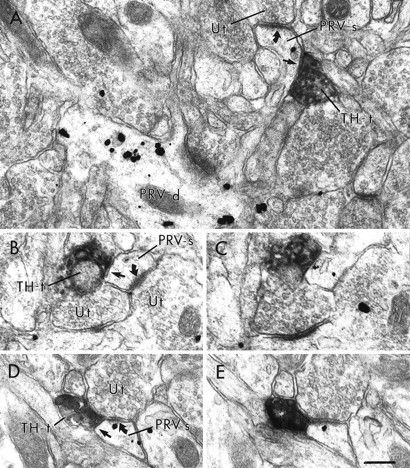
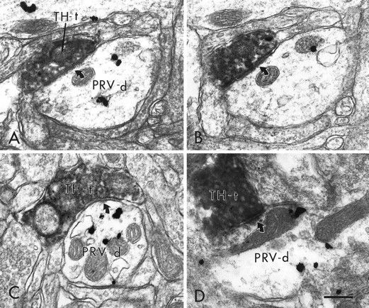
Afferents to the prefrontal cortex (PFC) from dopamine neurons in the ventral tegmental area have been implicated in working memory processes and in the pathogenesis of schizophrenia. Previous anatomical investigations have demonstrated that dopamine terminals synapse on dendritic spines and shafts of pyramidal cells in the PFC. Moreover, neurochemical and physiological studies suggest that dopamine modulates the activity of PFC neurons that project to the nucleus accumbens. However, whether this modulation involves direct synaptic input to cortico-accumbens projection neurons has not been determined. To address this question, retrograde transport of an attenuated strain of pseudorabies virus (PRV) from the nucleus accumbens was combined with immunoperoxidase labeling of tyrosine hydroxylase (TH) to identify dopamine terminals in the PFC. At survival times <48 hr, extensive dendritic distribution of immunogold labeling for PRV was observed in cortico-accumbens neurons. However, evidence consistent with trans-synaptic passage of PRV within this timeframe was observed only rarely. When examined at the electron microscopic level, immunogold labeling for PRV was localized to neuronal somata, proximal and distal dendrites, and dendritic spines. Some of these dendritic processes received symmetric synaptic input from TH-immunoreactive terminals. These data represent the first demonstration of dopamine synaptic contacts onto an identified population of pyramidal cells in the PFC. The findings have important implications for understanding how dopamine modulates cortical outflow to limbic regions in normal brain and pathological states such as schizophrenia.


Figures

Fig. 1.
Fig. 1.
Light micrographs of a representative PRV injection in the NAc (A) and the resulting labeling of pyramidal neurons within the PFC (B). In A, the PRV injection is centered between the anterior commissure (ac) and the medial surface of the hemisphere. In B, many pyramidal neurons and their apical and basilar dendrites are extensively labeled after an injection of PRV into the NAc. Medial is toward the top, and dorsal is toward the left. Scale bar: A, 160 μm; B, 40 μm.
Fig. 2.
Fig. 2.
Fluorescence micrographs of the same coronal section through the PFC showing the distribution of neurons labeled for either PRV (A) or GABA (B) 48 hr after an injection of PRV into the NAc. Within this field, most neurons contain labeling for either PRV or GABA (small arrows), but not both markers. One neuron adjacent to a small blood vessel (asterisk) contains labeling for both markers (open arrow). Note the lighter fluorescence signal and limited dendritic labeling of the dual-labeled neuron compared with other PRV-labeled neurons within the same field. Scale bar, 20 μm.
Fig. 3.
Fig. 3.
Electron micrographs of the rat PFC showing the distribution of immunogold-silver labeling for PRV within the somata of a cortico-accumbens neuron. In A, immunogold-silver particles are distributed throughout the nucleus (Nu) and cytoplasm. The area outlined corresponds to the field shown at higher magnification in B. At this higher magnification, immunogold-silver particles can be seen within the nucleus and in association with the membrane envelope of virions (open arrows) and other membranous organelles. Scale bar:A, 1 μm; B, 0.5 μm.
Fig. 4.
Fig. 4.
Electron micrographs showing dendritic shafts and spines containing gold–silver immunoreactivity for PRV. InA, two PRV-labeled dendrites (PRV-d) receive asymmetric synapses (curved arrows) from unlabeled terminals (Ut). In B, a labeled dendritic spine (PRV-s) emanates from a PRV-labeled dendrite. The labeled spine receives an asymmetric synapse from an unlabeled terminal. Two other PRV-labeled dendrites are also present in this field. Scale bar, 0.25 μm.
Fig. 5.
Fig. 5.
Electron micrographs showing the distribution of PRV immunoreactivity within distal dendrites and dendritic spines within the PFC. In A, viral immunolabeling is present in two small caliber dendritic shafts (PRV-d) and a dendritic spine (PRV-s). The labeled spine receives an asymmetric synapse (curved arrow) from an unlabeled terminal (Ut). In B, an unlabeled terminal forms an asymmetric synapse on a dendritic spine that contains both an immunogold particle and a virion (open arrow). In C and D, a PRV-labeled dendritic spine shown in serial sections contains several immunogold-silver particles and receives an asymmetric synapse from an unlabeled terminal. Scale bar, 0.25 μm.
Fig. 6.
Fig. 6.
Electron micrographs of PRV-labeled dendrites contacted (straight arrows) by TH-immunoreactive terminals (TH-t) within the PFC. In A andB, PRV-labeled dendrites are contacted by TH-labeled terminals without forming distinct synaptic specializations. The PRV-labeled dendrite in B also receives an asymmetric synapse (curved arrow) from an unlabeled terminal (Ut). Scale bar, 0.25 μm.
Fig. 7.
Fig. 7.
Electron micrographs showing TH-labeled terminals within the PFC directly apposed to PRV-labeled dendritic spines without forming distinct synaptic specializations. In A, a PRV-labeled spine (PRV-s) emanates from a dendrite that also contains immunogold labeling for PRV (PRV-d). The labeled spine receives an asymmetric synapse (curved arrow) from an unlabeled terminal (Ut) and is also contacted (straight arrow) by a TH-labeled terminal (TH-t). Serial sections (B, C; D, E) illustrate two PRV-labeled spines that receive asymmetric synaptic contact from unlabeled terminals as well as appositional contact from TH-labeled terminals. In D andE, the TH-labeled terminal is directly apposed to the unlabeled terminal. Scale bar, 0.25 μm.
Fig. 8.
Fig. 8.
Electron micrographs in the PFC showing TH-labeled terminals that form distinct synaptic contacts with PRV-immunoreactive dendrites. In A and B, a TH-labeled terminal (TH-t), shown in serial sections, forms a symmetric synapse (curved arrows) on a PRV-labeled dendrite (PRV-d). C and Dshow two additional examples of PRV-labeled dendrites that receive symmetric synaptic contact (curved arrows) from TH-labeled terminals. Scale bar, 0.25 μm.

Similar articles

Cited by

References

    1. Alexander G, Crutcher M, DeLong M. Basal ganglia-thalamocortical circuits: parallel substrates for motor, oculomotor, “prefrontal”, and “limbic” functions. Prog Brain Res. 1990;85:119–146. - PubMed
    1. Audet MA, Doucet G, Oleskevich S, Descarries L. Quantified regional and laminar distribution of the noradrenaline innervation in the anterior half of adult rat cerebral cortex. J Comp Neurol. 1988;274:307–318. - PubMed
    1. Bartha A. Experimental reduction of virulence of Aujeszky's disease virus. Magy Allatorv Lapja. 1961;16:43–45.
    1. Berger B, Thierry AM, Tassin JP, Moyne MA. Dopaminergic innervation of the rat prefrontal cortex: a fluorescence histochemical study. Brain Res. 1976;106:133–145. - PubMed
    1. Brog J, Salyapongse A, Deutch A, Zahm D. The patterns of afferent innervation of the core and shell in the “accumbens” part of the rat ventral striatum: immunohistochemical detection of retrogradely transported fluoro-gold. J Comp Neurol. 1993;338:255–278. - PubMed

Publication types

MeSH terms

LinkOut - more resources