. 1999 Dec;118(3):451-7.
doi: 10.1046/j.1365-2249.1999.01069.x.

Gammadelta T cells in Behçet's disease (BD) and recurrent aphthous stomatitis (RAS)

Affiliations

Gammadelta T cells in Behçet's disease (BD) and recurrent aphthous stomatitis (RAS)

J Freysdottir et al. Clin Exp Immunol. 1999 Dec.

Abstract

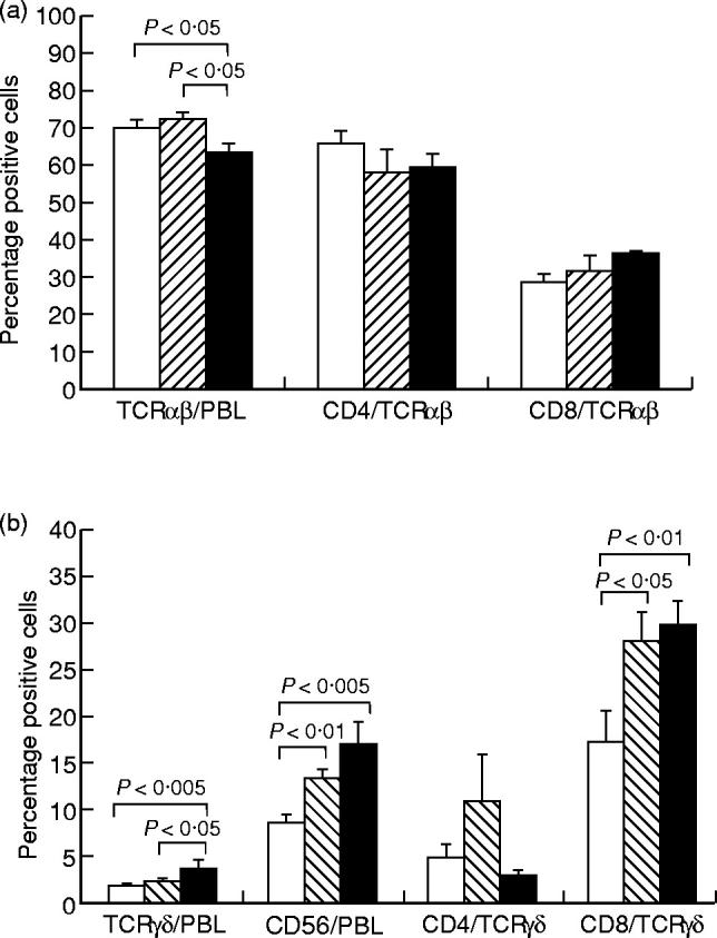
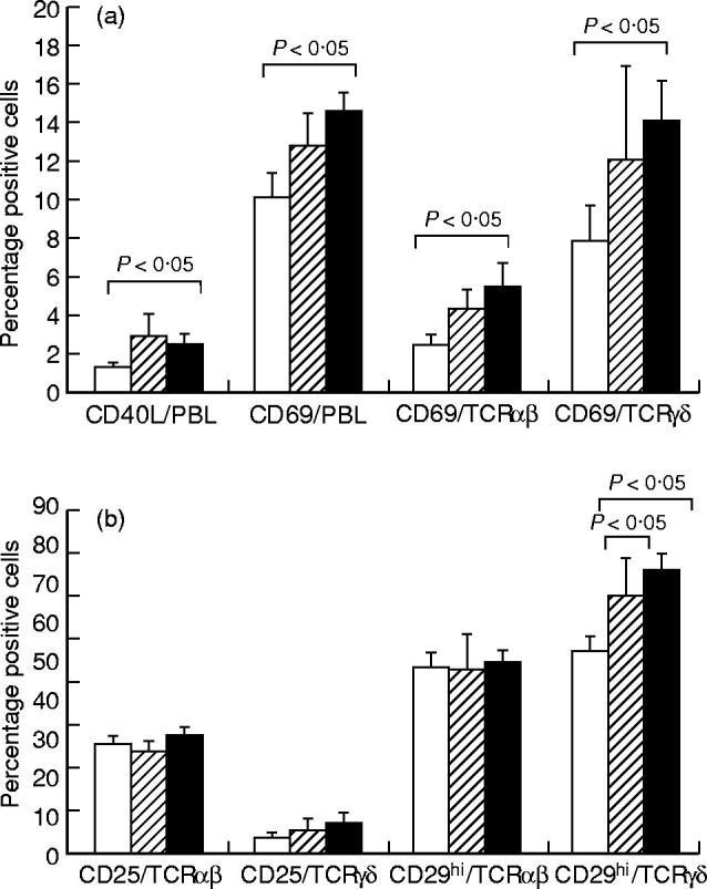
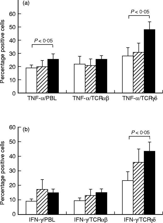
The immunopathogenesis of BD is believed to be T cell-mediated. The objective of this study was to characterize the activation stage and cytokine profile of peripheral blood lymphocytes (PBL), with particular emphasis on gammadelta T cells. Venous blood was collected from 20 patients with BD, and for comparison, from 11 patients with RAS and from 15 healthy controls. Both the expression of activation markers (CD25, CD29, CD40 ligand, CD69 and HLA-DR) on freshly isolated PBL and T cell subsets, and the expression of intracellular cytokines (IL-4, IL-10, interferon-gamma (IFN-gamma) and tumour necrosis factor-alpha (TNF-alpha)) on mitogen-stimulated PBL and T cell subsets were analysed by double immunofluorescent staining and flow cytometry. Significantly decreased proportion of alphabeta T cells and increased proportion of gammadelta T cells, CD56+ cells and CD8+ gammadelta T cells were found in BD patients compared with healthy controls. This was also seen to a lesser extent in patients with RAS. Furthermore, in BD a significantly increased proportion of the gammadelta T cell population expressed CD69 and high levels of CD29 and were induced to produce IFN-gamma and TNF-alpha compared with healthy controls. In contrast, an increased percentage of gammadelta T cells from RAS patients was induced to produce IFN-gamma, but not TNF-alpha. These results indicate that in BD, activated gammadelta T cells, capable of producing IFN-gamma and TNF-alpha, are present in peripheral blood, suggesting that gammadelta T cells are dynamic and may be regulating immunopathogenic events.


Figures

Fig. 1
Fig. 1
Clinical pattern of disease activity of the BD patients. □, Male; ▪, female.
Fig. 2
Fig. 2
The expression of αβ T cell receptor (TCR) on peripheral blood lymphocytes (PBL) and expression of CD4 or CD8 on αβ T cells (a) and the expression of γδ TCR or CD56 on PBL and expression of CD4 or CD8 on γδ T cells (b), analysed by double immunofluorescent staining and flow cytometry. Results are expressed as percentage positive cells, shown as mean ± s.e.m. for 20 BD patients (▪), 11 RAS patients (hatched bars) and 20 healthy controls (□). Statistically significant differences between BD patients and controls, BD patients and RAS patients, and RAS patients and controls are indicated.
Fig. 3
Fig. 3
The expression of CD40 ligand (CD40L) or CD69 on peripheral blood lymphocytes (PBL) and expression of CD69 on αβ T cells or γδ T cells (a) and the expression of CD25 and CD29hi on αβ T cells and γδ T cells (b), analysed by double immunofluorescent staining and flow cytometry. Results are expressed as percentage positive cells, shown as the mean ± s.e.m. for 20 BD patients (▪), 11 RAS patients (hatched bars) and 20 healthy controls (□). Statistically significant differences between BD patients and controls, BD patients and RAS patients, and RAS patients and controls are indicated.
Fig. 4
Fig. 4
The expression of tumour necrosis factor-alpha (TNF-α) on peripheral blood lymphocytes (PBL), αβ T cells or γδ T cells (a) and the expression of IFN-γ on PBL, αβ T cells or γδ T cells (b), analysed by double immunofluorescent staining and flow cytometry. Results are expressed as percentage positive cells, shown as the mean ± s.e.m. for 20 BD patients (▪), 11 RAS patients (hatched bars) and 20 healthy controls (□). Statistically significant differences between BD patients and controls, BD patients and RAS patients, and RAS patients and controls are indicated.

References

    1. Embil JA, Stephens RG, Manuel FR. Prevalence of recurrent herpes labialis and aphthous ulcers among young adults on six continents. Can Med Assoc J. 1975;113:627–30. - PMC - PubMed
    1. Axell T, Henricsson V. The occurrence of recurrent aphthous ulcers in an adult Swedish population. Acta Odontol Scand. 1985;43:121–5. - PubMed
    1. Fortune F, Walker J, Lehner T. The expression of γδ T cell receptor and the prevalence of primed, activated and IgA-bound T cells in Behçet's syndrome. Clin Exp Immunol. 1990;82:326–32. - PMC - PubMed
    1. Valesini G, Pivetti-Pezzi P, Mastrandrea F, et al. Evaluation of T cell subsets in Behçet's syndrome using anti-T cell monoclonal antibodies. Clin Exp Immunol. 1985;60:55–60. - PMC - PubMed
    1. Savage NW, Mahanonda R, Seymour GJ, et al. The proportion of suppressor-inducer T-lymphocytes is reduced in recurrent aphthous stomatitis. J Oral Pathol. 1988;17:293–7. - PubMed

MeSH terms