. 1999 Dec;98(4):510-9.
doi: 10.1046/j.1365-2567.1999.00926.x.

The intercellular adhesion molecule type-1 is required for rapid activation of T helper type 1 lymphocytes that control early acute phase of genital chlamydial infection in mice

Affiliations

The intercellular adhesion molecule type-1 is required for rapid activation of T helper type 1 lymphocytes that control early acute phase of genital chlamydial infection in mice

J U Igietseme et al. Immunology. 1999 Dec.

Abstract

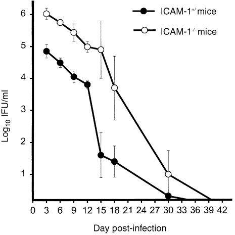
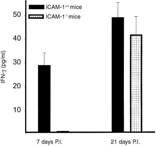
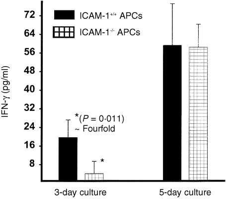
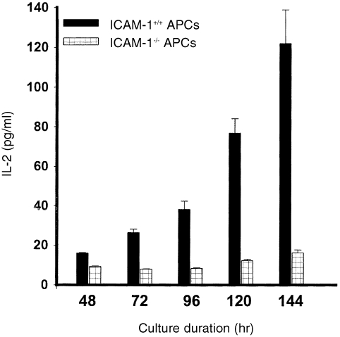
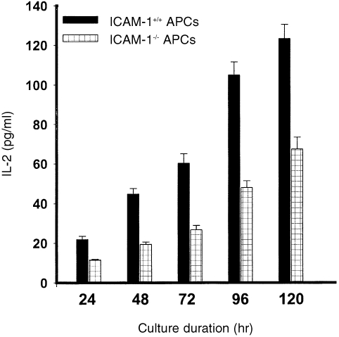
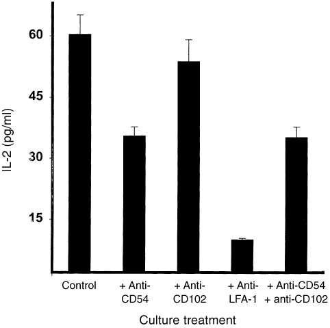
Recent studies in animal models of genital chlamydial disease revealed that early recruitment of dendritic cells and specific T helper type-1 (Th1) cells into the genital mucosae is crucial for reducing the severity of the acute phase of a cervico-vaginal infection and arresting ascending disease. These immune effectors are therefore important for preventing major complications of genital chlamydial infection. Other in vitro studies showed that intercellular adhesion molecule-1 (ICAM-1) plays a role in the antichlamydial action of specific CD4+ and CD8+ T cells. In the present study, we investigated the clinicopathological consequences of ICAM-1 deficiency during chlamydial genital infection in ICAM-1 knockout (ICAM-1KO) mice, and analysed the cellular and molecular immunological bases for any observed pathology or complication. Following a primary genital infection of female ICAM-l-/- and ICAM-1+/+ mice, the intensity of the disease during the first 3 weeks (as assessed by shedding of chlamydiae in the genital tract) was significantly greater in ICAM-1KO mice than in ICAM-1+/+ mice (P < 0.0001), although both ICAM-l-/- and ICAM-1+/+ mice subsequently cleared the primary infection. There was greater ascending disease during the initial stage of the infection, and a higher incidence of tubal disease (hydrosalpinx formation) after multiple infections in ICAM-l-/- mice. Analysis of the cellular and molecular bases for the increased acute and ascending disease in ICAM-l-/- mice revealed that the high affinity of ICAM-1 for leucocyte function antigen type-1 is a property that promotes rapid activation of specific Th1 cells, as well as their early recruitment into the genital mucosa. Moreover, ICAM-1 was more important for naive T-cell activation than primed Th1 cells, although its absence delayed or suppressed immune T-cell activation by at least 50%. Taken together, these results indicated that ICAM-1 is crucial for rapid T-cell activation, early recruitment and control of genitally acquired Chlamydia trachomatis.


Figures

Figure 1
Figure 1
Course of genital chlamydial infection in ICAM‐1–/– and ICAM‐1+/+ mice. Female ICAM‐1–/– and ICAM‐1+/+ mice were infected intravaginally with MoPn. The course of the infection was monitored by periodic (every 3 days) cervico‐vaginal swabbing of individual animal. Chlamydia was isolated from the swabs in tissue culture according to standard methods and inclusions were visualized and enumerated by immunofluorescence. Results are expressed in IFU/ml. Experiments were repeated to give l0 or l2 animals per experimental group.
Figure 2
Figure 2
Recruitment of chlamydia‐reactive Th1 cells after chlamydial genital infection of ICAM‐1–/– and ICAM‐1+/+ mice. Nylon wool‐purified T cells were isolated at the indicated time‐points from genital tract tissues of infected female ICAM‐l–/– and ICAM‐1+/+ mice. The T cells were stimulated with APCs and chlamydial antigen for 5 days and the amounts of IFN‐γ in the culture supernatants were measured by ELISA as described in the Materials and Methods section. The concentrations of IFN‐γ are expressed as the mean (pg/ml) of results from different experiments. Control cultures that contained T cells and APCs but without chlamydial antigen did not show any measurable amounts of IFN‐γ and so the data are not presented in the results shown.
Figure 3
Figure 3
Role of ICAM‐1 in rapid T‐cell activation. Nylon wool‐purified immune splenic T cells (2 × 10⁵/well) were isolated from chlamydia‐infected ICAM‐1+/+ mice and stimulated with 10 µg/ml of chlamydial antigen and l05 cells/well of APCs from either ICAM‐1–/– or ICAM‐1+/+ mice. After 3 or 5 days of incubation the amounts of IFN‐γ in the culture supernatants were measured by ELISA method. The concentrations of IFN‐γ are expressed as the mean (pg/ml) of results from different experiments. Cultures containing T cells and APCs but without chlamydial antigen did not show any measurable amounts of IFN‐γ and so the data are not presented.
Figure 4
Figure 4
Role of ICAM‐1 in the activation of naive T cells. Nylon wool‐purified T cells were isolated from the spleens of naive ICAM‐1+/+ mice. The T cells (2 × l05/well) were stimulated with 10 µg/ml of chlamydial antigen and 2 × l05 cells/well of APCs from either ICAM‐1–/– or ICAM‐1+/+ mice. At the indicated time of incubation the amounts of IL‐2 in the culture supernatants were measured by ELISA as described in the Materials and Methods section. The concentrations of IL‐2 are expressed as the mean (pg/ml) of results from different experiments. Cultures containing T cells and APCs but without chlamydial antigen did not show any measurable amounts of IL‐2 and so the data are not presented.
Figure 5
Figure 5
Role of ICAM‐1 in the activation of immune T cells. Nylon wool‐purified T cells were isolated from the spleens of genitally chlamydial‐infected ICAM‐1+/+ mice. The T cells (2 × l05/well) were stimulated with 10 µg/ml of chlamydial antigen and 2 × l05 cells/well of APCs from either ICAM‐1–/– or ICAM‐1+/+ mice. At the indicated times of incubation the amounts of IL‐2 in the culture supernatants were measured by ELISA as described in the Materials and Methods section. The concentrations of IL‐2 are expressed as the mean (pg/ml) of results from different experiments. Cultures containing T cells and APCs but without chlamydial antigen did not show any measurable amounts of IL‐2 and so the data are not presented.
Figure 6
Figure 6
Contribution of LFA‐1 and its ligands to immune T‐cell activation. Nylon wool‐purified T cells were isolated from the spleens of genitally infected ICAM‐1+/+ mice. The T cells (2 × 10⁵/well) were stimulated with 10 µg/ml of chlamydial antigen, APCs from ICAM‐1+/+ mice, in the presence of neutralizing mAbs directed against LFA‐1, CD54 (ICAM‐1), CD102 (ICAM‐2) or a combination of anti‐CD54 and CD102. After 72 hr of incubation, the amounts of IL‐2 in the culture supernatants were measured by ELISA as described in the Materials and Methods section. The concentrations of IL‐2 are expressed as the mean (pg/ml) of results from different experiments. Cultures containing T cells and APCs but without chlamydial antigen did not show any measurable amounts of IL‐2 and so the data are not presented in the results shown.

Similar articles

Cited by

References

    1. WHO (World Health Organization) Global Prevalence and Incidence of Selected Curable Sexually Transmitted Diseases: Overview and Estimates. Geneva: WHO; 1996.
    1. Schachter J, Grayston JT. Epidemiology of human chlamydial infections. In: Stephens RS, Byrne GI, Christiansen, G, et al., editors. Chlamydial Infections. San Franciso, CA: ICS; p. 3.
    1. D.C. & P.H.S Chlamydia trachomatis Genital Infections – United States, 1995. Morbidity Mortality Weekly Report. 1997;46:193. - PubMed
    1. Paavonen J, Wolner‐hanssen P. Chlamydia trachomatis: a major threat to reproduction. Hum Reprod. 1989;4:111. - PubMed
    1. Scholes D, Stergachis A, Heidrich FE, Andrilla H, Holmes KK, Stamm WE. Prevention of pelvic inflammatory disease by screening for cervical chlamydial infection. N Engl J Med. 1996;334:1362. - PubMed

Publication types

MeSH terms

Substances