. 1999 Dec;98(4):584-9.
doi: 10.1046/j.1365-2567.1999.00898.x.

Some Bence-Jones proteins enter cultured renal tubular cells, reach nuclei and induce cell death

Affiliations

Some Bence-Jones proteins enter cultured renal tubular cells, reach nuclei and induce cell death

K Matsuura et al. Immunology. 1999 Dec.

Abstract

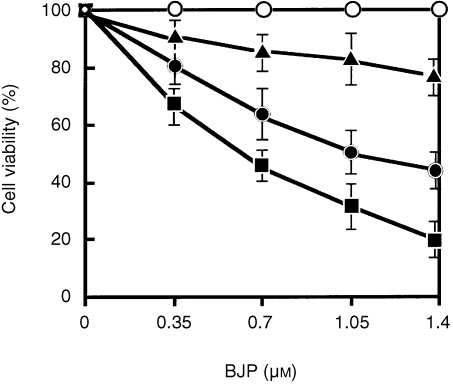
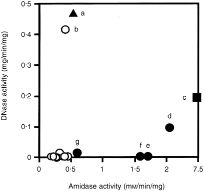
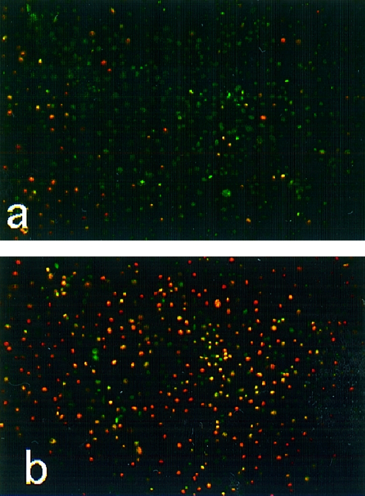
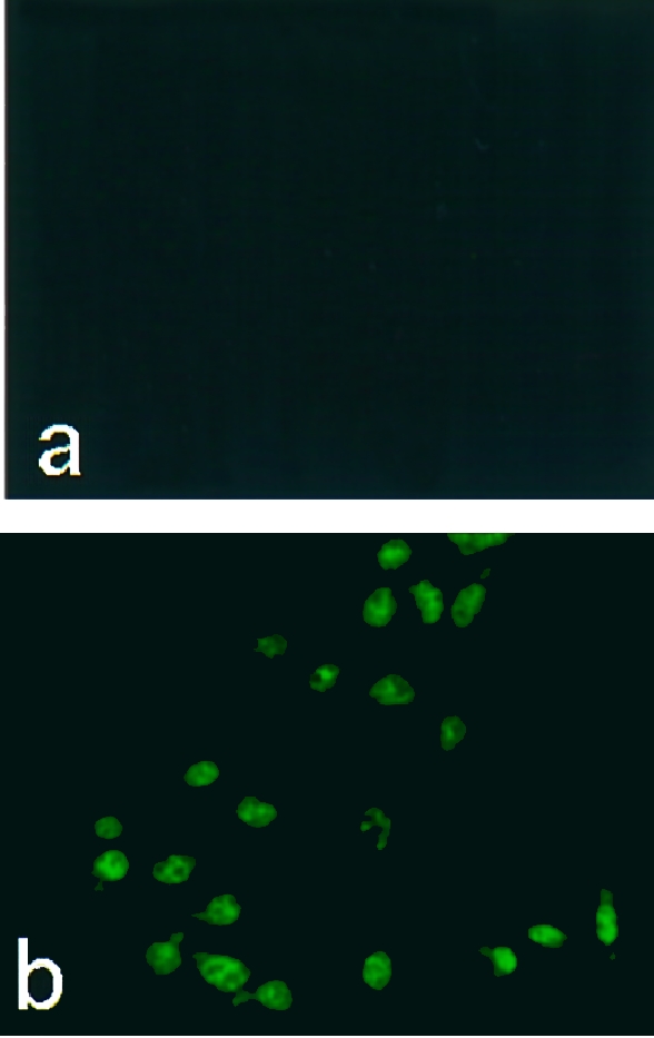
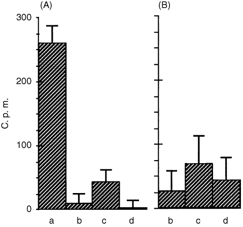
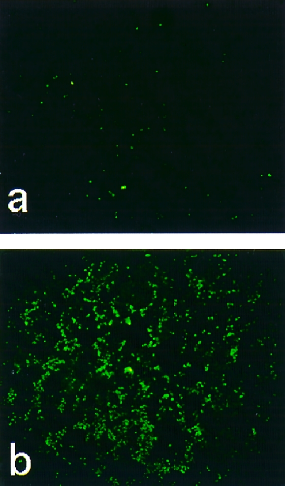
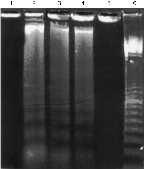
Eighteen monoclonal Bence-Jones proteins (BJPs) were examined for their effects on cultured LLC-PK1 (porcine kidney proximal tubule) cells as well as for their amidase and DNase activities. Five proteins were found to enter the cell and to gain access to the nucleus without degradation of epitopes. Intranuclear BJPs ultimately induced DNA fragmentation and cell death. BJPs with relatively high amidase activity were cytotoxic. On the other hand, three of four BJPs with DNase activity had a cytocidal effect on cultured cells; the remaining BJP, which had a relatively high DNase activity but a very low amidase activity, failed to enter the cell and was not cytotoxic in vitro. These results suggest that catalytic and cytotoxic activities of some BJPs may make a significant contribution, in a substantial proportion of myeloma patients, to the development and/or deterioration of the disease.


Figures

Figure 1
Figure 1
Effects of Bence–Jones proteins (BJPs) on cell viability. LLC‐PK1 cells were preincubated with different concentrations of BJP, and the cell viability was determined as described in the text. Amidase and DNase activities of individual BJPs are shown in Fig. 7, and the same symbols are used for the same BJPs in both figures:▪, patient c in Fig. 7; •, patients d–f in Fig. 7; ▴, patient a in Fig. 7; ○, patient b and all other BJPs that are not indicated by alphabetical characters in Fig. 7. Vertical bars indicate standard deviations of the mean values.
Figure 7
Figure 7
Relationship between amidase, DNase and in vitro cytotoxicity of Bence–Jones proteins (BJPs). Cell viability was determined as described in the text: ≈ 25% (▪), 50% (•), 75% (▴) and 100% (○) viable after 1 hr of incubation with 1 μm of BJP. The symbols correspond to those given in Fig. 1.
Figure 2
Figure 2
Cytocidal effect of Bence–Jones proteins (BJPs). After incubation of LLC‐PK1 cells with 1·0 μm of non‐cytotoxic (a) or cytotoxic (b) BJP for 1 hr as described in the text, the cells were double stained with Hoechst 33342 (greenish blue) and propidium iodide (reddish yellow), essentially as described previously.
Figure 3
Figure 3
Immunofluorescence staining of intracellular Bence–Jones proteins (BJPs). After incubation of LLC‐PK1 cells with 1·0 μm of non‐cytotoxic (a) or cytotoxic (b) BJP, the cells were stained with fluorescein isothiocyanate (FITC)‐labelled anti‐human κ‐chain goat immunoglobulin G (IgG), essentially as described.
Figure 4
Figure 4
Binding of radiolabelled cytotoxic BJP to LLC‐PK1 cells in the absence (a) and presence of 1000‐fold (A) and 10‐fold (B) concentrations of non‐cytotoxic BJP (b), BSA (c) and cytotoxic non‐labelled BJP (d). c.p.m., counts per minute.
Figure 5
Figure 5
Cytochemical detection of DNA fragmentation. After incubation of LLC‐PK1 cells with 1·0 μm of non‐cytotoxic (a) or cytotoxic (b) Bence–Jones proteins (BJPs), the cells were subjected to terminal deoxynucleotidyltransferase‐mediated dUTP‐biotin nick‐end labelling (TUNEL) staining, as described previously.
Figure 6
Figure 6
DNA fragmentation in LL‐CPK1 cells after incubation with Bence–Jones proteins (BJPs). LL‐CPK1 cells were incubated with BJPs and the DNA fragments present in the lysate were detected by the method of Hockenbery et al. Lane 1, 2 μm of bovine serum albumin (BSA); lanes 2–4, 2, 1 and 0·25 μm of cytotoxic BJP, respectively; lane 5, 2 μm of non‐cytotoxic BJP; lane 6, 100‐bp DNA ladder marker (Amersham Pharmacia Biotech, Tokyo, Japan).

References

    1. Paul S. Natural catalytic antibodies. Mol Biotechnol. 1996;5:197. - PubMed
    1. Paul S. Antibody catalysis. In: Paul P, editor. Contemporary Immunology: Autoimmune Diseases. Totowa, NJ: Humana Press; 1998. p. 221.
    1. Paul S, Li L, Kalaga RA, et al. Natural catalytic antibodies: peptide‐hydrolyzing activities of Bence Jones proteins and VL fragment. J Biol Chem. 1995;270:15257. - PubMed
    1. Li L, Paul S, Tyutyulkova S, et al. Catalytic activity of anti‐thyroglobulin antibodies. J Immunol. 1995;154:3328. - PubMed
    1. Shuster AM, Gololobov GV, Kvashuk OA, et al. DNA hydrolyzing autoantibodies. Science. 1992;256:665. - PubMed

MeSH terms