

Genetic differences in endocrine pancreatic tumor subtypes detected by comparative genomic hybridization
- PMID: 10595906
- PMCID: PMC1866934
- DOI: 10.1016/S0002-9440(10)65495-8
Genetic differences in endocrine pancreatic tumor subtypes detected by comparative genomic hybridization
Abstract
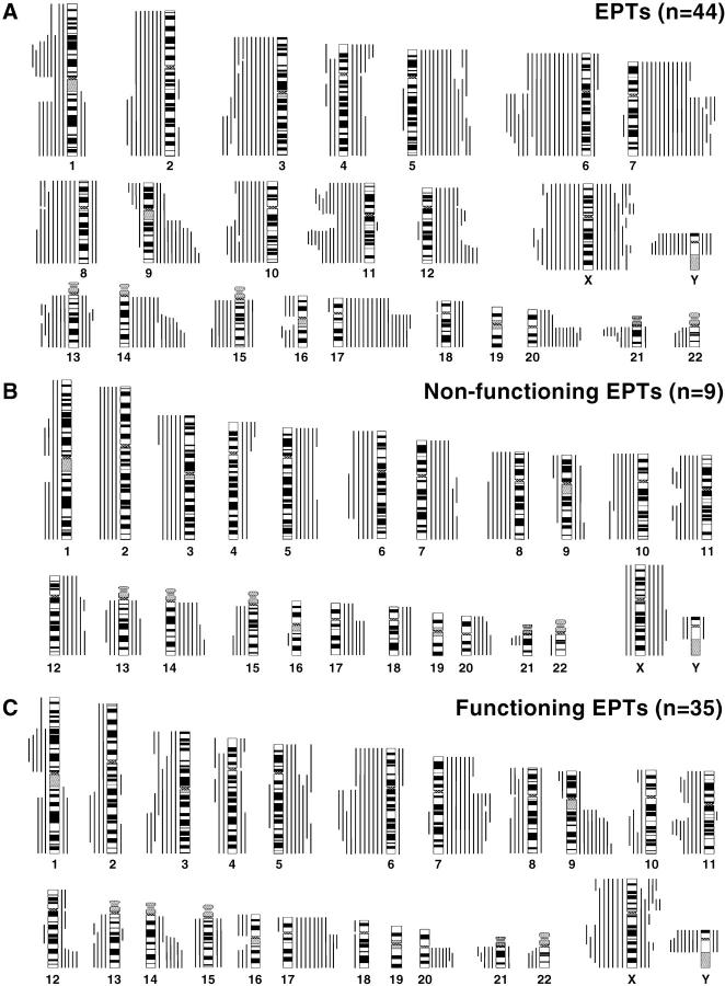
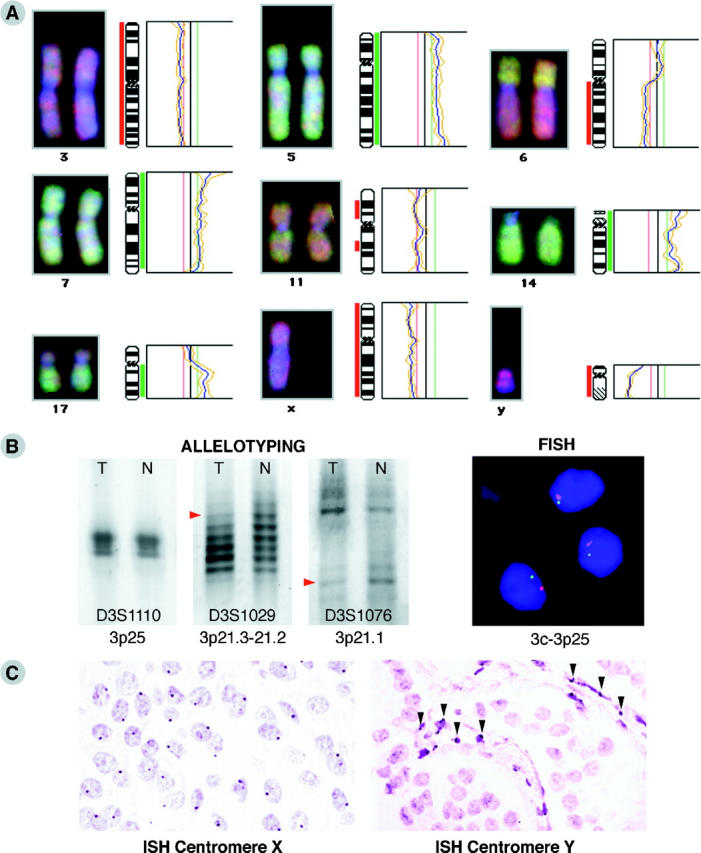
The molecular pathogenesis as well as histogenesis of endocrine pancreatic tumors (EPTs) is not well understood, and the clinical behavior of EPTs is difficult to predict using current morphological criteria. Thus, more accurate markers of risk and better understanding of tumor initiation and progression are needed to allow a precise classification of EPTs. We have studied 44 benign and malignant EPTs by comparative genomic hybridization to correlate the overall number of genetic alterations with clinical and histopathological parameters and to identify chromosomal regions which might harbor genes involved in EPT pathogenesis and progression. Aberrations were found in 36 EPTs, and chromosomal losses (mean, 5.3) were slightly more frequent than gains (mean, 4. 6). The most frequent losses involved Y (45% of male EPTs), 6q (39%), 11q (36%), 3p, 3q, 11p (each 30%), 6p (27%), and 10q and Xq (each 25%), whereas most common gains included 7q (43%), 17q (41%), 5q and 14q (each 32%), 7p, 9q, 17p, 20q (each 27%), and 12q and Xp (each 25%). A correlation was found between the total number of genetic changes per tumor and both tumor size and disease stage. In particular, losses of 3p and 6 and gains of 14q and Xq were found to be associated with metastatic disease. Furthermore, characteristic patterns of genetic changes were found in the various EPT subtypes, eg, 6q loss in malignant insulinomas, indicating that these groups might evolve along genetically different pathways. The highlighted genetic aberrations, including the newly found involvement of 6q losses and sex chromosome alterations, should stimulate the further analysis of these chromosomal regions, which may lead to the discovery of novel genes important in the tumorigenesis and evolution of EPTs.
Figures
References
-
- Klöppel G, In ’t Veld PA, Komminoth P, Heitz PU: The endocrine pancreas. Kovacs K Asa SL eds. Functional Endocrine Pathology. 1998, :pp 415-487 Blackwell Scientific Publications, Oxford
-
- Chandrasekharappa SC, Guru SC, Manickam P, Olufemi S-E, Collins FS, Emmert-Buck MR, Debelenko LV, Zhuang Z, Lubensky IA, Liotta LA, Crabtree JS, Wang Y, Roe BA, Weisemann J, Boguski MS, Agarwal SK, Kester MB, Kim YS, Heppner C, Dong Q, Spiegel AM, Burns AL, Marx S: Positional cloning of the gene for multiple endocrine neoplasia-type 1. Science 1997, 276:404-407 - PubMed
-
- Latif F, Tory K, Gnarra J, Yao M, Duh FM, Orcutt ML, Stackhouse T, Kuzmin I, Modi W, Geil L: Identification of the von Hippel-Lindau disease tumor suppressor gene. Science 1993, 260:1317-1320 - PubMed
-
- Zhuang Z, Vortmeyer AO, Pack S, Huang S, Pham TA, Wang C, Park W-S, Agarwal SK, Debelenko LV, Kester M, Guru SC, Manickam P, Olufemi S-E, Yu F, Heppner C, Crabtree JS, Skarulis MC, Venzon DJ, Emmert-Buck MR, Spiegel AM, Chandrasekharappa SC, Collins FS, Burns AL, Marx SJ, Jensen RT, Liotta LA, Lubensky IA: Somatic mutations of the MEN 1 tumor suppressor gene in sporadic gastrinomas and insulinomas. Cancer Res 1997, 57:4682-4686 - PubMed
-
- Görtz B, Roth J, Krähenmann A, De Krijger RR, Muletta-Feurer S, Rütimann K, Saremaslani P, Speel EJM, Heitz PU, Komminoth P: Mutations and allelic deletions of the MEN 1 gene are associated with a subset of sporadic endocrine pancreatic and neuroendocrine tumors and not restricted to foregut neoplasms. Am J Pathol 1999, 154:429-436 - PMC - PubMed
Publication types
MeSH terms
Substances
LinkOut - more resources
Full Text Sources
Medical
