. 1999 Dec;155(6):1941-51.
doi: 10.1016/S0002-9440(10)65513-7.

Altered centrosome structure is associated with abnormal mitoses in human breast tumors

Affiliations

Altered centrosome structure is associated with abnormal mitoses in human breast tumors

W L Lingle et al. Am J Pathol. 1999 Dec.

Abstract

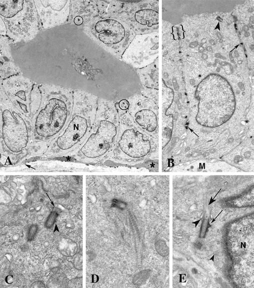
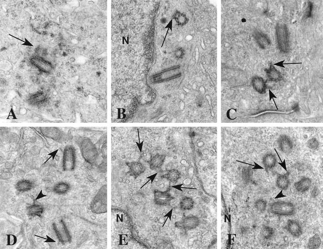
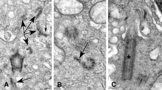
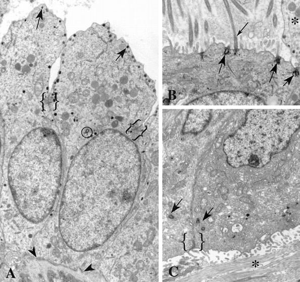
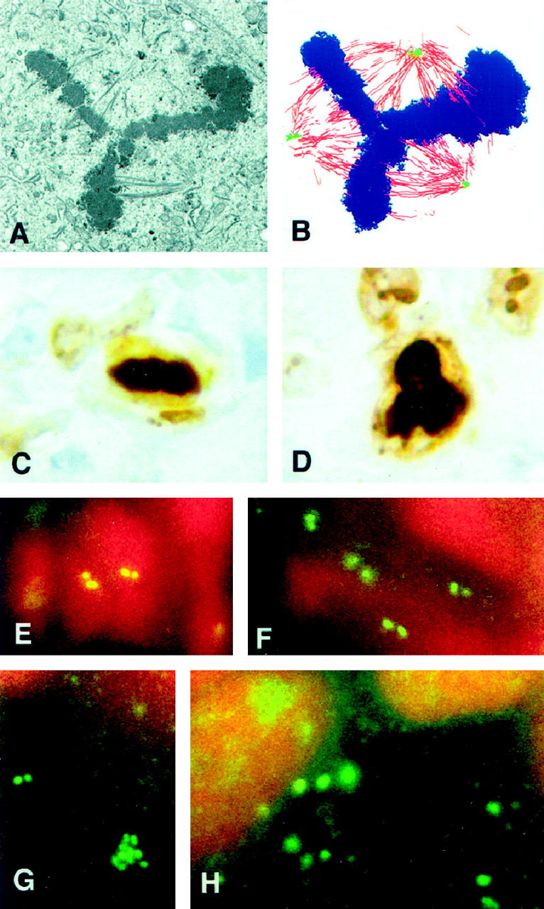
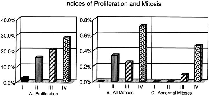
Centrosomes are the major microtubule organizing center in mammalian cells and establish the spindle poles during mitosis. Centrosome defects have been implicated in disease and tumor progression and have been associated with nullizygosity of the p53 tumor suppressor gene. In the present ultrastructural analysis of 31 human breast tumors, we found that centrosomes of most tumors had significant alterations compared to centrosomes of normal breast tissue. These alterations in included 1) supernumerary centrioles, 2) excess pericentriolar material, 3) disrupted centriole barrel structure, 4) unincorporated microtubule complexes, 5) centrioles of unusual length, 6) centrioles functioning as ciliary basal bodies, and 7) mispositioned centrosomes. These alterations are associated with changes in cell polarity, changes in cell and tissue differentiation, and chromosome missegregation through multipolar mitoses. Significantly, the presence of excess pericentriolar material was associated with the highest frequency of abnormal mitoses. Centrosome abnormalities may confer a mutator phenotype to tumors, occasionally yielding cells with a selective advantage that emerge and thrive, thus leading the tumor to a more aggressive state.


Figures

Figure 1.
Figure 1.
Normal breast epithelium. A: The normal breast ductal epithelium consists of a high cuboidal layer of luminal cells subtended by a discontinuous layer of myoepithelial cells (*) and basement membrane (arrow). The nuclei (N) are basal and the centrosomes (circled) are apical. B: Adjacent luminal epithelial cells are joined by lateral junctional complexes (brackets) near the apical membrane and desmosomes (arrows) between their lateral membranes. A single centriole (arrowhead) is located at the apex next to a junctional complex. A portion of a myoepithelial cell (M) is seen at the base of the luminal epithelial cell. C: The mature centriole of this nonorthogonal diplosome bears a short primary cilium (arrow) at its distal end in this luminal epithelial cell. A small subdistal appendage (arrowhead) is present on the mature centriole, whereas the immature centriole lacks appendages. Although very little pericentriolar material is present, the centrioles do have a coating of fine fibers. D: A striated rootlet extends from the proximal end of this mature centriole toward the base of the luminal epithelial cell. E: Fine fibers (small arrowhead) extend between the diplosome and the nearby nucleus (N) in this myoepithelial cell. Distal appendages (large arrowhead) extend between the centriole and the plasma membrane at the site of primary cilium (large arrow) emergence. Subdistal appendages (small arrow) are prominent on the mature centriole. The immature centriole is seen in oblique section. Original magnifications, ×3500 (A), ×8850 (B), ×27,500 (C), ×25,600 (D), ×21,200 (E).
Figure 2.
Figure 2.
Supernumerary centrioles in breast tumors. A: A procentriole (arrow) is present at the proximal end of one of the two centrioles in this section. This procentriole is identifiable by its orthogonal orientation relative to the full length centriole and by the width of its lumen. Notice the electron opaque pericentriolar satellites surrounding the centrioles. B: Two centrioles are seen in cross section and a third is in longitudinal section. One centriole has subdistal appendages (arrow). All three are close to the nucleus (N). There is no orthogonal relationship between any of the three centrioles. C: At least two of these four centrioles have subdistal appendages (arrows). D: The barrels of these five centrioles are coated with a fine electron opaque material. Two centrioles have distal appendages (arrows) and at least one also has subdistal appendages (arrowhead). E: This group of six centrioles is linked by fine fibers between their subdistal appendages (arrows). The group is next to the nucleus (N). F: At least nine centriole profiles are present in this thin section. Subdistal (arrows) and distal (arrowhead) appendages are seen on many of the centrioles. The nucleus (N) is adjacent to this cluster of centrioles. Original magnifications, ×27,500 (A and B), ×32,300 (C and F), ×31,000 (D), ×34,150 (E).
Figure 3.
Figure 3.
Excess pericentriolar material in breast tumors. A: Centrosomes in two adjacent cells are seen. Desmosomes (small arrows) tether the plasma membranes. All of the centriole profiles include subdistal appendages that are characteristic of mature centrioles (large arrows). Electron opaque fibrogranular material is present around both centrosomes. B: The barrels of these centrioles are coated with a dark granular material and pericentriolar satellites are present. One centriole has distal and subdistal appendages (arrow) while the other has a procentriole (arrowhead) associated with it. C: Fine electron opaque fibers coat the five centriole profiles seen in this section. Two orthogonal centrioles are connected by a dense parallel array of fibers (arrow). D: Two centrioles with numerous dark granules are present in this section. E: This centrosome contains one centriole and several masses (arrows) similar to generative complexes visible in this section. Original magnifications, ×17,900 (A), ×31,650 (B), ×28,000 (C), ×28,700 (D), ×27,650 (E).
Figure 4.
Figure 4.
Abnormal centriole structure in breast tumors. A: Subdistal appendages are seen in this oblique section through a centriole. Numerous microtubule complexes (large arrows) are seen in various planes of section throughout the cytoplasm near the centriole. As is seen in cross section of the complexes, the individual microtubules share a portion of the wall of the neighbor microtubules (small arrow). B: The open-ring configuration of this centriole is shown in cross section. Two of the nine triplet microtubule complexes are splayed away from the centriole barrel (arrow). C: This centriole bearing a primary cilium (*) is nearly twice as long as normal centrioles. Original magnifications, ×54,500 (A), ×59,625 (B), ×47,700 (C).
Figure 5.
Figure 5.
Positional centrosomal anomalies in breast tumors. A: Secretory granules (arrows) are present at the apical membrane of these cells displaying apocrine metaplasia. Junctional complexes (brackets) mark the transition from lateral to apical membrane domains. Apocrine beaks extend into the lumen of the duct. Notice the centriole (circled) near the apical end of the nucleus. These cells have apical/basal polarity and rest on a basement membrane (arrowheads). B: Extra centrioles in this cell are inserted at the apical plasma membrane where they function as basal bodies (large arrows) for cilia (small arrow). Microvilli and cilia project into the lumen. The beak of an adjacent apocrine cell (*) is visible. The ciliated cell does not protrude into the lumen, as does the apocrine cell; but like its apocrine neighbor, it has apical/basal polarity and rests on a basement membrane (not visible in this figure). C: The two centrosomes (arrows) seen in adjacent cells are located near the junctional complex between these polarized cells (bracket). However the apical membrane domain with microvilli faces collagen (*) of the stromal tissue rather than the lumen of a duct. This invasive group of cells has ramified through the breast stroma and is not subtended by a basement membrane. The polarity of these cells is inverted, with the basal domains abutting the basal domains of other cells and the apical domains facing the stroma rather than a lumen. Original magnifications, ×8150 (A), ×10,000 (B), ×7900 (C).
Figure 6.
Figure 6.
Multipolar mitoses and centrin immunofluorescence. A: This section through a symmetrical tripolar mitotic cell shows part of the metaphase plate and portions of the tripolar spindle. B: Tracings of microtubules (red), spindle poles (green), and condensed chromosomes (blue) from six nonadjacent serial sections through the cell shown in A are shown in this overlay. The upper spindle pole appears to contain two separate, but adjacent, microtubule foci that have coalesced. C: A normal metaphase plate is shown in this Ki-67 immunostained paraffin section of a breast tumor. D: A tripolar metaphase cell immunolabeled with Ki-67 is shown in this tumor section. E: In normal breast epithelium, the centrosomes appear as distinct pairs of spots when labeled with antibodies against centrin. Centrosomes of two adjacent cells are shown in this cryosection. F: In this tumor characterized with normal centrosome ultrastructure, the centrosomes are similar to those of normal tissue when immunolabeled using antibodies against centrin. G: Centrin immunofluorescence of the same tumor shown in Figure 2, E and F ▶ , reveals a cluster of centriole-sized spots as well as a normal looking pair of spots. By transmission electron microscopy this tumor had up to 9 centrioles in a single thin section, but no excess pericentriolar material. H: Centrin immunofluorescence of the same tumor as shown in Figures 3D and 6A ▶ ▶ reveals numerous large, amorphous spots. By transmission electron microscopy, centrosomes of this tumor contain excess pericentriolar material and extra centrioles. Original magnifications, ×160 (A and B), × 925 (C and D), ×2050 (E-H).
Figure 7.
Figure 7.
Indices of proliferation and mitosis. Tissues were placed in categories as follows: I (solid black bars), six normal tissues from reduction mammoplasties with normal centriole/centrosome structure (all normal tissues examined fell in this category); II (solid gray bars), nine tumor tissues with normal centrioles/centrosomes; III (striped bars), twelve tumor tissues with abnormal centrioles (this includes tissues with supernumerary centrioles and those with centriole defects, but excludes those with excess pericentriolar material); and IV (stippled bars), seven tumor tissues with excess pericentriolar material, regardless of centriole defects. The Wilcoxon Rank Sum Test was used to determine statistical significance. Median values are plotted. A: The proliferation index of category I (normal tissue) is significantly lower than the other three categories and that of category IV (tumors with excess pericentriolar material) is significantly greater than categories I and II, but not III. Categories II and III are not significantly different from each other. B and C: Category IV has significantly higher frequencies of mitosis and abnormal mitosis than the other three categories. The other categories were not significantly different from each other.

Similar articles

Cited by

References

    1. Hartwell LH, Weinert TA: Checkpoints: controls that ensure the order of cell cycle events. Science 1989, 246:629-634 - PubMed
    1. Boveri T: Zur Frage der Emtstehung Maligner Tumoren. Fischer Verlag, Jena, 1914. English translation by Boveri M, The Origin of Malignant Tumors. Baltimore, Waverly Press, 1929
    1. Levine DS, Sanchez CA, Rabinovitch PS, Reid BJ: Formation of the tetraploid intermediate is associated with the development of cells with more than four centrioles in the elastase-simian virus 40 tumor antigen transgenic mouse model of pancreatic cancer. Proc Natl Acad Sci USA 1991, 88:6427-6431 - PMC - PubMed
    1. Balczon R, Bao L, Zimmer WE, Brown K, Zinkowski RP, Brinkley BR: Dissociation of centrosome replication events from cycles of DNA synthesis and mitotic division in hydroxyurea-arrested Chinese hamster ovary cells. J Cell Biol 1995, 130:105-115 - PMC - PubMed
    1. Cross SM, Sanchez CA, Morgan CA, Schimke MK, Ramel S, Idzerda RL, Raskind WH, Reid BJ: A p53-dependent mouse spindle checkpoint. Science 1995, 267:1353-1356 - PubMed

Publication types