. 1999 Dec;155(6):1953-65.
doi: 10.1016/S0002-9440(10)65514-9.

Human vascular adhesion protein-1 in smooth muscle cells

Affiliations

Human vascular adhesion protein-1 in smooth muscle cells

K Jaakkola et al. Am J Pathol. 1999 Dec.

Abstract

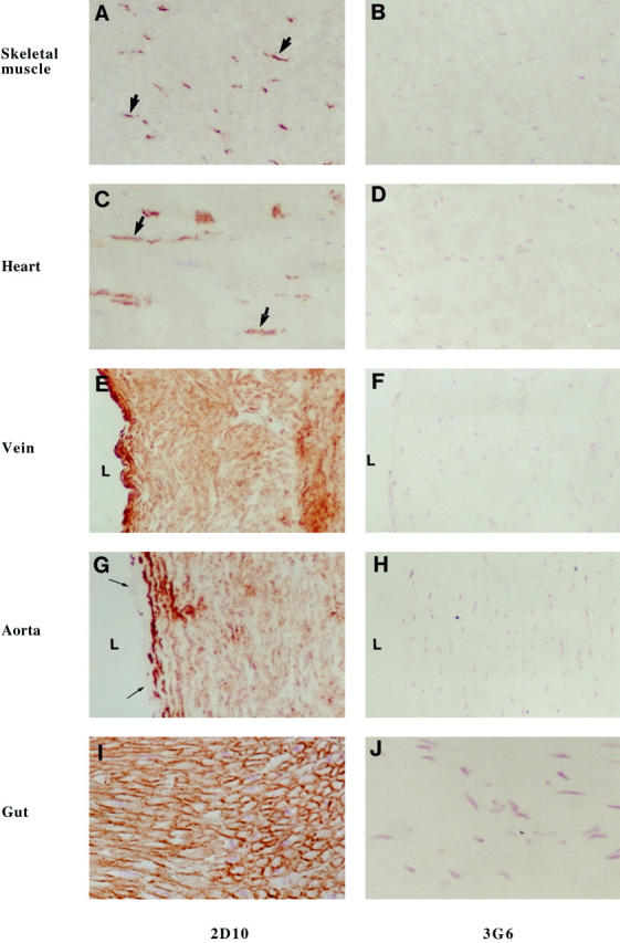
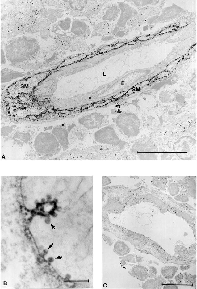
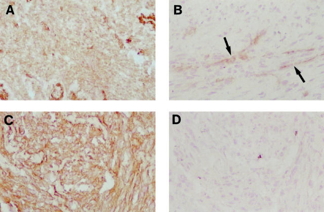
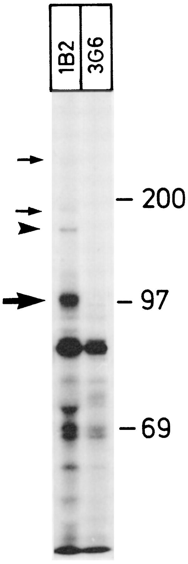
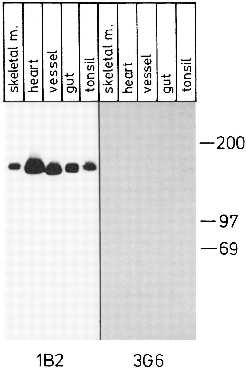
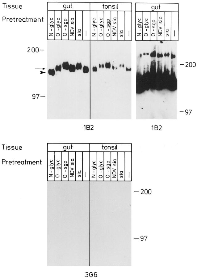
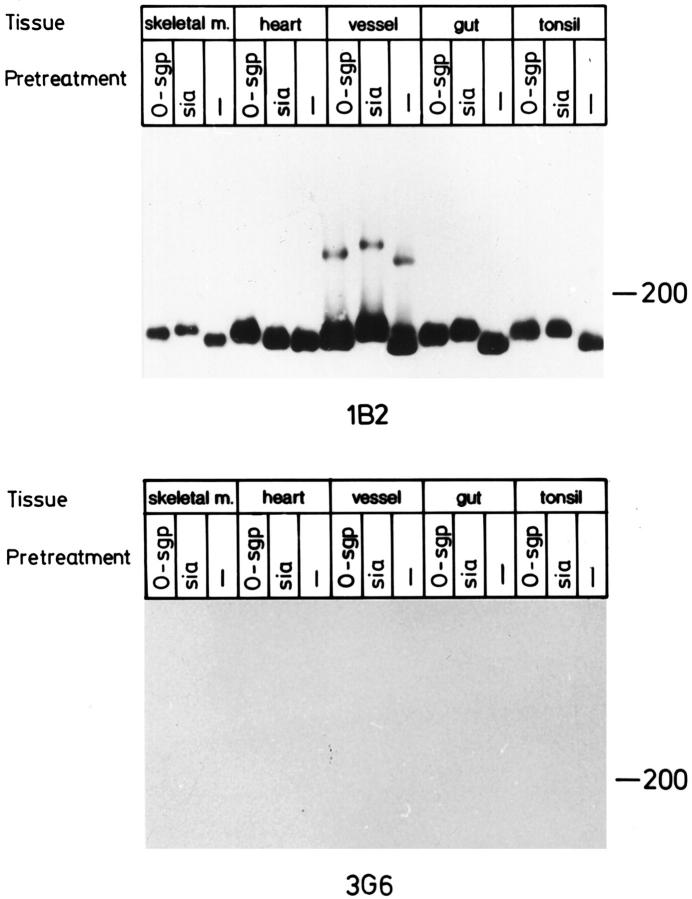
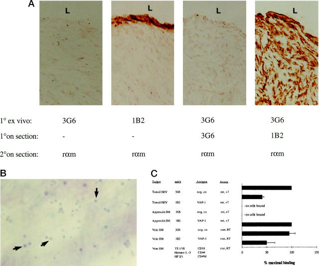
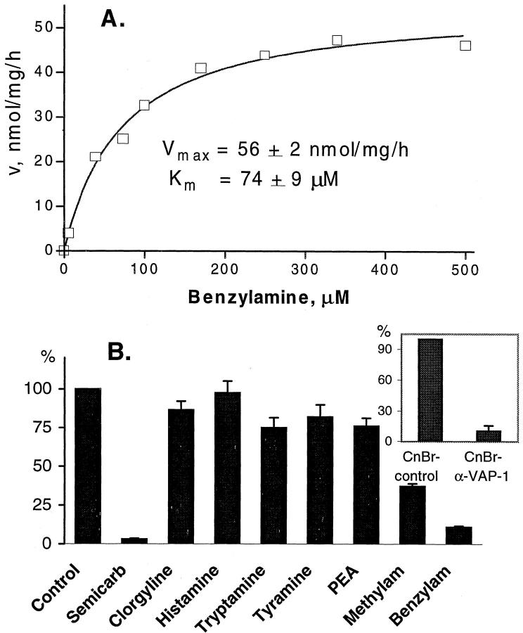
Human vascular adhesion protein-1 (VAP-1) is a dual-function molecule with adhesive and enzymatic properties. In addition to synthesis in endothelial cells, where it mediates lymphocyte binding, VAP-1 is expressed in smooth muscle cells. Here we studied the expression, biochemical structure, and function of VAP-1 in muscle cells and compared it to those in endothelial cells. VAP-1 is expressed on the plasma membrane of all types of smooth muscle cells, but it is completely absent from cardiac and skeletal muscle cells. In tumors, VAP-1 is retained on all leiomyoma cells, whereas it is lost in half of leiomyosarcoma samples. In smooth muscle VAP-1 predominantly exists as a approximately 165-kd homodimeric glycoprotein, but a trimeric (approximately 250 kd) form of VAP-1 is also found. It contains N-linked oligosaccharide side chains and abundant sialic acid decorations. In comparison, in endothelial cells dimeric VAP-1 is larger, no trimeric forms are found, and VAP-1 does not have N-glycanase-sensitive oligosaccharides. Unlike endothelial VAP-1, VAP-1 localized on smooth muscle cells does not support binding of lymphocytes. Instead, it deaminates exogenous and endogenous primary amines. In conclusion, VAP-1 in smooth muscle cells is structurally and functionally distinct from VAP-1 present on endothelial cells.


Figures

Figure 1.
Figure 1.
VAP-1 is selectively found in smooth muscle cells but not in striated or cardiac muscle cells. Immunocytochemical reactions on frozen sections from specimens from the indicated tissues were made using mAbs against VAP-1 (2D10) and against a negative control antigen (3G6) as the first-stage antibodies. Thick arrows point to positively stained endothelial cells in skeletal and heart muscle samples, and thin arrows to the VAP-1-negative endothelial cell layer in aorta. L, lumen of the vessel. Magnifications: A–H, ×200; I and J, ×400.
Figure 2.
Figure 2.
VAP-1 localizes to the plasma membrane in smooth muscle and is present in caveolae. A: In immunolabeling electron microscopy an anti-VAP-1 MAb reacts with vascular smooth muscle cells in tonsil. In this particular vessel VAP-1 is absent from the endothelial cells (E), and only the subendothelial smooth muscle cells (SM) are VAP-1 positive. L, lumen of the vessel. B: At high magnification the localization of VAP-1 into plasma membrane and caveolae (arrows) is readily apparent. C: A control reaction with normal mouse serum. Nonspecific grains apparently resulting from the very sensitive silver intensification of DAB used in A and C are seen in the background. Scale bars: A and C, 10 μm; B, 0.5 μm.
Figure 3.
Figure 3.
VAP-1 expression is modulated in tumors. VAP-1 expression in leiomyomas (A) and leiomyosarcomas (B and C) was analyzed by immunoperoxidase staining. D: The specimen in C, stained with a negative control MAb. In B, VAP-1-positive vessels are indicated by arrows. Magnification, ×200.
Figure 4.
Figure 4.
Dimeric and multimeric forms of VAP-1 are present in smooth muscle cells. [35S]Methionine/cysteine-labeled fragments of smooth muscle were precipitated with an anti-VAP-1 mAb 1B2 and with a negative control mAb 3G6. Under reducing conditions, the monomeric 90–100-kd form of VAP-1 (thick arrow) and the dimeric VAP-1 (arrowhead) are most prominent. Fainter but specific VAP-1 bands are also visible at higher molecular mass (thin arrows). Molecular mass standards in kd are shown on the right.
Figure 5.
Figure 5.
VAP-1 dimer in smooth muscle is smaller than that in endothelial cells. Lysates from the indicated tissues were prepared, separated in SDS-PAGE under nonreducing conditions, and subjected to immunoblotting with an anti-VAP-1 mAb 1B2 and with a negative control mAb 3G6. Samples from skeletal muscle, heart, and tonsil contain mostly endothelial VAP-1, whereas vessel and gut samples are of smooth muscle origin. All samples were analyzed in the same gel, which is a representative of three independent analyses. Note that the negative control was exposed 4 times longer than the 1B2 filter.
Figure 6.
Figure 6.
VAP-1 is an N-glycosylated sialoglycoprotein in smooth muscle cells. Tissue lysates from the indicated sources were digested with the enzymes as described in Materials and Methods and analyzed by immunoblotting with an anti-VAP-1 mAb 1B2 and with a negative control mAb 3G6. All digests were analyzed in the parallel lanes of the same gel. The arrow points to untreated smooth muscle VAP-1 and the arrowhead to the N-glycanase-digested smooth muscle VAP-1. An intentionally overexposed film from the gut is also included to demonstrate the effects of the glycosidase digestions on the trimeric form of VAP-1. No signal was seen from molecules of the same size in prolonged exposures of tonsil samples or from the strip probed with the negative control mAb. These data are representative of three independent experiments with similar results.
Figure 7.
Figure 7.
Sialoglycoprotein nature of VAP-1. VAP-1 from different sources was analyzed with enzymatic digestions and immunoblotting with an anti-VAP-1 mAb 1B2 and with a negative control mAb 3G6. To allow direct comparison of the molecular masses, all samples were analyzed in parallel lanes of one gel. The results are representative of two independent experiments.
Figure 8.
Figure 8.
Different function of VAP-1 in endothelial and smooth muscle cells. A: Tissue explants used for the smooth muscle cell adhesion assays. After the endothelial layer was denuded from the luminal aspect (L) of the vein, the tissue was incubated with the primary mAbs 1B2 against VAP-1 and 3G6 as a negative control (1° ex vivo), washed, snap-frozen, sectioned, and subjected to immunoperoxidase reactions with the indicated mAbs (1° on section), and with the second-stage HRP-conjugated rabbit anti-mouse Ig (rαm, 2° on section). Note that the anti-VAP-1 MAb penetrates only about 10 μm from the surface of the specimen (second panel), even though the antigen is present throughout the tissue (fourth panel). B: Binding of PBLs to smooth muscle cells. PBLs were incubated with the de-endothelized vascular explant, the nonadherent cells were removed by 1× gravitation, and the adherent cells (some pointed out by arrows) were fixed to the sections and counterstained. Magnification, ×100. C: VAP-1 mediates PBL binding to HEV but not to smooth muscle cells. The results of the in vitro HEV and smooth muscle (SM) cell binding assays and of the ex vivo vascular explant (vein) assays after mAb blocking of the indicated molecules are presented as mean ± SEM of four or more independent experiments. Analyses were made under rotatory (rot) or static (stat) conditions at +7°C or at room temperature (RT).
Figure 9.
Figure 9.
Smooth muscle VAP-1 is an amine oxidase. A: Kinetics of smooth muscle monoamine oxidase activity. The rate of [14C]benzylamine deamination (nanomoles of substrate oxidized by mg lysate protein per hour) by smooth muscle lysate versus substrate concentration is plotted. Data are the mean of duplicate assays from a representative experiment that was repeated twice. B: Substrate specificity of smooth muscle monoamine oxidase. The enzyme activity in smooth muscle lysate is arbitrarily set at 100% (control). PEA, β-phenylethylamine. Inset: The smooth muscle lysate was precleared with cyanogen bromide-activated Sepharose beads to which a control mAb (CnBr-contr, set as 100%) or anti-VAP-1 MAb (CnBr-α-VAP-1) was coupled before the enzyme assay was performed. Data represent the mean ± SEM of three experiments performed in duplicate.

Similar articles

Cited by

References

    1. Salmi M, Jalkanen S: A 90-kilodalton endothelial cell molecule mediating lymphocyte binding in humans. Science 1992, 257:1407-1409 - PubMed
    1. Salmi M, Kalimo K, Jalkanen S: Induction and function of vascular adhesion protein-1 at sites of inflammation. J Exp Med 1993, 178:2255-2260 - PMC - PubMed
    1. Salmi M, Jalkanen S: Human vascular adhesion protein-1 (VAP-1) is a unique sialoglycoprotein that mediates carbohydrate-dependent binding of lymphocytes to endothelial cells. J Exp Med 1996, 183:569-579 - PMC - PubMed
    1. Smith DJ, Salmi M, Bono P, Hellman J, Leu T, Jalkanen S: Cloning of vascular adhesion protein-1 reveals a novel multifunctional adhesion molecule. J Exp Med 1998, 188:17-27 - PMC - PubMed
    1. Lyles GA: Mammalian plasma and tissue-bound semicarbazide-sensitive amine oxidases: biochemical, pharmacological and toxicological aspects. Int J Biochem Cell Biol 1996, 28:259-274 - PubMed

Publication types

MeSH terms