

Expression of angiogenesis stimulators and inhibitors in human thyroid tumors and correlation with clinical pathological features
- PMID: 10595926
- PMCID: PMC1866941
- DOI: 10.1016/S0002-9440(10)65515-0
Expression of angiogenesis stimulators and inhibitors in human thyroid tumors and correlation with clinical pathological features
Abstract
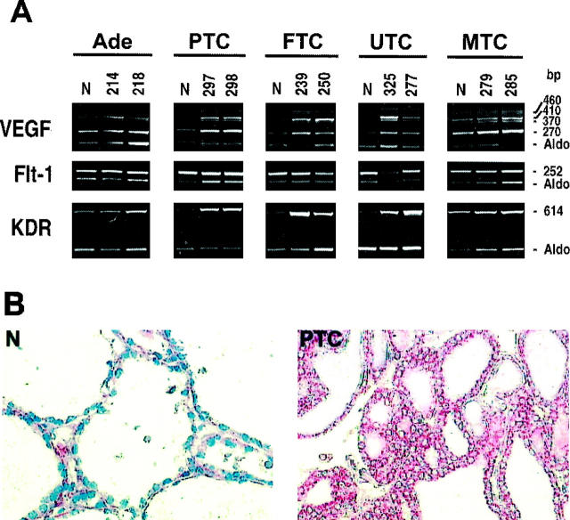
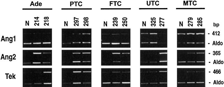
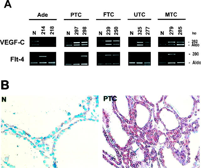
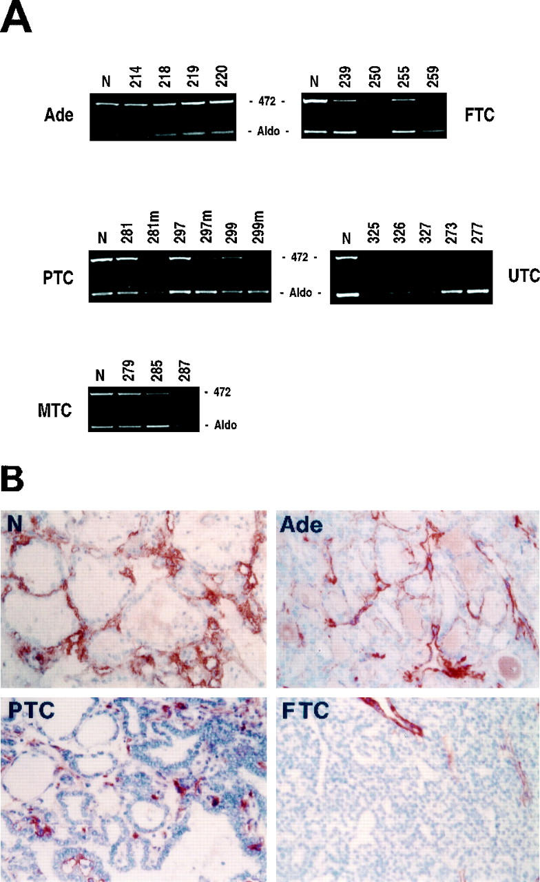
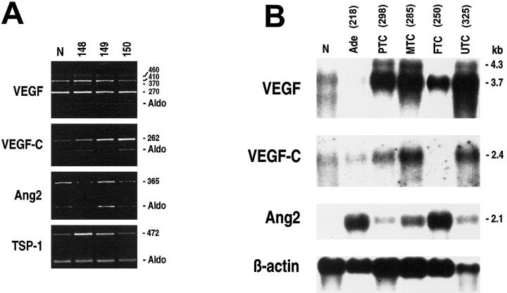
Experimental evidence has shown, both in vitro and in animal models, that neoplastic growth and subsequent metastasis formation depend on the tumor's ability to induce an angiogenic switch. This requires a change in the balance of angiogenic stimulators and inhibitors. To assess the potential role of angiogenesis factors in human thyroid tumor growth and spread, we analyzed their expression by semiquantitative RT-PCR and immunohistochemistry in normal thyroid tissues, benign lesions, and different thyroid carcinomas. Compared to normal tissues, in thyroid neoplasias we observed a consistent increase in vascular endothelial growth factor (VEGF), VEGF-C, and angiopoietin-2 and in their tyrosine kinase receptors KDR, Flt-4, and Tek. In particular, we report the overexpression of angiopoietin-2 and VEGF in thyroid tumor progression from a prevascular to a vascular phase. In fact, we found a strong association between tumor size and high levels of VEGF and angiopoietin-2. Furthermore, our results show an increased expression of VEGF-C in lymph node invasive thyroid tumors and, on the other hand, a decrease of thrombospondin-1, an angioinhibitory factor, in thyroid malignancies capable of hematic spread. These results suggest that, in human thyroid tumors, angiogenesis factors seem involved in neoplastic growth and aggressiveness. Moreover, our findings are in keeping with a recent hypothesis that in the presence of VEGF, angiopoietin-2 may collaborate at the front of invading vascular sprouts, serving as an initial angiogenic signal that accompanies tumor growth.
Figures
Similar articles
-
Expression of the Ets-1 transcription factor in human astrocytomas is associated with Fms-like tyrosine kinase-1 (Flt-1)/vascular endothelial growth factor receptor-1 synthesis and neoangiogenesis.Cancer Res. 1999 Nov 1;59(21):5608-14. Cancer Res. 1999. PMID: 10554042
-
Modulation of in vivo growth of thyroid tumor-derived cell lines by sense and antisense vascular endothelial growth factor gene.Oncogene. 1999 Aug 26;18(34):4860-9. doi: 10.1038/sj.onc.1202869. Oncogene. 1999. PMID: 10490819
-
Analysis of blood vessel maturation processes during cyclic ovarian angiogenesis.Lab Invest. 1998 Nov;78(11):1385-94. Lab Invest. 1998. PMID: 9840613
-
Mechanisms of angiogenesis and their use in the inhibition of tumor growth and metastasis.Oncogene. 2000 Dec 11;19(53):6122-9. doi: 10.1038/sj.onc.1203969. Oncogene. 2000. PMID: 11156525 Review.
-
Biomedical significance of endothelial cell specific growth factor, angiopoietin.Exp Mol Med. 2002 Mar 31;34(1):1-11. doi: 10.1038/emm.2002.1. Exp Mol Med. 2002. PMID: 11989972 Review.
Cited by
-
Posttranslational Modifications in Thyroid Cancer: Implications for Pathogenesis, Diagnosis, Classification, and Treatment.Cancers (Basel). 2022 Mar 22;14(7):1610. doi: 10.3390/cancers14071610. Cancers (Basel). 2022. PMID: 35406382 Free PMC article. Review.
-
Study of single nucleotide polymorphism of vascular endothelium factor in patients with differentiated thyroid cancer.Clin Diabetes Endocrinol. 2022 Dec 15;8(1):9. doi: 10.1186/s40842-022-00146-x. Clin Diabetes Endocrinol. 2022. PMID: 36517920 Free PMC article.
-
Tumor-associated macrophages express lymphatic endothelial growth factors and are related to peritumoral lymphangiogenesis.Am J Pathol. 2002 Sep;161(3):947-56. doi: 10.1016/S0002-9440(10)64255-1. Am J Pathol. 2002. PMID: 12213723 Free PMC article.
-
Novel treatments for anaplastic thyroid carcinoma.Gland Surg. 2020 Jan;9(Suppl 1):S28-S42. doi: 10.21037/gs.2019.10.18. Gland Surg. 2020. PMID: 32055496 Free PMC article. Review.
-
Slit2N and Robo4 regulate lymphangiogenesis through the VEGF-C/VEGFR-3 pathway.Cell Commun Signal. 2014 Apr 7;12:25. doi: 10.1186/1478-811X-12-25. Cell Commun Signal. 2014. PMID: 24708522 Free PMC article.
References
-
- Risau W, Flamme I: Vasculogenesis. Annu Rev Cell Dev Biol 1995, 11:73-91 - PubMed
-
- Folkman J, D’Amore PA: Blood vessel formation: what is its molecular basis? Cell 1996, 87:1153-1155 - PubMed
-
- Risau W: Mechamisms of angiogenesis. Nature 1997, 386:671-674 - PubMed
-
- Klagsbrun M, D’Amore PA: Regulation of angiogenesis. Annu Rev Physiol 1991, 53:217-239 - PubMed
-
- Ferrara N, Davis-Smyth T: The biology of vascular endothelial growth factor. Endocr Rev 1997, 18:4-25 - PubMed
Publication types
MeSH terms
Substances
LinkOut - more resources
Full Text Sources
Other Literature Sources
Medical
Miscellaneous
