

A short isoform of Col9a1 supports alveolar bone repair
- PMID: 10595929
- PMCID: PMC1866927
- DOI: 10.1016/S0002-9440(10)65518-6
A short isoform of Col9a1 supports alveolar bone repair
Abstract
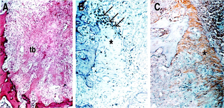
Bone wound created in intramembranous alveolar bone heals without the formation of cartilage precursor tissue. However, the expression of cartilage collagen mRNAs has been suggested. In this report, we examined the expression and the potential role of type IX collagen in bone restoration and remodeling. The sequence specific polymerase chain reaction demonstrated the exclusive expression of short transcriptional isoform of alpha1(IX) collagen (Col9a1) in alveolar bone wound healing, while the long isoform of Col9a1 transcript was absent. Type IX collagen was immunolocalized in the preliminary matrix organized in granulation tissue before trabecular bone formation in tooth extraction socket. In Col9a1-null mutant mice, there were considerable variations in alveolar bone wound healing with the absence of or abnormally organized trabecular bone. Occasionally, unusual apposition of cortical-bone-like layers in bone marrow space was observed. The Col9a1-null mice indicated no growth retardation, and their facial and long bones maintained the normal size and shape. However, the primary spongiosa region of adult Col9a1 mutant mice showed an abnormal trabecular bone structure associated with abnormal immunostaining with the hypertrophic cartilage specific type X collagen antibody. These data suggest that type IX collagen short transcriptional variant is involved in the restoration and remodeling processes of trabecular bone.
Figures


Similar articles
-
Intramembranous bone healing process subsequent to tooth extraction in mice: micro-computed tomography, histomorphometric and molecular characterization.PLoS One. 2015 May 29;10(5):e0128021. doi: 10.1371/journal.pone.0128021. eCollection 2015. PLoS One. 2015. PMID: 26023920 Free PMC article.
-
Trabecular bone deterioration in col9a1+/- mice associated with enlarged osteoclasts adhered to collagen IX-deficient bone.J Bone Miner Res. 2008 Jun;23(6):837-49. doi: 10.1359/jbmr.080214. J Bone Miner Res. 2008. PMID: 18251701 Free PMC article.
-
Altered cartilage phenotype expressed during intramembranous bone formation.J Bone Miner Res. 1993 Nov;8(11):1377-87. doi: 10.1002/jbmr.5650081112. J Bone Miner Res. 1993. PMID: 8266829
-
Trabecular bone formation in the healing of the rodent molar tooth extraction socket.J Bone Miner Res. 1997 Dec;12(12):2061-7. doi: 10.1359/jbmr.1997.12.12.2061. J Bone Miner Res. 1997. PMID: 9421238
-
Collagen IX is indispensable for timely maturation of cartilage during fracture repair in mice.Matrix Biol. 2007 Mar;26(2):85-95. doi: 10.1016/j.matbio.2006.09.010. Epub 2006 Oct 3. Matrix Biol. 2007. PMID: 17112713
Cited by
-
Intramembranous bone healing process subsequent to tooth extraction in mice: micro-computed tomography, histomorphometric and molecular characterization.PLoS One. 2015 May 29;10(5):e0128021. doi: 10.1371/journal.pone.0128021. eCollection 2015. PLoS One. 2015. PMID: 26023920 Free PMC article.
-
The triple helix of collagens - an ancient protein structure that enabled animal multicellularity and tissue evolution.J Cell Sci. 2018 Apr 9;131(7):jcs203950. doi: 10.1242/jcs.203950. J Cell Sci. 2018. PMID: 29632050 Free PMC article. Review.
-
Genetic networks in osseointegration.J Dent Res. 2013 Dec;92(12 Suppl):109S-18S. doi: 10.1177/0022034513504928. Epub 2013 Oct 24. J Dent Res. 2013. PMID: 24158334 Free PMC article. Review.
-
Trabecular bone deterioration in col9a1+/- mice associated with enlarged osteoclasts adhered to collagen IX-deficient bone.J Bone Miner Res. 2008 Jun;23(6):837-49. doi: 10.1359/jbmr.080214. J Bone Miner Res. 2008. PMID: 18251701 Free PMC article.
-
A role for suppressed bone formation favoring catch-up fat in the pathophysiology of catch-up growth after food restriction.Eur J Nutr. 2011 Dec;50(8):645-55. doi: 10.1007/s00394-011-0174-7. Epub 2011 Feb 19. Eur J Nutr. 2011. PMID: 21336871
References
-
- Einhorn TA: The bone organ system: form and function. Marcus R Feldman D Kelsey J eds. Osteoporosis. 1996, :pp 3-22 Academic Press, San Diego
-
- Savontaus M, Vuorio E, Metsaranta M: Growth retardation in transgenic mice harboring a type II collagen mutation. Ann NY Acad Sci 1996, 785:328-330 - PubMed
-
- Suva LJ, Seedor G, Endo N, Quartuccio HA, Thompson DD, Bab I, Rodan GA: Pattern of gene expression following rat tibial marrow ablation. J Bone Miner Res 1993, 8:379-388 - PubMed
-
- Ting K, Petropulos LA, Iwatsuki M, Nishimura I: Altered cartilage phenotype expressed during intramembranous bone formation. J Bone Miner Res 1993, 8:1377-1387 - PubMed
-
- Devlin H, Hoyland J, Newall JF, Ayad S: Trabecular bone formation in the healing of the rodent molar tooth extraction socket. J Bone Miner Res 1997, 12:2061-2067 - PubMed
Publication types
MeSH terms
Substances
Grants and funding
LinkOut - more resources
Full Text Sources
Molecular Biology Databases
