. 1999 Dec;155(6):2067-76.
doi: 10.1016/S0002-9440(10)65525-3.

Gastrin-releasing peptide receptors in non-neoplastic and neoplastic human breast

Affiliations

Gastrin-releasing peptide receptors in non-neoplastic and neoplastic human breast

M Gugger et al. Am J Pathol. 1999 Dec.

Abstract

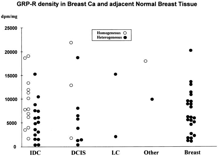
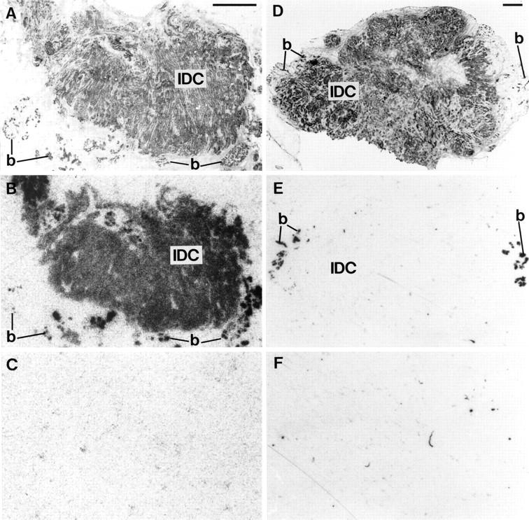
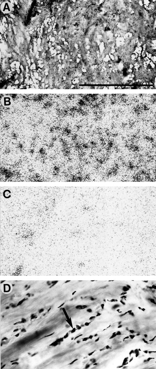
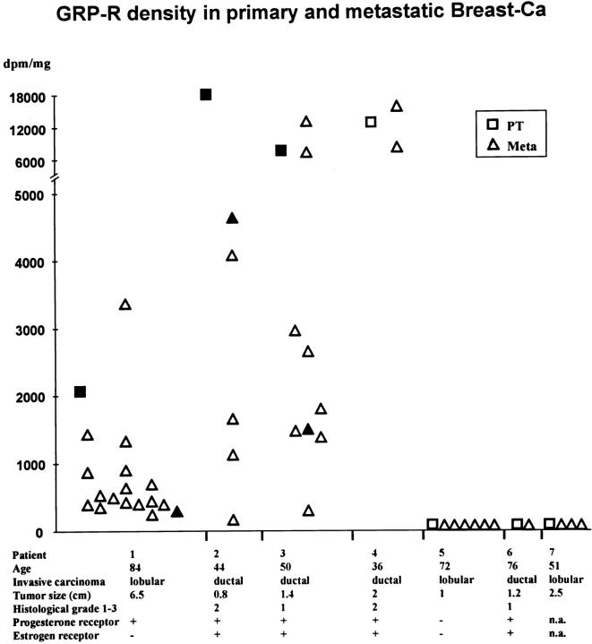
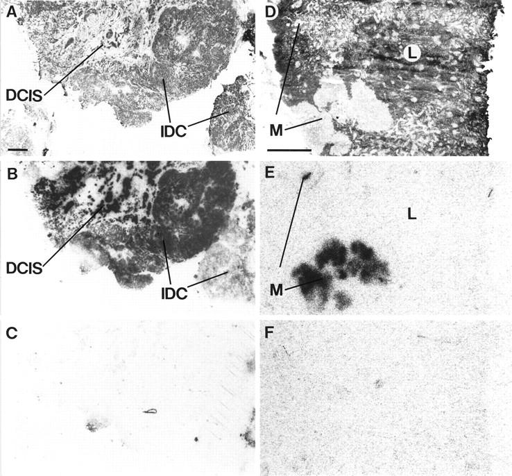
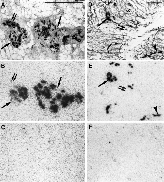
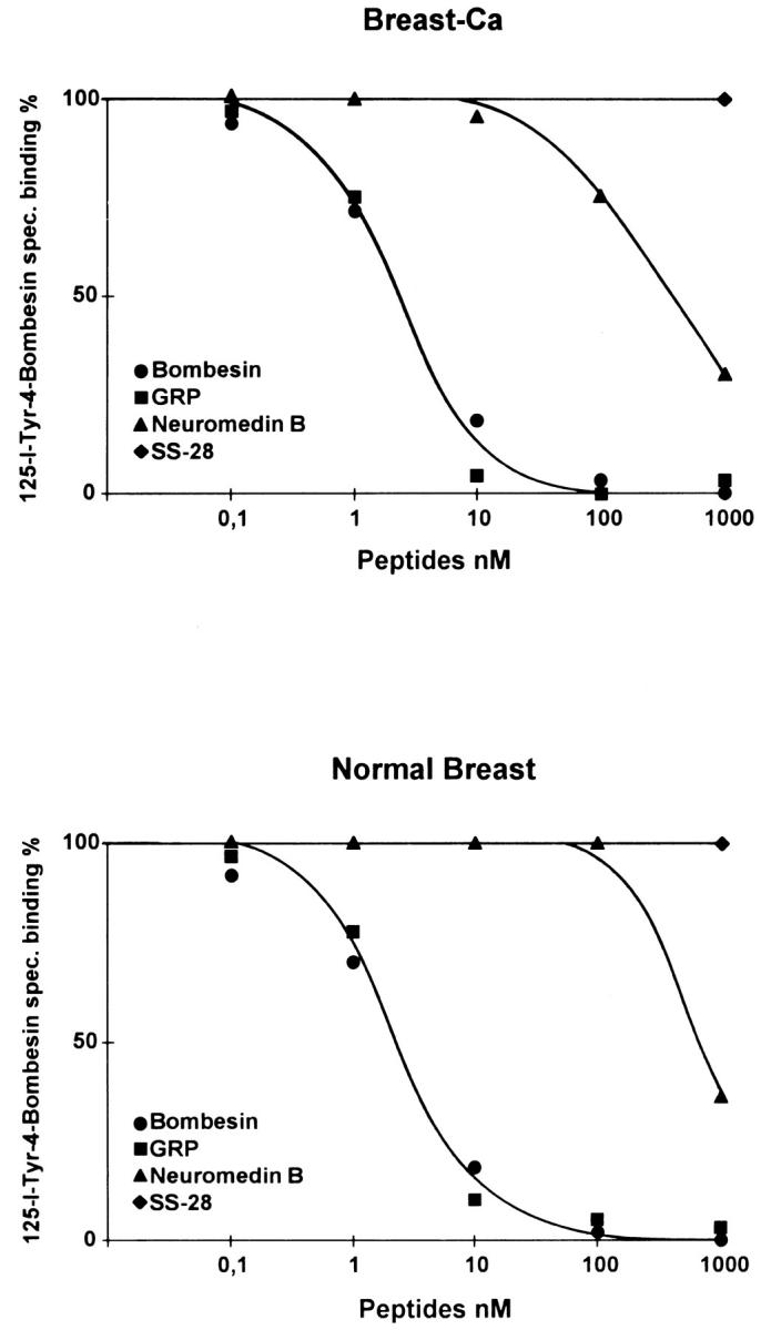
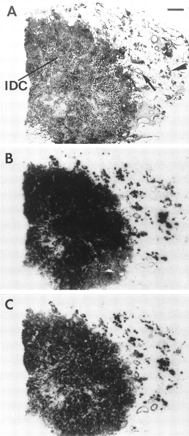
The regulatory peptide gastrin-releasing peptide (GRP) may play a role in human cancer as a stimulatory growth factor. To understand the potential role of GRP in human breast cancer, we have evaluated GRP receptor expression in human non-neoplastic and neoplastic breast tissues and in axillary lymph node metastases, using in vitro receptor autoradiography on tissue sections with [(125)I]Tyr(4)-bombesin and with [(125)I]D-Tyr(6), beta Ala(11), Phe(13), Nle(14)-bombesin(6-14) as radioligands. GRP receptors were detected, often in high density, in neoplastic epithelial mammary cells in 29 of 46 invasive ductal carcinomas, in 11 of 17 ductal carcinomas in situ, in 1 of 4 invasive lobular carcinomas, in 1 of 2 lobular carcinomas in situ, and in 1 mucinous and 1 tubular carcinoma. A heterogeneous GRP receptor distribution was found in the neoplastic tissue samples in 32 of 52 cases with invasive carcinoma and 12 of 19 cases with carcinoma in situ. The lymph node metastases (n = 33) from those primary carcinomas expressing GRP receptors were all positive, whereas surrounding lymphoreticular tissue was negative. GRP receptors were also present in high density but with heterogeneous distribution in ducts and lobules from all available breast tissue samples (n = 23). All of the receptors corresponded to the GRP receptor subtype of bombesin receptors, having high affinity for GRP and bombesin and lower affinity for neuromedin B. All tissues expressing GRP receptors were identified similarly with both radioligands. These data describe not only a high percentage of GRP receptor-positive neoplastic breast tissues but also for the first time a ubiquitous GRP receptor expression in nonneoplastic human breast tissue. Apart from suggesting a role of GRP in breast physiology, these data represent the molecular basis for potential clinical applications of GRP analogs such as GRP receptor scintigraphy, radiotherapy, or chemotherapy.


Figures

Figure 1.
Figure 1.
GRP receptor density (dpm/mg of tissue) in breast carcinomas and adjacent normal breast tissue, using [125I]Tyr4-bombesin as radioligand. IDC, invasive ductal carcinoma; DCIS, ductal carcinoma in situ; LC, lobular invasive and in situ carcinoma; Other, mucinous and tubular carcinoma. The GRP receptor density encompasses low (417 dpm/mg) to very high (21,940 dpm/mg) values for carcinomas (IDC, DCIS, LC, and Other) and moderate (1144 dpm/mg) to very high (20,282 dpm/mg) values for the adjacent normal breast tissue (Breast), which shows without exception a heterogeneous distribution.
Figure 2.
Figure 2.
GRP receptors in two examples of invasive ductal carcinomas, containing adjacent normal human breast tissue. A: H&E-stained section. IDC, Invasive ductal carcinoma; b, normal breast tissue.Scale bar = 1 mm. B: Autoradiogram showing total binding of [125I]Tyr4-bombesin. There is strong and homogeneous labeling of the carcinoma (IDC). Normal breast tissue (b) is also strongly but heterogeneously labeled. C: Autoradiogram showing nonspecific binding of [125I]Tyr4-bombesin (in the presence of 10−6 mol/L bombesin). D: H&E-stained section. IDC, invasive ductal carcinoma; b, normal breast tissue. Scale bar = 1 mm. E: Autoradiogram with completely negative carcinoma tissue. b, normal breast tissue. F: Autoradiogram showing nonspecific binding of [125I]Tyr4-bombesin (in the presence of 10−6 mol/L bombesin).
Figure 3.
Figure 3.
GRP receptors in an invasive lobular carcinoma. A: H&E-stained section. Invasive lobular carcinoma is arranged in many Indian files. Scale bar = 1 mm. B: Autoradiogram showing total binding of [125I]Tyr4-bombesin. Diffusely and moderately labeled dots and lines represent the receptor-positive Indian files of the carcinoma tissue. C: Autoradiogram showing nonspecific binding. D: Detail of A at high magnification, showing carcinoma cells lined up in an Indian file-pattern (arrow).
Figure 4.
Figure 4.
GRP receptor density (dpm/mg of tissue) in primary breast carcinomas and corresponding axillary metastases of seven patients, using [125I]Tyr4-bombesin. Primary tumor is with either homogeneous (□) or heterogeneous (▪) GRP receptor distribution. Axillary metastases are with either homogeneous (▵) or heterogeneous (▴) GRP receptor distribution.
Figure 5.
Figure 5.
High density (dpm/mg of tissue) of GRP receptors in an invasive ductal carcinoma (IDC) adjacent to carcinoma in situ (DCIS) (A–C) and a GRP receptor-positive axillary lymph node metastasis (D–F) from the same patient. A: H&E-stained section.Scale bar = 1 mm. B: Autoradiogram showing total binding of [125I]Tyr4-bombesin. The carcinoma (IDC) labels strongly and homogeneously with the exception of the part in the lower right. The carcinoma in situ (DCIS) shows a strong and homogeneous labeling throughout the sample. C: Autoradiogram showing nonspecific binding. D: H&E-stained section. L, Lymphoreticular tissue; M, carcinoma metastasis. Scale bar = 1 mm. E: Autoradiogram showing total binding of [125I]Tyr4-bombesin. Shown is strong labeling of the metastasis but completely negative lymphoreticular tissue. F: Autoradiogram showing nonspecific binding.
Figure 6.
Figure 6.
High density (dpm/mg of tissue) of GRP receptors in normal breast tissue in a cancer-bearing (A–C) or normal (D–F) breast. A: H&E-stained section, showing terminal duct-lobular unit (arrows and double arrow). Scale bar = 1 mm. B: Autoradiogram showing total binding of [125I]Tyr4-bombesin. Epithelial cells of the terminal duct-lobular unit label strongly in most (arrow) but not all (double arrow) parts. C: Autoradiogram showing nonspecific binding. D: H&E-stained section, showing lobules (arrow and double arrow) and subsegmental duct (arrowhead). Scale bar = 1 mm. E: Autoradiogram showing total binding of [125I]Tyr4-bombesin, showing strong labeling of the lobules in most (arrow) but not all (double arrow) parts. Also the subsegmental duct is labeled in part (arrowhead). F: Autoradiogram showing nonspecific binding.
Figure 7.
Figure 7.
High affinity and specificity of the [125I]Tyr4-bombesin binding in displacement experiments. Sections of tissue samples with breast carcinoma (upper graph), and normal breast tissue (lower graph) were incubated with [125I]Tyr4-bombesin and increasing concentrations of unlabeled bombesin (•), GRP (▪), neuromedin B (▴), and 1000 nmol/L somatostatin (♦). High-affinity displacement of the trace is found with bombesin and GRP, whereas neuromedin B shows lower affinity. Somatostatin has no effect. Nonspecific binding was subtracted from all of the values. The observed rank order of potencies of these analogs is characteristic for the GRP receptor subtype.
Figure 8.
Figure 8.
Comparison of GRP receptor distribution in non-neoplastic and neoplastic breast tissues in a single case tested with [125I]Tyr4-bombesin (B) and [125I]d-Tyr6, β Ala11, Phe13, Nle14-bombesin(6–14) (C). A: H&E-stained section. IDC, invasive ductal carcinoma; arrow, duct; arrowhead, lobules. Scale bar = 1 mm. Nonspecific binding is negligible. Non-neoplastic breast tissues, including ducts and lobules as well as invasive ductal carcinoma, are similarly labeled with both radioligands.

Similar articles

Cited by

References

    1. Reubi JC: Neuropeptide receptors in health and disease: the molecular basis for in vivo imaging. J Nucl Med 1995, 36:1825-1835 - PubMed
    1. Reubi JC: In vitro identification of vasoactive intestinal peptide receptors in human tumors: implications for tumor imaging. J Nucl Med 1995, 36:1846-1853 - PubMed
    1. Reubi JC, Schaer JC, Waser B: Cholecystokinin(CCK)-A, and CCK-B/gastrin receptors in human tumors. Cancer Res 1997, 57:1377-1386 - PubMed
    1. Markwalder R, Reubi JC: Gastrin-releasing peptide receptors in the human prostate: relation to neoplastic transformation. Cancer Res 1999, 59:1152-1159 - PubMed
    1. Krenning EP, Kwekkeboom DJ, Pauwels S, Kvols LK, Reubi JC: Somatostatin Receptor Scintigraphy. Freeman LM eds. Nuclear Medicine Annual. 1995, :pp 1-50 Raven Press, New York

Substances