

Primary central nervous system lymphomas are derived from germinal-center B cells and show a preferential usage of the V4-34 gene segment
- PMID: 10595937
- PMCID: PMC1866926
- DOI: 10.1016/S0002-9440(10)65526-5
Primary central nervous system lymphomas are derived from germinal-center B cells and show a preferential usage of the V4-34 gene segment
Abstract
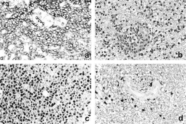
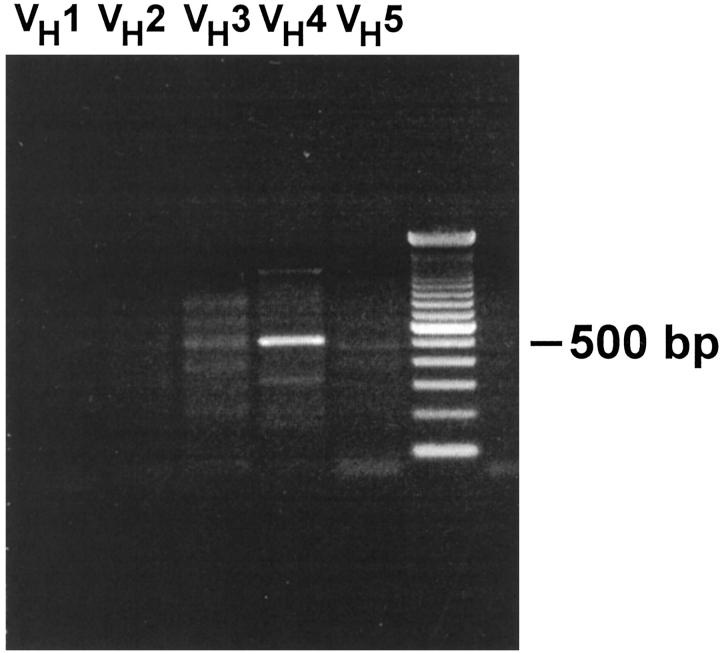
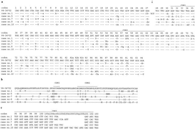
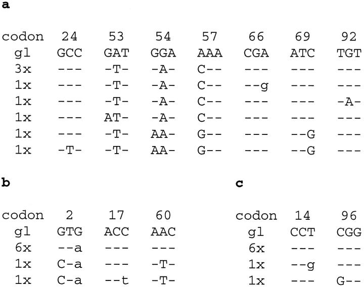
Primary central nervous system lymphomas (PCNSLs) have recently received considerable clinical attention due to their increasing incidence. To clarify the histogenetic origin of these intriguing neoplasms, PCNSLs from 10 HIV-negative patients were analyzed for immunoglobulin (Ig) gene rearrangements. All tumors exhibited clonal IgH gene rearrangements. Of the 10 cases, 5 used the V4-34 gene segment, and all of these lymphomas shared an amino acid exchange from glycine to aspartate due to a mutation in the first codon of the complementarity-determining region 1. No preferential usage of D(H), J(H), V(kappa), J(kappa), V(lambda), or J(lambda) gene segments was observed. All potentially functional rearrangements exhibited somatic mutations. The pattern of somatic mutations indicated selection of the tumor cells (or their precursors) for expression of a functional antibody. Mean mutation frequencies of 13. 2% and 8.3% were detected for the heavy and light chains, respectively, thereby exceeding other lymphoma entities. Cloning experiments of three tumors showed ongoing mutation in at least one case. These data suggest that PCNSLs are derived from highly mutated germinal-center B cells. The frequent usage of the V4-34 gene and the presence of a shared replacement mutation may indicate that the tumor precursors recognized a shared (super) antigen.
Figures
References
-
- Lassmann H, Rössler K, Zimprich F, Vass K: Expression of adhesion molecules and histocompatibility antigens at the blood-brain barrier. Brain Pathol 1991, 1:115-123 - PubMed
-
- Tontsch U, Rott O: Cortical neurons selectively inhibit MHC class II induction in astrocytes but not in microglial cells. Int Immunol 1993, 5:249-254 - PubMed
-
- Irani DN, Lin KI, Griffin DE: Regulation of brain-derived T cells during acute central nervous system inflammation. J Immunol 1997, 158:2318-2326 - PubMed
-
- Lantos PL, VandenBerg SR, Kleihues P: Tumours of the nervous system. Greenfield’s Neuropathology, vol 2, ed 6. Edited by DI Graham, PL Lantos. New York, Oxford University Press, 1997, pp 766–775
Publication types
MeSH terms
Substances
Associated data
- Actions
- Actions
- Actions
- Actions
- Actions
- Actions
- Actions
- Actions
- Actions
- Actions
- Actions
- Actions
- Actions
- Actions
- Actions
- Actions
- Actions
- Actions
- Actions
- Actions
- Actions
- Actions
- Actions
- Actions
- Actions
- Actions
- Actions
LinkOut - more resources
Full Text Sources
Medical
