. 1999 Dec;155(6):2153-65.
doi: 10.1016/S0002-9440(10)65533-2.

Prominent axonopathy in the brain and spinal cord of transgenic mice overexpressing four-repeat human tau protein

Affiliations

Prominent axonopathy in the brain and spinal cord of transgenic mice overexpressing four-repeat human tau protein

K Spittaels et al. Am J Pathol. 1999 Dec.

Abstract

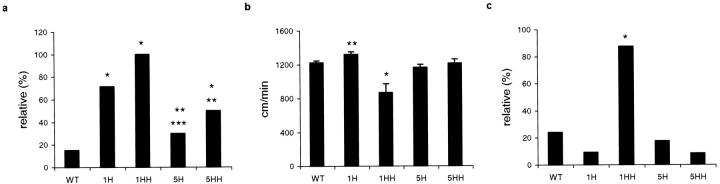
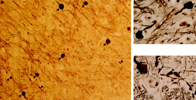
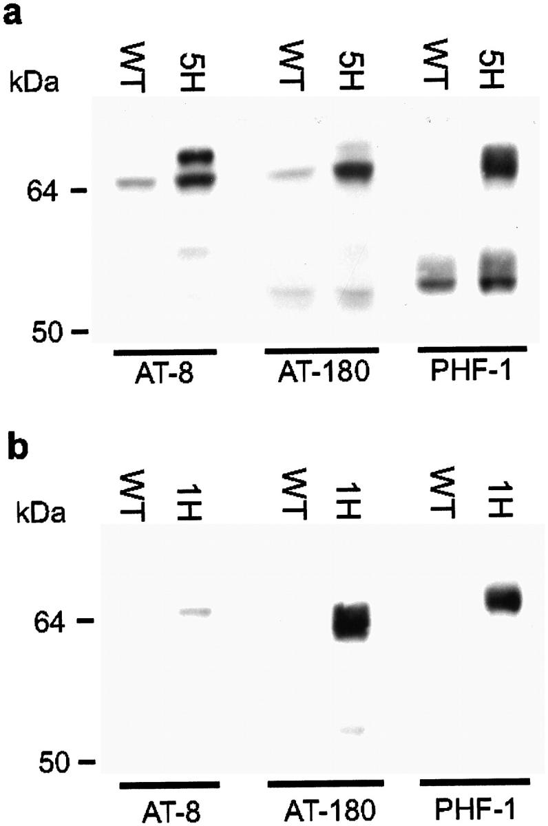
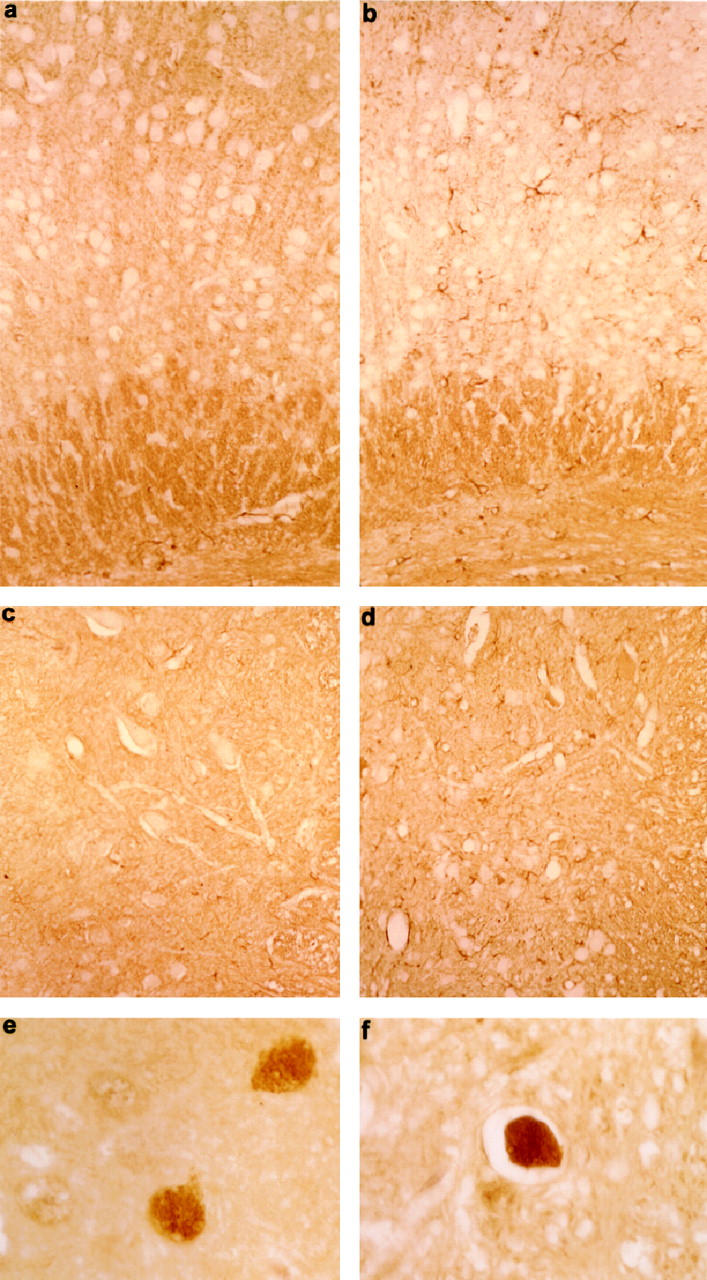
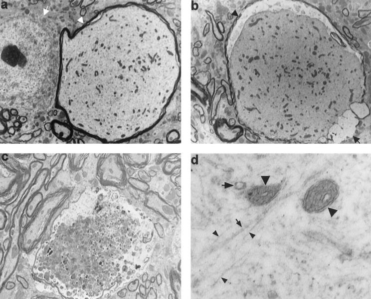
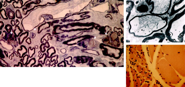
Mutations in the human tau gene cause frontotemporal dementia and parkinsonism linked to chromosome 17. Some mutations, including mutations in intron 10, induce increased levels of the functionally normal four-repeat tau protein isoform, leading to neurodegeneration. We generated transgenic mice that overexpress the four-repeat human tau protein isoform specifically in neurons. The transgenic mice developed axonal degeneration in brain and spinal cord. In the model, axonal dilations with accumulation of neurofilaments, mitochondria, and vesicles were documented. The axonopathy and the accompanying dysfunctional sensorimotor capacities were transgene-dosage related. These findings proved that merely increasing the concentration of the four-repeat tau protein isoform is sufficient to injure neurons in the central nervous system, without formation of intraneuronal neurofibrillary tangles. Evidence for astrogliosis and ubiquitination of accumulated proteins in the dilated part of the axon supported this conclusion. This transgenic model, overexpressing the longest isoform of human tau protein, recapitulates features of known neurodegenerative diseases, including Alzheimer's disease and other tauopathies. The model makes it possible to study the interaction with additional factors, to be incorporated genetically, or with other biological triggers that are implicated in neurodegeneration.


Figures

Figure 1.
Figure 1.
Recombinant DNA construct used to generate htau40 transgenic mice and Western blot analysis of expression of human tau40 protein in brain, spinal cord, spinal ganglia, and sciatic nerve of selected transgenic mouse strains. a: Structure of the authentic mouse thy1 gene and the modified mini-gene construct yielding neuron-specific expression of the embedded cDNA. Original exons are numbered and represented by black blocks. b: Western blotting of extracts of brain derived from wild-type and from transgenic htau40 mice with antibodies Tau-5 and HT-7. Approximately 10 μg of protein was loaded per lane. Samples were treated with alkaline phosphatase before application on the gel. c: Western blotting of extracts of spinal cord derived from wild-type and from transgenic htau40 mice with antibodies Tau-5 and HT-7. Approximately 10 μg of protein was loaded per lane. Samples were treated with alkaline phosphatase before loading on the gel. d: Numeric results from densitometric scanning and quantification of Western blots displayed in b and c. e: Western blotting of extracts of spinal ganglia (sp.g.) and sciatic nerves (sc.n.) derived from wild-type and htau40-1 transgenic mice with polyclonal antibody B19. Approximately 5 μg of protein was loaded per lane. WT, wild-type mice; H and HH, heterozygous and homozygous htau40 mice, respectively. The three different transgenic mouse strains are indicated as htau40-1, -2, and -5. All mice were about 3 months old (±1 week).
Figure 2.
Figure 2.
Performance of transgenic and wild-type mice in three sensorimotor tasks. a: Number of mice that fell off the walking rod during a 3-minute test period, expressed relative to the number of tested in each group. Compared to WT mice, significantly more 1H, 1HH, and 5HH mice fell down. *WT-1H: P < 0.001, O.R. 13.5 (40–4.6); WT-1HH: P < 0.001, O.R. 90 (1000–4.5); WT-5HH: P < 0.001, O.R. 5.4 (20.4–1.4). This motor impairment was shown to be gene dosage dependent. **5H-1HH: P < 0.001, O.R. 38.4 (1.8–1000); 5HH-1HH: P < 0.05, O.R. 17 (0.8–500); ***5H-1H: P < 0.001, O.R. 5.8 (1.6–20.8). b: Swimming speed defined as distance traveled in 1 minute. In the forced swimming test, 1HH mice (*) traversed a significantly shorter distance in 1 minute than wild-type and other transgenic mice (P < 0.001). 1H mice (**) performed even better than wild-type littermates (P < 0.05). c: Inverted wire grid hanging, expressed as number of mice that remained suspended for the entire 1-minute test period, relative to the number of mice tested in each group. Compared to wild-type mice, significantly more 1HH mice (*) failed to remain suspended on the inverted wire mesh grid. WT-1HH: P < 0.001, O.R. 22.2 (125–3.8). Number of mice tested is given in Materials and Methods. Asterisks do not denote significance levels, but indicate groups of mice to be compared. WT, wild-type mice; 1H and 1HH, heterozygous and homozygous htau40-1 mice, respectively; 5H and 5HH, heterozygous and homozygous htau40-5 mice, respectively. All mice were between 2 and 4 months old.
Figure 3.
Figure 3.
Silver staining of brain and spinal cord of homozygous htau40 transgenic mice at the age of 2.5 months. a: Multiple dilated axons or axonal spheroids (arrows) and some irregular dystrophic axons (arrowheads) in cortex (×360). b: Higher magnification of two dilated axons in thalamus. Note that the dilations approach the size of neuronal cell bodies (×920). c: Aspect of spinal cord gray matter (anterior horn) with a grossly dilated axon (arrow) and several irregularly thickened dystrophic axons (arrowheads) (×390).
Figure 4.
Figure 4.
Immunohistochemistry of brain and spinal cord of homozygous htau40 transgenic mice at 2.5 months. a: Low-power view of neocortex stained with monoclonal antibody AT-8, showing somatodendritic localization of tau protein in pyramidal neurons of layer V (×110). b: Reaction of monoclonal antibody MC-1 in neocortex with a neuron (arrowhead) and dystrophic neurites (arrows) (×800). c: Anterior horn of spinal cord, showing axonal dilations (arrows), dystrophic neurites (arrowheads), and a neuronal cell body staining with antibody MC-1 (×365). d: Neuron with axonal dilation (arrow), staining with the antibody SMI-32, directed to neurofilament (NF)-H. The proximal axon (small arrow) connecting the cell body (arrowhead) has a normal caliber (×600). e and f: Dilated axons (arrowheads) in thalamus immunostained for NF-L (e) and NF-M (f) (×305).
Figure 5.
Figure 5.
Western blotting of extracts of brain and spinal cord from wild-type and transgenic htau40 mice of 3 months with phosphorylation-dependent antibodies AT-8, AT-180, and PHF-1. Western blots of brain (a) and spinal cord (b) homogenates of wild-type (WT) and heterozygous htau40-5 (5H) and htau40-1 (1H) transgenic mice. Approximately 100 μg of protein was loaded per lane for incubation with AT-8 and AT-180, and 15 μg for detection with PHF-1.
Figure 6.
Figure 6.
Immunohistochemistry of brain and spinal cord of homozygous htau40 transgenic mice of 2.5 months. a–d: GFAP immunostaining of cortex and spinal cord of homozygous htau40-1 mice showing astrogliosis in the cortex (b) and anterior horn (d). Compare with cortex (a) and anterior horn (c) of a wild-type mouse. Magnification: a and b, ×210; c and d, ×190. e and f: Ubiquitin-positive dilated axons in cortex (e) and spinal cord (f) of a homozygous htau40-1 mouse (×700).
Figure 7.
Figure 7.
Ultrastructure of dilated axons with varying degrees of degeneration in spinal cord of 2- and 8-month-old homozygous htau40-1 mice. a: Dilated axon (arrowhead), larger in diameter than the neighboring neuronal cell body (arrow), is distended by accumulation of neurofilaments, microtubuli, mitochondria, and vesicles. Compare with several normal axons present in the lower left-hand corner of the section (×2915). b: Dilated axon with thinned (arrowhead) or absent (arrow) myelin sheath and with retracted axoplasm (×2915). c: Degenerating dystrophic axon filled with numerous dense and multivesicular bodies and with the myelin sheath thinned and disrupted (×4165). d: Higher magnification of dilated axon, showing neurofilaments (small arrowheads), microtubuli (small arrow), mitochondria (large arrowheads), and vesicles (large arrow) (×40,000).
Figure 8.
Figure 8.
Immunogold staining of a dilated axon in the spinal cord of a transgenic htau40-1 mouse (homozygous, 8 month). Preembedding immunogold staining with AT-8 (a) and Alz-50 (b). Note that the silver-enhanced gold particles are equally dispersed in the axoplasm. The irregularity of the particles is due to the silver enhancement and osmium postfixation. Magnification: a, ×25,200; b, ×35,700.
Figure 9.
Figure 9.
Peripheral nerve and skeletal muscle of htau40 transgenic mice at the age of 2 and 8 months. a: Semithin section of sciatic nerve displaying macrophages filled with myelin debris (arrows), indicative of Wallerian degeneration. Note also a slightly dilated degenerating axon with a thinned myelin sheath (arrowhead) (×1300). b: Electron micrograph of two axons with prominent axon-Schwann cell networks (arrows) (×3735) as opposed to a normal axon (arrowhead). c: Quadriceps muscle with grouping of atrophic fibers (left side of field) and normal fibers (right side of field) (×210).

Similar articles

Cited by

References

    1. Wiche G, Oberkanins C, Himmler A: Molecular structure and function of microtubule-associated proteins. Int Rev Cytol 1991, 124:217-273 - PubMed
    1. Billingsley ML, Kincaid RL: Regulated phosphorylation and dephosphorylation of tau protein: effects on microtubule interaction, intracellular trafficking and neurodegeneration. Biochem J 1997, 323:577-591 - PMC - PubMed
    1. Mandelkow EM, Mandelkow E: Tau in Alzheimer’s disease. Trends Cell Biol 1998, 8:125-127 - PubMed
    1. Goedert M, Spillantini MG, Crowther RA: Cloning of a big tau microtubule-associated protein characteristic of the peripheral nervous system. Proc Natl Acad Sci USA 1992, 89:1983-1987 - PMC - PubMed
    1. Andreadis A, Brown WM, Kosik KS: Structure and novel exons of the human tau gene. Biochemistry 1992, 31:10626-10633 - PubMed

Publication types

MeSH terms