

Magnetic resonance imaging of male and female genitals during coitus and female sexual arousal
- PMID: 10600954
- PMCID: PMC28302
- DOI: 10.1136/bmj.319.7225.1596
Magnetic resonance imaging of male and female genitals during coitus and female sexual arousal
Abstract
Objective: To find out whether taking images of the male and female genitals during coitus is feasible and to find out whether former and current ideas about the anatomy during sexual intercourse and during female sexual arousal are based on assumptions or on facts.
Design: Observational study.
Setting: University hospital in the Netherlands.
Methods: Magnetic resonance imaging was used to study the female sexual response and the male and female genitals during coitus. Thirteen experiments were performed with eight couples and three single women.
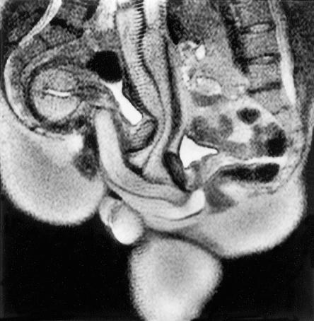
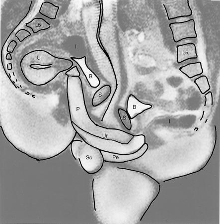
Results: The images obtained showed that during intercourse in the "missionary position" the penis has the shape of a boomerang and 1/3 of its length consists of the root of the penis. During female sexual arousal without intercourse the uterus was raised and the anterior vaginal wall lengthened. The size of the uterus did not increase during sexual arousal.
Conclusion: Taking magnetic resonance images of the male and female genitals during coitus is feasible and contributes to understanding of anatomy.
Figures

Similar articles
-
Female sexual arousal: genital anatomy and orgasm in intercourse.Horm Behav. 2011 May;59(5):780-92. doi: 10.1016/j.yhbeh.2010.12.004. Epub 2010 Dec 30. Horm Behav. 2011. PMID: 21195073 Free PMC article.
-
Magnetic resonance imaging (MRI) of sexual intercourse: second experience in missionary position and initial experience in posterior position.J Sex Marital Ther. 2002;28 Suppl 1:63-76. doi: 10.1080/00926230252851203. J Sex Marital Ther. 2002. PMID: 11898711
-
Magnetic resonance imaging of sexual intercourse: initial experience.J Sex Marital Ther. 2001 Oct-Dec;27(5):475-82. doi: 10.1080/713846807. J Sex Marital Ther. 2001. PMID: 11554209
-
Anatomic variation and orgasm: Could variations in anatomy explain differences in orgasmic success?Clin Anat. 2016 Jul;29(5):665-72. doi: 10.1002/ca.22703. Epub 2016 Apr 4. Clin Anat. 2016. PMID: 26916103 Review.
-
Sexual instrumentation.IEEE Trans Biomed Eng. 1983 Jun;30(6):309-19. doi: 10.1109/tbme.1983.325130. IEEE Trans Biomed Eng. 1983. PMID: 6347867 Review. No abstract available.
Cited by
-
Mechanisms and Evidence of Genital Coevolution: The Roles of Natural Selection, Mate Choice, and Sexual Conflict.Cold Spring Harb Perspect Biol. 2015 Jul 1;7(7):a017749. doi: 10.1101/cshperspect.a017749. Cold Spring Harb Perspect Biol. 2015. PMID: 26134314 Free PMC article. Review.
-
Penile reconstruction in a newborn following complicated circumcision: A case report.Int J Surg Case Rep. 2018;51:74-77. doi: 10.1016/j.ijscr.2018.08.003. Epub 2018 Aug 9. Int J Surg Case Rep. 2018. PMID: 30144715 Free PMC article.
-
Controlled dehydration, structural flexibility and gadolinium MRI contrast compound binding in the human plasma glycoprotein afamin.Acta Crystallogr D Struct Biol. 2019 Dec 1;75(Pt 12):1071-1083. doi: 10.1107/S2059798319013500. Epub 2019 Nov 19. Acta Crystallogr D Struct Biol. 2019. PMID: 31793901 Free PMC article.
-
Hearing, touching, and multisensory integration during mate choice.Front Neural Circuits. 2022 Sep 20;16:943888. doi: 10.3389/fncir.2022.943888. eCollection 2022. Front Neural Circuits. 2022. PMID: 36247731 Free PMC article. Review.
-
Female sexual arousal: genital anatomy and orgasm in intercourse.Horm Behav. 2011 May;59(5):780-92. doi: 10.1016/j.yhbeh.2010.12.004. Epub 2010 Dec 30. Horm Behav. 2011. PMID: 21195073 Free PMC article.
References
-
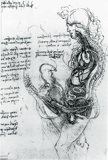
- Chianchi M. Leonardo, the anatomy. Florence: Giunti; 1998. p. 56.
-
- Clark K, Pedretti C. The drawings of Leonardo da Vinci in the collection of Her Majesty the Queen at Windsor Castle. London: Phaidon; 1968.
-
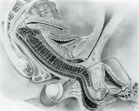
- Dickinson RL. Human sex anatomy, a topographical hand atlas. 2nd ed. London: Baillière, Tindall and Cox; 1949. pp. 84–109.
-
- Masters WH, Johnson VE. Human sexual response. Boston: Little, Brown; 1966.
-
- Johnson VE, Masters WH, Lewis KC. The physiology of intravaginal contraception failure. In: Calderone MS, editor. Manual of contraceptive practice. Baltimore: Williams and Wilkins; 1964. pp. 138–150.
MeSH terms
LinkOut - more resources
Full Text Sources
Other Literature Sources