. 1999 Dec 1;13(23):3092-105.
doi: 10.1101/gad.13.23.3092.

The homeodomain protein vax1 is required for axon guidance and major tract formation in the developing forebrain

Affiliations

The homeodomain protein vax1 is required for axon guidance and major tract formation in the developing forebrain

S Bertuzzi et al. Genes Dev. .

Abstract

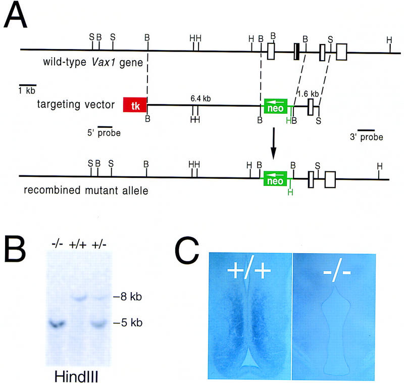
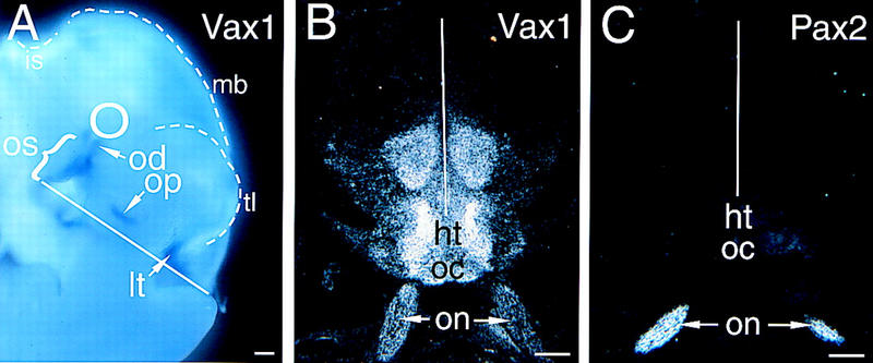
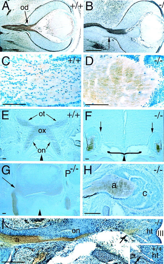
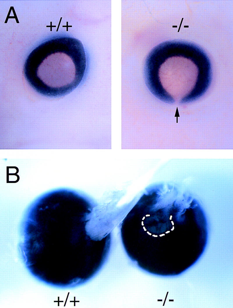
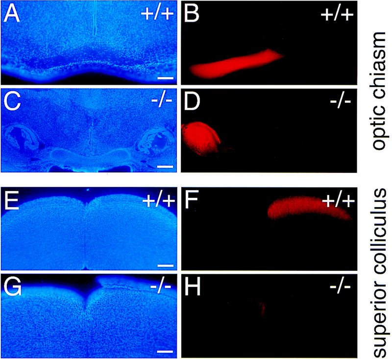
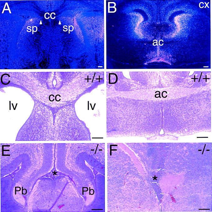
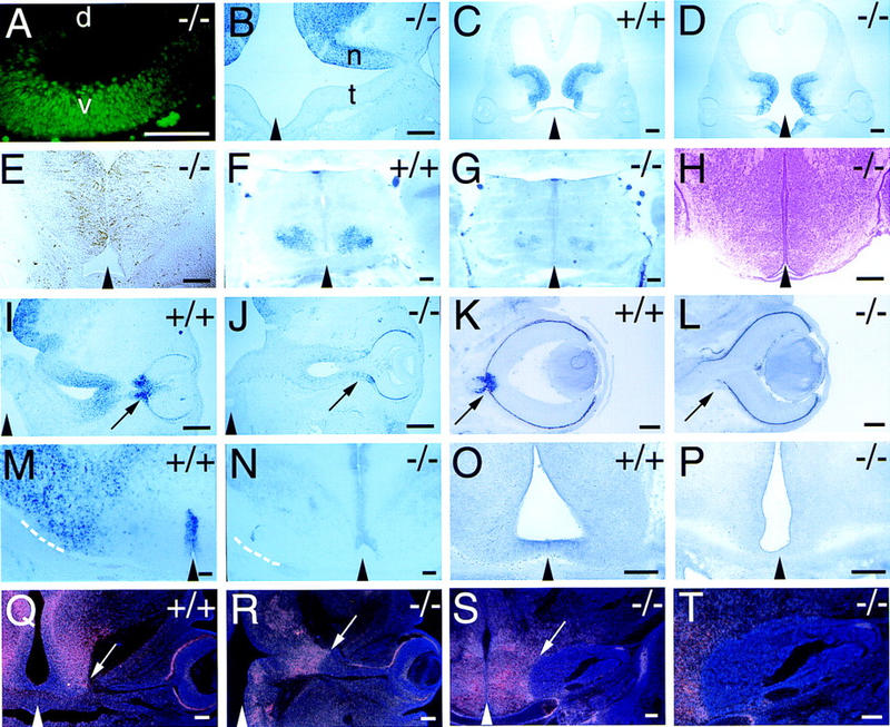
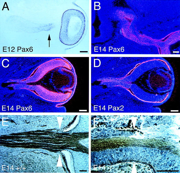
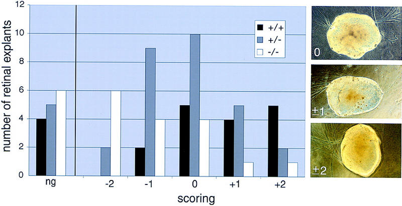
The homeodomain protein Vax1 is expressed in a highly circumscribed set of cells at the ventral anterior midline of the embryonic CNS. These cells populate the choroid fissure of the optic disk, the body of the optic stalk and nerve, the optic chiasm and ventral diencephalon, and the anterior midline zones that abut developing commissural tracts. We have generated mutant mice that lack Vax1. In these mice (1) the optic disks fail to close, leading to coloboma and loss of the eye-nerve boundary; (2) optic nerve glia fail to associate with and appear to repulse ingrowing retinal axons, resulting in a fascicle of axons that are completely segregated from optic nerve astrocytes; (3) retinal axons fail to penetrate the brain in significant numbers and fail to form an optic chiasm; and (4) axons in multiple commissural tracts of the anterior CNS, including the corpus callosum and the hippocampal and anterior commissures, fail to cross the midline. These axon guidance defects do not result from the death of normally Vax1(+) midline cells but, instead, correlate with markedly diminished expression of attractive guidance cues in these cells. Vax1 therefore regulates the guidance properties of a set of anterior midline cells that orchestrate axon trajectories in the developing mammalian forebrain.


Figures

Figure 1
Figure 1
Inactivation of the mouse Vax1 gene. (A) Structure of the mouse Vax1 gene, the targeting vector for inactivation, and the recombined allele. Sequences that encode the Vax1 homeodomain are contained in the second and third exons diagrammed, and are indicated in black. The first diagrammed exon contains the Vax1 initiator ATG. The first and second exons are deleted and replaced with a G418 resistance cassette (neo) in the mutant allele, and a thymidine kinase cassette (tk) was used to select against nonhomologous recombinants (see Materials and Methods). The positions of the 5′ and 3′ probes used to assess homologous recombination of the left and right arms are indicated. (B) BamHI; (H) HindIII; (S) SacI. (B) Southern blot of HindIII-digested genomic DNA from wild-type (+/+), heterozygous mutant (+/−), and homozygous mutant (−/−) mice, hybridized with the 3′ probe indicated in A. (C) Lack of Vax1 transcript in the subventricular zone of the ventral diencephalon of E13.5 mutant mice (−/−), as detected by in situ hybridization (see Material and Methods).
Figure 2
Figure 2
Vax1 and Pax2 mRNA expression in the developing brain. (A) Vax1 mRNA in an E10.5 mouse embryo, detected by whole-mount in situ hybridization, is evident in the optic stalk (os, bracket), optic disk (od), olfactory pit (op), and lamina terminalis (lt). The position of the neural retina, which does not express Vax1, is indicated by the circle. The surface boundaries of the telencephalon (tl), midbrain (mb), and isthmus (is) are indicated by the broken lines. The CNS midline is indicated by the solid white line in this and subsequent panels. (B) Vax1 mRNA (33P-radiolabeled in situ), in a frontal section through the CNS of an E18.5 embryo, is expressed by the glial cells of the two optic nerves (on, arrows) and by midline cells at the optic chiasm (oc) and in the hypothalamus (ht). Note that there is high Vax1 expression at the junction between the optic nerve and the hypothalamus, and little Vax1 mRNA in the ventricular zone immediately adjacent to the third ventricle. (C) Pax2 mRNA, in a frontal section adjacent to that of B, is evident in the same optic nerve cells (on, arrows) that express Vax1, but not at the optic chiasm or in the hypothalamus. Scale bars, 100 μm.
Figure 3
Figure 3
Defects in RGC axon pathfinding in Vax1 mutants. (A) Frontal section through an E13.5 wild-type eye (facing right). Retinal ganglion cell axons in the optic nerve (brown) are visualized with an antibody to L1. The optic disc (od) is indicated by the arrow. (B) Section through an E13.5 Vax1 mutant eye, similar to that in A. L1+ axons fasciculate in one restricted domain of the developing optic nerve, completely avoiding the Vax1−/− glial precursors of the nerve. Note that there is no longer a well-defined border between the eye and the optic nerve (apparent in A) and that aberrant retinal pigment epithelial cells are present at the periphery of the nerve (arrow). (C) High-power view of an E18.5 wild-type optic nerve, showing glial cells (toluidine blue stained, dark blue nuclei) interspersed among and regularly associating with L1+ retinal axons (light brown). (D) Similar section to that in C, but in a Vax1−/− optic nerve. Naked L1+ axons entirely avoid and displace the glial and retinal cells of the nerve. (E) L1+ RGC axons entering the wild-type E18.5 brain in the hypothalamus and crossing to the contralateral side of the brain at the optic chiasm (ox). The optic nerves (on) and the optic tract (ot) are indicated. (F) L1+ retinal axons in Vax1 mutants (also at E18.5) do not form an optic chiasm and moreover, do not penetrate the brain but, instead, terminate in encapsulated bundles. The empty space beneath the floor of the hypothalamus is contiguous with the nasal cavity and is due to the cleft palate routinely seen in Vax1−/− mice (bracket). Arrows indicate the pronounced cellular barrier separating the cap of RGC axons and the brain proper. (G) Frontal section through the hypothalamus of E18.5 Pax2−/− mouse embryo. Unlike in the Vax1 mutant mice, retinal axons, identified by L1 staining, readily penetrate the brain (arrow), even though the Pax2 mutants lack an organized optic chiasm. Compare the difference at the insertion points (arrows) of the optic nerves between Vax1 mutants (F) and Pax2 mutants (G). (H) High-power image of the E18.5 Vax1 mutant cell cap, formed by optic nerve astrocyte precursors and displaced retinal cells, which encapsulate the RGC axons (a). At this stage, the thick cell cap (c) is a physical barrier preventing RGC axons from contact with the base of the brain. Note the presence of dark retinal pigment epithelial cells at the lower periphery of the distal end of the stunted optic nerve. (I) Tracing of the pioneer RGC axons that reach the hypothalamus in Vax1 mutants (−/−) and in normal mice (+/+, inset) at E13.0. As shown in B, axons (a) and glial cells are separated in the optic nerve (on). Note that at this stage the cell cap at the distal end of the mutant optic nerve is not yet formed. As RGC axons reach the hypothalamus (ht), they splay out at the insertion point (black arrow), failing to fasciculate or to take the normal ventral pathway (white arrows in both montage and inset) toward the optic chiasm below the third ventricle (III). Black triangles at the bottom of EG indicate the position of the midline. Scale bars, 100 μm.
Figure 4
Figure 4
Homozygous Vax1 mutant mice show bilateral coloboma and disruption of the eye/optic nerve boundary. (A) Coloboma is evident at the pigmented back of an E13 mutant eye (arrow in −/−), due to the failure of the optic fissure to close. (B) Persistence of coloboma (broken line), together with poorly organized retinal axons in the optic nerve, in a dissected P12 mutant eye (−/−) with attached disordered nerve, as compared with the closed eye and well-formed optic nerve evident in P12 wild type (+/+).
Figure 5
Figure 5
Growth of retinal ganglion cell axons into the brains of newborn wild-type (+/+) and Vax1 mutant (−/−) mice. The right eyes of newborn mice were completely filled with DiI to label the axons of all RGC neurons projecting from the right retina, and mice were analyzed 1 day later. (Similar injections were also performed and analyzed at mid-embryogenesis in utero; see Materials and Methods). (A, B) Coronal section at the level of the optic chiasm in a wild-type newborn, stained with DAPI to label nuclei (A) and visualized under rhodamine fluorescence to display DiI-labeled axons (B). Axons transit through the optic chiasm normally. (C,D) Coronal section similar to A and B but in a Vax1−/− newborn. Axons fail to enter the brain at this level and terminate in an encapsulated, light-bulb-shaped knot; the optic chiasm is completely absent. (E,F) Coronal section, caudal to that of A and B at the level of the superior colliculus, in a wild-type newborn. Approximately 95% of labeled RGC axons cross to the opposite side of the brain at the chiasm and terminate in the contralateral superior colliculus (F), as is normal in the mouse. (G,H) Coronal section similar to E and F but in a Vax1−/− newborn. The rare axons that escape the knot in D project through lateral regions of the ventral hypothalamus that are normally Vax1 negative (see Fig. 2B), and terminate exclusively in the ipsilateral superior colliculus (H). Scale bars, 100 μm.
Figure 6
Figure 6
Anterior commissural fiber tracts fail to form in Vax1 mutant mice. (A) High-power view of in situ hybridization in neonatal brain (coronal plane) showing that Vax1 expression at the dorsal midline in the region of the septum (sp) and of the corpus callosum (cc) persists until birth. White arrowheads denote residual expression near the position of the midline glial sling of the embryo (Silver 1993). (B) Low-power view of a coronal section of an E16.5 mouse brain at the level of the anterior commissure (ac), showing Vax1 expression in midline cells surrounding the commissural tract. Note the lack of Vax1 mRNA in neurons of the developing cortex (cx). (C) Coronal section through the corpus callosum (cc) of a wild-type neonatal mouse brain. At this stage, most of the axons responsible for the interhemispheric connections have crossed the midline, and a thick bundle of axons (cc) crossing the midline is visualized by hematoxylin and eosin staining. Note the open lateral ventricles (lv). (D) Coronal section through a neonatal wild-type mouse brain at the level of the anterior commissure (ac), illustrating the normal midline crossing of axons in this commissure. (E) In Vax1 mutants, the corpus callosum fails to form. No axons cross the midline but, instead, turn away into the lateral ventricles and accumulate in Probst-bundles (Pb)— thick knots of tangled axons adjacent to the dorsal midline. The point at which callosal axons would normally cross the midline is indicated by the asterisk. (F) Same plane of sectioning as in D, but in a Vax1 mutant embryo in which axons fail to cross the midline in the anterior commissure. The normal midline crossing point is again indicated by the asterisk. Scale bars, 100 μm.
Figure 7
Figure 7
Cell fate and neural patterning in wild-type and Vax1 mutant embryos. Black triangles at the bottom of selected panels indicate the position of the midline. (A) High-power view of the E11 optic stalk of a Vax1 mutant (−/−), illustrating a normal distribution of Pax2 expression in the ventral (v), but not dorsal (d), stalk. (B) Section of the E12 optic stalk of a Vax1 mutant (eye at extreme right), illustrating a normal distribution of BF1 expression in the nasal (n), but not temporal (t), stalk. (C,D) Nkx2.1 expression in frontal sections of the ventral diencephalon (presumptive hypothalamus) of a wild-type (C, +/+) and Vax1 mutant (D, −/−) mouse at E12. (E) RC2 expression in a frontal section of the hypothalamus of a Vax1 mutant mouse at E13. Glial cells expressing the RC2 antigen are still present in a normal fashion in the mutant ventral diencephalon. (F,G) Nkx2.2 expression in frontal sections of the hypothalamus of a wild-type (F, +/+) and a Vax1 mutant (G, −/−) mouse at E19.5. (H) Coronal section of a E17.5 Vax1 mutant ventral diencephalon stained with hematoxylin and eosin showing normal histology in the area of Vax1 expression. (I,J) Netrin-1 expression at the optic fissure and stalk of a wild-type (I, +/+) and Vax1 mutant (J, −/−) mouse at E12 sectioned in a frontal orientation. Eyes are pointing to the right; arrows indicate the optic fissure, which fails to close in the mutant. Note marked reduction of Netrin-1 in the mutant. (K,L) Netrin-1 expression in the optic disk and proximal stalk of a wild-type (K, +/+) and Vax1 mutant (L, −/−) mouse at E14.5. Note complete absence of Netrin-1 in the mutant. Arrow in K points to the expression of Netrin-1 in the closed optic disc, whereas the arrow in L indicates the lack of Netrin-1 expression and the open fissure left at the eye/nerve boundary in the mutant embryos. (M,N) High-power view of Netrin-1 expression in the hypothalamus of wild-type (M, +/+) and Vax1 mutant (N, −/−) mouse at E18.5, shown in a coronal cut. Note that in N, Netrin-1 expression in the lateral hypothalamus, at the entry point of the optic nerve (broken lines in M and N), is greatly reduced; surrounding the third ventricle, in which there is no overlap with Vax1 expression, this Netrin-1 expression is maintained. (O,P) EphB3 receptor tyrosine kinase expression in wild-type (O, +/+) and Vax1 mutant mouse (P, −/−) in the ventral diencephalon at E13.5, shown in a coronal section. Expression of EphB3 is lost in the mutant embryo. (Q,R) Coronal sections of Slit-1 mRNA expression, detected by radioactive in situ hybridization, in wild-type (Q, +/+) and Vax1 mutant (R, −/−) mice at E13.5. Arrows indicate the optic nerve/hypothalamus boundary, which coincides with a pronounced border of Slit-1 expression. Note that in R, this sharp Slit-1 is maintained. (S,T) Medium power (S) and higher power (T) view of Slit-1 expression, detected by radioactive in situ hybridization, in Vax1 mutant mice (−/−) at E18.5. The Slit-1 boundary barrier (arrowed in S) is maintained into late embryogenesis, at which point the capped bundle of RGC axons becomes directly wedged against it. Scale bars, 100 μm.
Figure 8
Figure 8
Pax6+ cells are ectopically present in the Vax1−/− optic nerve. (A) At E12, Pax6 mRNA (visualized by digoxigenin in situ hybridization) is detected in the mutant neural retina, as expected, but also in the optic stalk, immediately adjacent to the retina (arrow). The remainder of the mutant stalk (nearer the brain) is Pax6 at E12. (B) By E14, ectopic Pax6+ cells (detected by 33P-labeled in situ hybridization) are present throughout the optic nerve. (C,D) In the E14 mutant optic nerve, both ectopic Pax6+ (C) and resident Pax2+ (D) cells are present. The sections in C and D are adjacent. (E) In the E14 wild-type embryo, a defined histological border (white arrowheads) is present between the eye and the optic nerve. Cells are stained with toluidine blue and axons immunostained with L1 antibody. (F) In E14 Vax1 mutants, the eye/nerve border is missing (white arrowheads indicate the normal location). Note that there is a continuum of histologically indistinguishable cells from the retina into the nerve, and also that pigment epithelium, which does not express either Vax1 or Pax6, streams into the nerve. Scale bars, 100 μm.
Figure 9
Figure 9
Collagen gel assay of retinal axon guidance by cells of wild-type and Vax1 mutant optic stalk; qualitative scoring of results. Wild-type retinae were positioned close to embryonic optic stalk explants from wild-type (+/+, black bars), heterozygous (+/−, gray bars), or homozygous (−/−, white bars) Vax1 mutant mice, and retinal axon outgrowth was scored after 4 days, as described in Materials and Methods. The histogram displays the number of retinal explants scored in six different categories: no growth (ng), strong away (−2), weak away (−1), unbiased (0), weak toward (+1), and strong toward (+2). Retinal axon outgrowth is biased away from homozygous mutant optic stalk explants (categories −2 and −1), whereas growth of these same axons is inversely biased toward wild-type optic stalks (+2 and +1). The outgrowth scores for the heterozygous explants exhibit an intermediate distribution. The three panels at right illustrate examples of retinal explants scored as unbiased (0), weakly biased (either toward or away) (±1), and strongly biased (±2).

References

    1. Anderson MG, Perkins GL, Chittick P, Shrigley RJ, Johnson WA. drifter, a Drosophila POU-domain transcription factor, is required for correct differentiation and migration of tracheal cells and midline glia. Genes & Dev. 1995;9:123–137. - PubMed
    1. Bartsch U, Kirchhoff F, Schachner M. Immunohistological localization of the adhesion molecules L1, N-CAM, and MAG in the developing and adult optic nerve of mice. J Comp Neurol. 1989;284:451–462. - PubMed
    1. Battye R, Stevens A, Jacobs JR. Axon repulsion from the midline of the Drosophila CNS requires slit function. Development. 1999;126:2475–2481. - PubMed
    1. Braisted JE, McLaughlin T, Wang HU, Friedman GC, Anderson DJ, O'Leary DDM. Graded and lamina-specific distributions of ligands of EphB receptor tyrosine kinases in the developing retinotectal system. Dev Biol. 1997;191:14–28. - PubMed
    1. Brose K, Bland KS, Wang KH, Arnott D, Henzel W, Goodman CS, Tessier-Lavigne M, Kidd T. Slit proteins bind Robo receptors and have an evolutionarily conserved role in repulsive axon guidance. Cell. 1999;96:795–806. - PubMed

Publication types

MeSH terms