. 1999 Dec 1;13(23):3149-59.
doi: 10.1101/gad.13.23.3149.

Wnt signaling in Xenopus embryos inhibits bmp4 expression and activates neural development

Affiliations

Wnt signaling in Xenopus embryos inhibits bmp4 expression and activates neural development

J C Baker et al. Genes Dev. .

Abstract

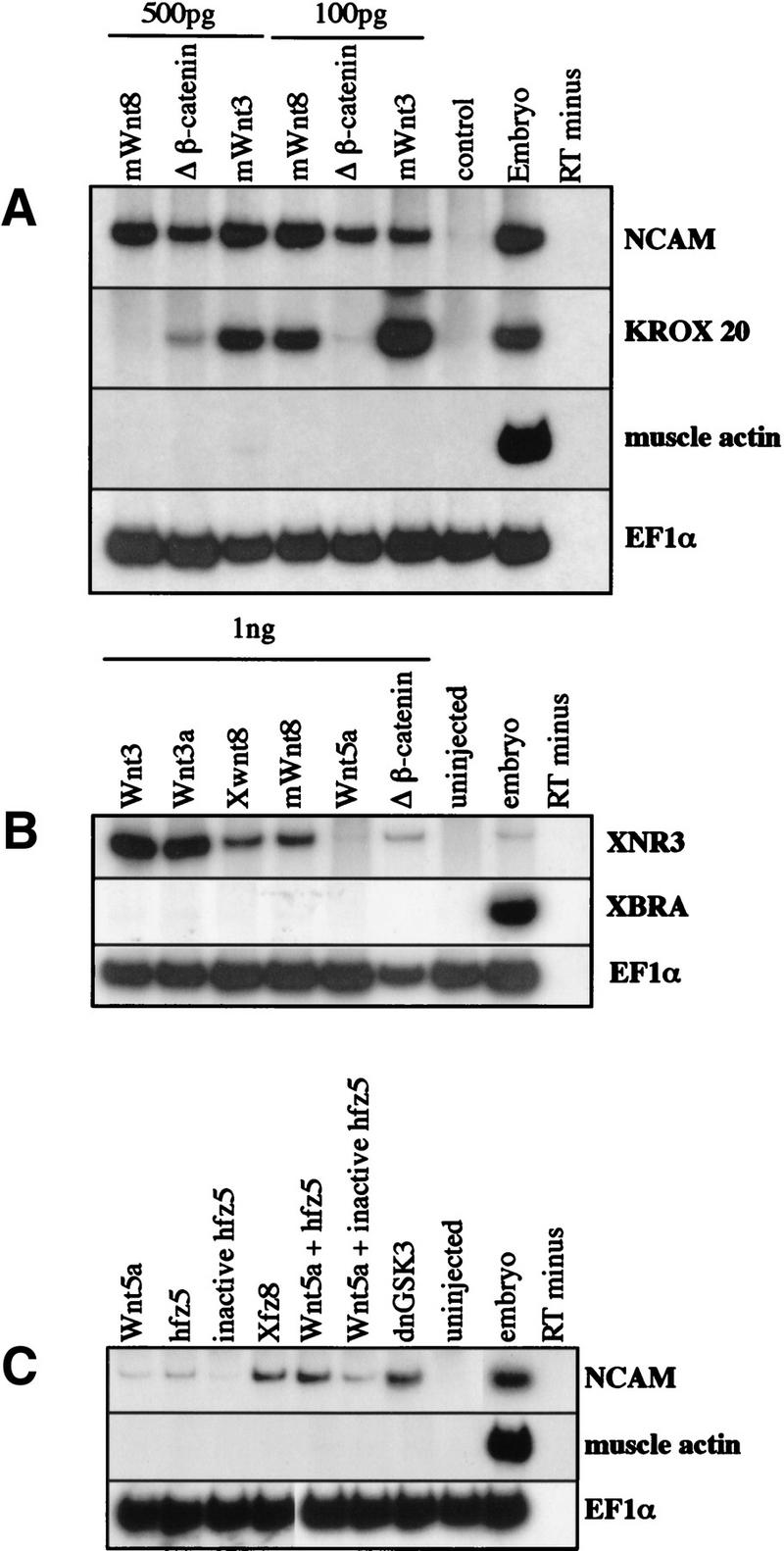
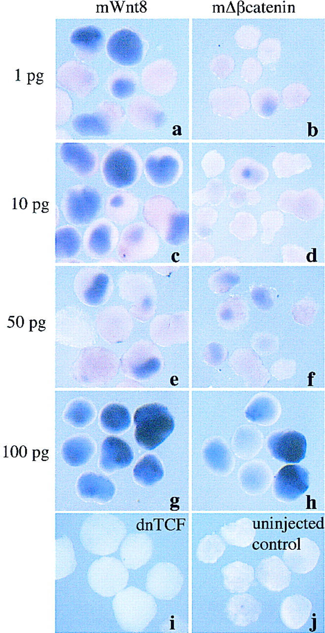
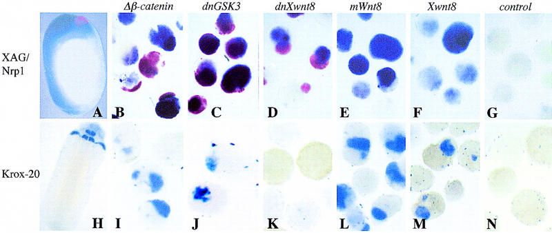
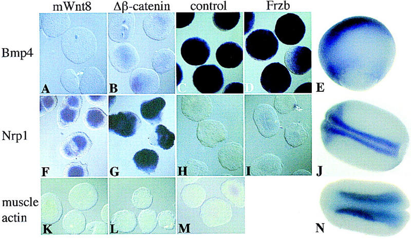
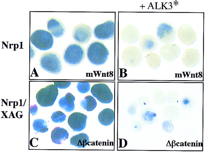
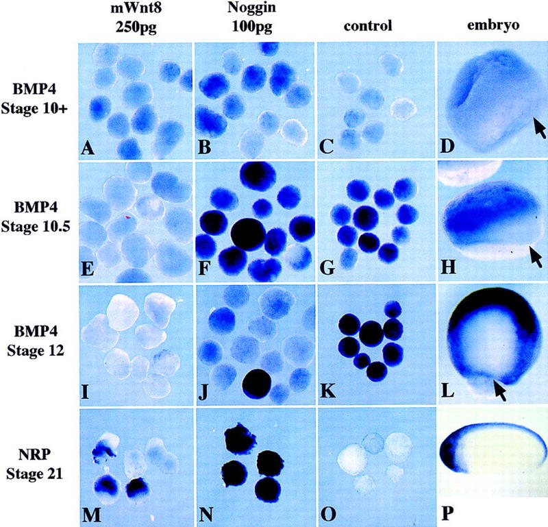
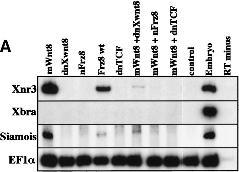
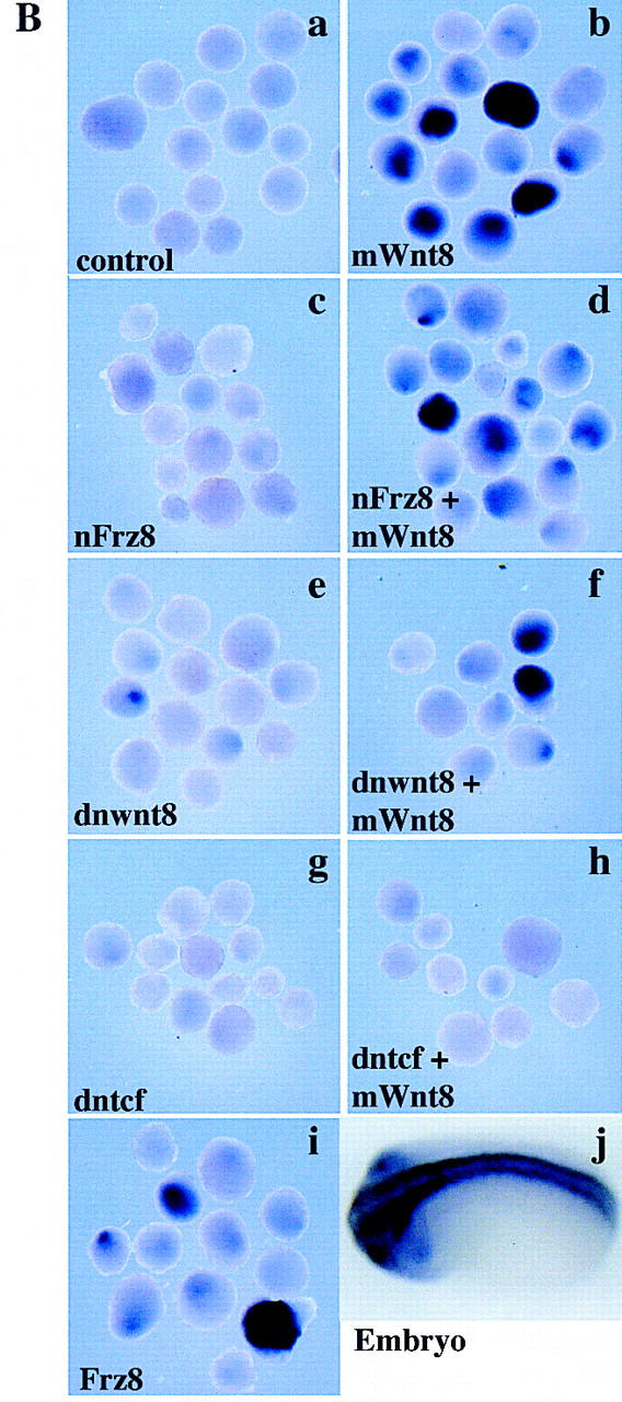
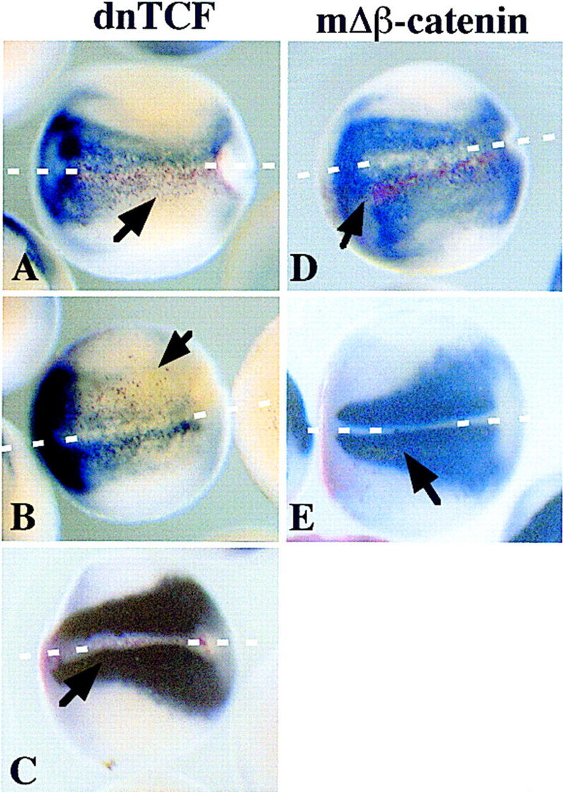
We report a new role for Wnt signaling in the vertebrate embryo: the induction of neural tissue from ectoderm. Early expression of mouse wnt8, Xwnt8, beta-catenin, or dominant-negative GSK3 induces the expression of neural-specific markers and inhibits the expression of Bmp4 in Xenopus ectoderm. We show that Wnt8, but not the BMP antagonist Noggin, can inhibit Bmp4 expression at early gastrula stages. Furthermore, inhibition of beta-catenin activity in the neural ectoderm of whole embryos by a truncated TCF results in a decrease in neural development. Therefore, we suggest that a cleavage-stage Wnt signal normally contributes to an early repression of Bmp4 on the dorsal side of the embryo and sensitizes the ectoderm to respond to neural inducing signals from the organizer. The Wnt targets Xnr3 and siamois have been shown previously to have neuralizing activity when overexpressed. However, antagonists of Wnt signaling, dnXwnt8 and Nxfrz8, inhibit Wnt-mediated Xnr3 and siamois induction, but not neural induction, suggesting an alternative mechanism for Bmp repression and neuralization. Conversely, dnTCF blocks both Wnt-mediated Xnr3 and neural induction, suggesting that both pathways require this transcription factor.


Figures

Figure 1
Figure 1
Wnt ligands, Frizzled receptors, and signaling components induce neural tissue in Xenopus ectodermal explants. Either mWnt3, Xwnt3a, Xwnt8, mWnt8, or mΔβ-catenin (500 pg or 100 pg) were injected into one-cell Xenopus embryos. Ectoderm was removed at blastula stage, and RNA extracted at late neurula stage (20) (A,C) gastrula stage (10.5) (B) or for analysis by RT-PCR. (A) Injected ectoderm analyzed at neurula stage for expression of the general neural marker, NCAM, the hindbrain marker, krox-20, the muscle-specific marker, muscle actin, and the ubiquitously expressed internal control, EF1α. (B) Injected ectoderm analyzed at the gastrula stage for the expression of the Wnt inducible gene, Xnr3, the general mesodermal marker, Xbra, and EF1α. (C) Ectoderm injected with 1 ng each of the following: Xwnt5a; human frizzled 5 (hfz5), a mutant human frizzled 5; Xenopus frizzled 8 (Xfz8); dnGSK3; and combinations of Xwnt5a, with either the wild-type hfz5 or mutant hfz5. In A–C, RNA from noninjected ectoderm, from whole embryos treated with reverse transcriptase, and from whole embryos treated without reverse transcriptase were used as controls. (D) Ectoderm injected with 1, 10, 50, or 100 pg of either mWnt8 (a,c,e,g) or mΔβ-catenin (b,d,f,h) aged until stage 20 and analyzed by in situ hybridization for Nrp1. Controls in this experiment include ectoderm injected with 1 ng of dnTCF (i) and uninjected ectoderm (j).
Figure 1
Figure 1
Wnt ligands, Frizzled receptors, and signaling components induce neural tissue in Xenopus ectodermal explants. Either mWnt3, Xwnt3a, Xwnt8, mWnt8, or mΔβ-catenin (500 pg or 100 pg) were injected into one-cell Xenopus embryos. Ectoderm was removed at blastula stage, and RNA extracted at late neurula stage (20) (A,C) gastrula stage (10.5) (B) or for analysis by RT-PCR. (A) Injected ectoderm analyzed at neurula stage for expression of the general neural marker, NCAM, the hindbrain marker, krox-20, the muscle-specific marker, muscle actin, and the ubiquitously expressed internal control, EF1α. (B) Injected ectoderm analyzed at the gastrula stage for the expression of the Wnt inducible gene, Xnr3, the general mesodermal marker, Xbra, and EF1α. (C) Ectoderm injected with 1 ng each of the following: Xwnt5a; human frizzled 5 (hfz5), a mutant human frizzled 5; Xenopus frizzled 8 (Xfz8); dnGSK3; and combinations of Xwnt5a, with either the wild-type hfz5 or mutant hfz5. In A–C, RNA from noninjected ectoderm, from whole embryos treated with reverse transcriptase, and from whole embryos treated without reverse transcriptase were used as controls. (D) Ectoderm injected with 1, 10, 50, or 100 pg of either mWnt8 (a,c,e,g) or mΔβ-catenin (b,d,f,h) aged until stage 20 and analyzed by in situ hybridization for Nrp1. Controls in this experiment include ectoderm injected with 1 ng of dnTCF (i) and uninjected ectoderm (j).
Figure 2
Figure 2
The secreted Wnt ligands Xwnt8 and mWnt8 induce neural tissue of posterior character, whereas Wnt signal transduction components induce neural tissue of both anterior and posterior character. One-cell Xenopus embryos were injected into the animal hemisphere with 0.5 ng of mΔβ-catenin (B,I), 1 ng of dnGSK3 (C,J), 1 ng of dnXwnt8 (D,K), 0.5 ng of mWnt8 (E,L), and 0.5 ng of Xwnt8 (F,M). At blastula stage ectoderm was explanted and allowed to develop until late-neurula stage (20). Aged ectoderm and whole embryos were then double stained by in situ hybridization for the cement gland marker, XAG (red), and the general neural marker, Nrp1 (blue) (A–G) or single stained by in situ hybridization for the hindbrain marker, krox-20 (blue) (H–N). A and H are whole-embryo controls; G and N are uninjected ectodermal explants.
Figure 3
Figure 3
Wnt signaling in ectoderm inhibits the expression of Bmp4. One-cell Xenopus embryos were injected into the animal hemisphere with 0.5 ng of mWnt8 (A,F,K), 0.5 ng of mΔβ-catenin (B,G,L), and 1 ng of Frzb (D,I). At blastula stage ectoderm was explanted and aged until gastrula stage (10.5) or neurula stage (20). Ectoderm was subsequently probed with digoxigenin-labeled antisense-Bmp4 (A–D), Nrp1 (F–I), or muscle actin (K–M), by in situ hybridization. E, J, and N are whole embryos probed with Bmp4, Nrp1, and muscle actin, respectively. C, H, and M are uninjected control ectodermal explants.
Figure 4
Figure 4
Activated ALK3 inhibits neural induction by mWnt8 and mΔβ-catenin. Ectoderm was injected with 100 pg of mWnt8 (A) or 500 pg of mΔβ-catenin (C) or coinjected along with 2 ng of constitutively active ALK3 (B,D). Injected ectoderm was aged until stage 20 and probed with either digoxigenin-labeled Nrp1 (A,B) or both digoxigenin-labeled Nrp1 and fluorescein-labeled XAG (C,D).
Figure 5
Figure 5
mWnt8, but not Noggin, represses Bmp4 expression during gastrulation. Ectoderm was injected with 250pg of mWnt8 (A,E,I,M) or 100 pg of noggin (B,F,J,N), aged to stage 10 (AC), stage 10.5 (E,F,G), stage 12 (IK), or stage 21 (MO), and probed with either digoxigenin-labeled Bmp4 (AL) or digoxigenin-labeled Nrp1 (MP). Uninjected ectoderm provides a negative control (C,G,K,O) and whole embryos indicate the normal pattern of expression (D,H,L,P). Arrows in D, H and L indicate the dorsal lip of the blastopore.
Figure 6
Figure 6
Neuralization without induction of β-catenin targets. One-cell Xenopus embryos were injected into the animal hemisphere with 250 pg of mWnt8, 1 ng of Nxfrz8, 500 pg of dnwnt8, 1 ng of dntcf, 1 ng of Frz8, or combinations of 250 pg of mWnt8 with either 1 ng of Nxfrz8, 500 pg of dnwnt8, or 1 ng of dntcf. At blastula stage ectoderm was explanted and aged until gastrula or late-neurula stages. Ectoderm aged until gastrula stage (10.5) was analyzed by RT-PCR for Xnr3, Xbra, EF1α and siamois (A); ectoderm aged until neurula stage (20) was probed with digoxigenin-labeled antisense-Nrp1 by in situ hybridization to detect general neural tissue (B). In B, a represents uninjected control ectodermal explants; j is a whole-embryo control.
Figure 6
Figure 6
Neuralization without induction of β-catenin targets. One-cell Xenopus embryos were injected into the animal hemisphere with 250 pg of mWnt8, 1 ng of Nxfrz8, 500 pg of dnwnt8, 1 ng of dntcf, 1 ng of Frz8, or combinations of 250 pg of mWnt8 with either 1 ng of Nxfrz8, 500 pg of dnwnt8, or 1 ng of dntcf. At blastula stage ectoderm was explanted and aged until gastrula or late-neurula stages. Ectoderm aged until gastrula stage (10.5) was analyzed by RT-PCR for Xnr3, Xbra, EF1α and siamois (A); ectoderm aged until neurula stage (20) was probed with digoxigenin-labeled antisense-Nrp1 by in situ hybridization to detect general neural tissue (B). In B, a represents uninjected control ectodermal explants; j is a whole-embryo control.
Figure 7
Figure 7
β-Catenin is necessary for normal neural development. dnTCF or mΔβ-catenin (200 pg), along with 200 pg of lacZ mRNA, were injected into one of two dorsal blastomeres of eight-cell-staged Xenopus embryos. At neural-plate stage (14), the embryos were stained for the presence of β-galactosidase activity with Red Gal and subsequently probed with digoxigenin-labeled antisense-Nrp1 (A,B,D) or digoxigenin-labeled antisense muscle actin (C,E) by in situ hybridization. A, B, and C are embryos that have been injected with dnTCF; D and E have been injected with mΔβ-catenin. Blue indicates the presense of neural tissue (A,B,D) or muscle (C,E); red indicates which cells are expressing the injected proteins. Cells expressing Red Gal were exclusively ectodermal. Arrows point to domains of β-galactosidase activity. White broken lines delineate the midline in all panels.

References

    1. Axelrod JD, Miller JR, Shulman JM, Moon RT, Perrimon N. Differential recruitment of Dishevelled provides signaling specificity in the planar cell polarity and Wingless signaling pathways. Genes & Dev. 1998;12:2610–2622. - PMC - PubMed
    1. Baker JC, Harland RM. A novel mesoderm inducer, Madr2, functions in the activin signal transduction pathway. Genes & Dev. 1996;10:1880–1889. - PubMed
    1. Bejsovec A, Wieschaus E. Signaling activities of the Drosophila wingless gene are separately mutable and appear to be transduced at the cell surface. Genetics. 1995;139:309–320. - PMC - PubMed
    1. Belo JA, Bouwmeester T, Leyns L, Kertesz N, Gallo M, Follettie M, De Robertis EM. Cerberus-like is a secreted factor with neutralizing activity expressed in the anterior primitive endoderm of the mouse gastrula. Mech Dev. 1997;68:45–57. - PubMed
    1. Bhanot P, Brink M, Samos CH, Hsieh JC, Wang Y, Macke JP, Andrew D, Nathans J, Nusse R. A new member of the frizzled family from Drosophila functions as a Wingless receptor. Nature. 1996;382:225–230. - PubMed

Publication types

MeSH terms

Substances

LinkOut - more resources