. 1999 Dec;181(24):7414-20.
doi: 10.1128/JB.181.24.7414-7420.1999.

Localization of synthesis of beta1,6-glucan in Saccharomyces cerevisiae

Affiliations

Localization of synthesis of beta1,6-glucan in Saccharomyces cerevisiae

R C Montijn et al. J Bacteriol. 1999 Dec.

Abstract

Beta1,6-Glucan is a key component of the yeast cell wall, interconnecting cell wall proteins, beta1,3-glucan, and chitin. It has been postulated that the synthesis of beta1,6-glucan begins in the endoplasmic reticulum with the formation of protein-bound primer structures and that these primer structures are extended in the Golgi complex by two putative glucosyltransferases that are functionally redundant, Kre6 and Skn1. This is followed by maturation steps at the cell surface and by coupling to other cell wall macromolecules. We have reinvestigated the role of Kre6 and Skn1 in the biogenesis of beta1,6-glucan. Using hydrophobic cluster analysis, we found that Kre6 and Skn1 show significant similarities to family 16 glycoside hydrolases but not to nucleotide diphospho-sugar glycosyltransferases, indicating that they are glucosyl hydrolases or transglucosylases instead of genuine glucosyltransferases. Next, using immunogold labeling, we tried to visualize intracellular beta1,6-glucan in cryofixed sec1-1 cells which had accumulated secretory vesicles at the restrictive temperature. No intracellular labeling was observed, but the cell surface was heavily labeled. Consistent with this, we could detect substantial amounts of beta1,6-glucan in isolated plasma membrane-derived microsomes but not in post-Golgi secretory vesicles. Taken together, our data indicate that the synthesis of beta1, 6-glucan takes place largely at the cell surface. An alternative function for Kre6 and Skn1 is discussed.


Figures

FIG. 1
FIG. 1
HCA plots of selected members of family 16 glycoside hydrolases and transglycosidases (18, 20). From top to bottom: Kre6 of Saccharomyces cerevisiae (SwissProt P32486), clotting factor G alpha subunit of Tachypleus tridentatus (GenBank D16622), β1,3-glucanase II of Oerskovia xanthineolytica (GenBank AF052745), and β1,3-1,4-glucanase of Bacillus macerans (SwissProt P23904; PDB 1BYH). The HCA plots were made, edited, and analyzed as described elsewhere (15, 19). To facilitate visual inspection of the plots, the symbols ∗, ⧫, ⊡, and □ are used for proline, glycine, serine, and threonine, respectively. Vertical lines show correspondences between proteins. The two catalytic glutamate residues are shown in white on black circles. The secondary structure elements of 1BYH are shown as open (β strand) and grey (α helix) boxes under the corresponding plot. The two insertions found in Kre6 are marked A and B.
FIG. 2
FIG. 2
Schematic 3-D structure of the β1,3-1,4-glucanase of B. macerans (PDB 1BYH). The two glutamate residues that belong to the catalytic site are shown in ball-and-stick representation. The two loops which carry the insertions in Kre6 are labeled A and B. The location of the resulting putative protuberance in Kre6 is shown by dashed lines. The figure was prepared with the program MOLSCRIPT (30).
FIG. 3
FIG. 3
Pustulan affinity-purified antibodies recognize protein-bound gentiobiose. Lane 1, 1 μg of BSA; lane 2, 1 μg of gentiobiose-BSA; lane 3, 5 μg of gentiobiose-BSA. The blot was developed with ECL for 1 min. Sizes are indicated in kilodaltons.
FIG. 4
FIG. 4
Immunogold labeling of β1,6-glucan in sec1-1 cells. (A) To induce accumulation of post-Golgi secretory vesicles, sec1-1 cells were kept at the restrictive temperature for 2 h. A representative cell is shown. About 360 gold particles are visible at the cell surface, whereas intracellular labeling is negligible. (B) The vesicles were visualized by cross-linking and staining of the carbohydrate cargo by the methods of McLean and Nakane (35) and Shinji et al. (49), respectively. Bar = 250 nm.
FIG. 5
FIG. 5
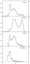
β1,6-Glucan colocalizes with plasma membrane-derived vesicles. To induce accumulation of post-Golgi secretory vesicles, sec1-1 cells were kept at the restrictive temperature for 2 h. The cells were spheroplasted and gently lysed. The resulting homogenate was fractionated by differential centrifugation, and the microsomal fraction was separated by gel filtration on a Sephacryl S-1000 column (55, 56). Aliquots of each column fraction (x axis) were assayed for protein content, invertase, plasma membrane ATPase, and β1,6-glucan content. y axes represent, from top to bottom, protein (micrograms), invertase (micromoles of Glc per minute), plasma membrane ATPase (micromoles of Pi per minute), and β1,6-glucan (arbitrary densitometric units).

References

    1. Barbeyron T, Gerard A, Potin P, Henrissat B, Kloareg B. The kappa-carrageenase of the marine bacterium Cytophaga drobachiensis. Structural and phylogenetic relationships within family-16 glycoside hydrolases. Mol Biol Evol. 1998;15:528–537. - PubMed
    1. Bickle M, Delley P A, Schmidt A, Hall M N. Cell wall integrity modulates RHO1 activity via the exchange factor ROM2. EMBO J. 1998;17:2235–2245. - PMC - PubMed
    1. Boone C, Sommer S S, Hensel A, Bussey H. Yeast KRE genes provide evidence for a pathway of cell wall β-glucan assembly. J Cell Biol. 1990;110:1833–1843. - PMC - PubMed
    1. Bowman B J, Slayman C W. The effect of vanadate on the plasma membrane ATPase of Neurospora crassa. J Biol Chem. 1979;254:2928–2934. - PubMed
    1. Brown J L, Bussey H. The yeast KRE9 gene encodes an O-glycoprotein involved in cell surface β-glucan assembly. Mol Cell Biol. 1993;13:6346–6356. - PMC - PubMed

Publication types

MeSH terms