. 1999 Dec;181(24):7421-9.
doi: 10.1128/JB.181.24.7421-7429.1999.

Characterization of a novel, antifungal, chitin-binding protein from Streptomyces tendae Tü901 that interferes with growth polarity

Affiliations

Characterization of a novel, antifungal, chitin-binding protein from Streptomyces tendae Tü901 that interferes with growth polarity

C Bormann et al. J Bacteriol. 1999 Dec.

Abstract

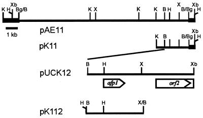
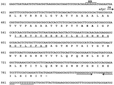
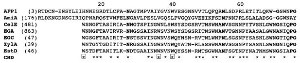
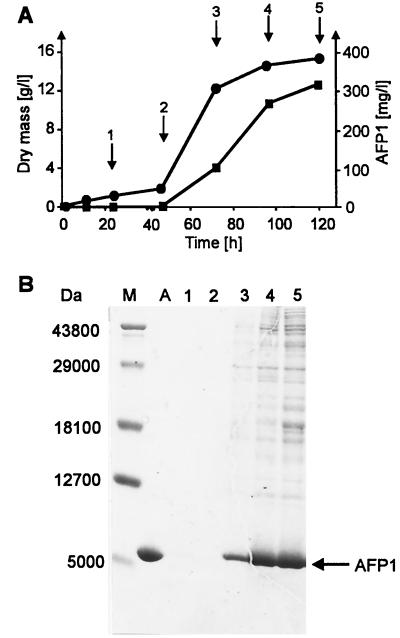
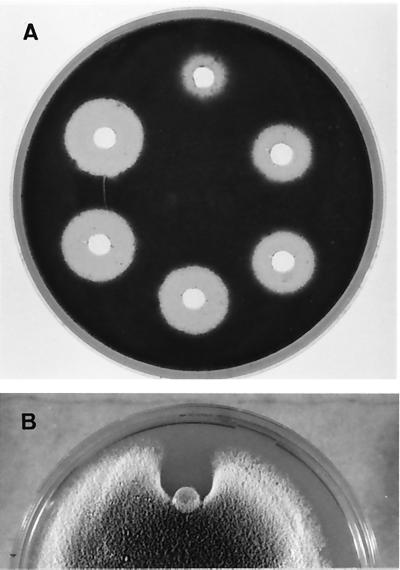
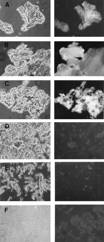
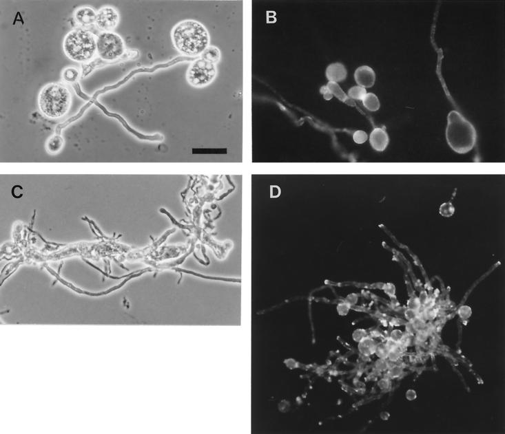
The afp1 gene, which encodes the antifungal protein AFP1, was cloned from nikkomycin-producing Streptomyces tendae Tü901, using a nikkomycin-negative mutant as a host and screening transformants for antifungal activity against Paecilomyces variotii in agar diffusion assays. The 384-bp afp1 gene has a low G+C content (63%) and a transcription termination structure with a poly(T) region, unusual attributes for Streptomyces genes. AFP1 was purified from culture filtrate of S. tendae carrying the afp1 gene on the multicopy plasmid pIJ699. The purified protein had a molecular mass of 9,862 Da and lacked a 42-residue N-terminal peptide deduced from the nucleotide sequence. AFP1 was stable at extreme pH values and high temperatures and toward commercial proteinases. AFP1 had limited similarity to cellulose-binding domains of microbial plant cell wall hydrolases and bound to crab shell chitin, chitosan, and cell walls of P. variotii but showed no enzyme activity. The biological activity of AFP1, which represents the first chitin-binding protein from bacteria exhibiting antifungal activity, was directed against specific ascomycetes, and synergistic interaction with the chitin synthetase inhibitor nikkomycin inhibited growth of Aspergillus species. Microscopy studies revealed that fluorescein-labeled AFP1 strongly bound to the surface of germinated conidia and to tips of growing hyphae, causing severe alterations in cell morphogenesis that gave rise to large spherical conidia and/or swollen hyphae and to atypical branching.


Figures

FIG. 1
FIG. 1
Restriction map of the 14.5-kb BamHI insert of pAE11 and subcloned fragments containing the afp1 gene. Bold lines represent segments of the vector pIJ699. The nucleotide sequence of the 2.08-kb BamHI-XbaI insert of pUCK12 was determined. The size and location of the afp1 gene and the 5′ region of orf2 were deduced from the nucleotide sequence and are indicated by an arrow and a box, respectively. Abbreviations for restriction enzymes: B, BamHI; Bg, BglII; H, HindIII; K, KpnI; X, XhoI; Xb, XbaI.
FIG. 2
FIG. 2
Nucleotide sequence of the S. tendae Tü901 afp1 gene and the predicted amino acid sequence. A putative ribosome-binding site (SD) is overlined. The translational start site of the afp1 gene is indicated by an arrow, and the termination codon is marked by a dash. Amino acids determined experimentally by N-terminal sequencing are underlined. The proposed cleavage site for signal peptidase is in boldface. →← indicates an inverted repeat sequence, and the poly(T) region is underlined.
FIG. 3
FIG. 3
BLASTP sequence alignment of amino acid residues 3 to 75 of the extracellular AFP1 with segments of N-acetylmuramoyl-l-alanine amidase from Synechocystis sp. (AmiA) and cellulase CelE (CelE), endoglucanase A (EGA), cellodextrinase (EGC), xylanase A (XylA), and esterase D (EstD) from Pseudomonas fluorescens. The sequences were obtained from GenBank (AFP1, this work; CelE, X86798; AmiA, D90909; CelE, X86798; EGA, X12570; EstD, X15429), from the PIR database (EGC, S19652), and from Swiss-Prot (XylA, P14768). CBD indicates residues of cellulose-binding domains which are identical or similar in the aligned sequences of CelE, EGA, EGC, XylA, and EstD. Aromatic amino acid residues involved in protein-sugar binding (34) are boxed. Amino acid residues that are identical in six of seven sequences are in boldface.
FIG. 4
FIG. 4
Time course of AFP1 production in S. tendae NP51(pK11) cultivated in CRM medium. (A) AFP1 (■) in the culture filtrate determined by agar diffusion assays with P. variotii as the test fungus compared to growth as determined by dry weight (●). (B) SDS-PAGE of culture filtrates from S. tendae NP51(pK11) and purified AFP1. Lanes 1 to 5, culture filtrate (40 μl) taken at time points (1 to 5) indicated by arrows in panel A; lane A, purified AFP1 (5 μg); lane M, molecular mass markers. Proteins were stained with Coomassie blue. AFP1 is marked by an arrow.
FIG. 5
FIG. 5
Growth inhibition of P. variotii by purified AFP1. (A) Growth of spores in response to 4, 8, 10, 15, 20, and 30 μg of AFP1 per well (clockwise beginning at the top). (B) Growth of hyphae in response to 30 μg of AFP1 applied on the paper disk, placed at the growing front of a colony and incubated for further 48 h.
FIG. 6
FIG. 6
Binding of AFP1 to chitin, chitosan, and fungal cell walls. Fluorescein-labeled AFP1 was incubated overnight with crab shell chitin (A), crab shell chitosan (B), P. variotii cell wall (C), β-1,3-glucan (curdlan; D), Avicel (E), and xylan (F). After three washes with 100 mM Tris-HCl (pH 7.6)–150 mM NaCl, samples were analyzed by phase-contrast (left) and fluorescence (right) microscopy.
FIG. 7
FIG. 7
Effect of AFP1 on the morphology of P. variotii and A. fumigatus and its localization. P. variotii (A and B) and A. fumigatus (C and D) were grown in the presence of fluorescein-labeled AFP1 (A, B, and D, 50 μg ml−1; C, 75 μg ml−1) for 12 h and analyzed by phase-contrast (left) and fluorescence (right) microscopy. Scale bar represents 20 μm.

References

    1. Abad L R, D'Urzo M P, Liu D, Narasimhan M L, Reuveni M, Zhu J K, Niu X, K. S N, Hasegawa P M, Bressan R A. Antifungal activity of tobacco osmotin has specificity and involves plasma membrane permeabilization. Plant Sci. 1996;118:11–23.
    1. Altschul S F, Madden T L, Schäffer A A, Zhang J, Zhang Z, Miller W, Lipman D J. Gapped BLAST and PSI-BLAST: a new generation of protein database search programs. Nucleic Acids Res. 1997;25:3389–3402. - PMC - PubMed
    1. Bormann C, Aberle K, Fiedler H-P, Schrempf H. Genetic complementation of Streptomyces tendaedeficient in nikkomycin production. Appl Microbiol Biotechnol. 1990;32:424–430. - PubMed
    1. Bormann C, Mattern S, Schrempf H, Fiedler H P, Zähner H. Isolation of Streptomyces tendaemutants with an altered nikkomycin spectrum. J Antibiot. 1989;42:913–918. - PubMed
    1. Broekaert W F, Marien W, Terras F R G, De Brolle M F C, Proost P, Van Damme J, Dillen L, Claeys M, Rees S B, Vanderleyden J, Cammue B P A. Antimicrobial peptides from Amaranthus caudatusseeds with sequence homology to the cysteine/glycine-rich domain of chitin-binding proteins. Biochemistry. 1992;31:4308–4314. - PubMed

Publication types

MeSH terms

Associated data

LinkOut - more resources