. 1999 Dec 13;147(6):1129-36.
doi: 10.1083/jcb.147.6.1129.

A novel regulatory mechanism of MAP kinases activation and nuclear translocation mediated by PKA and the PTP-SL tyrosine phosphatase

Affiliations

A novel regulatory mechanism of MAP kinases activation and nuclear translocation mediated by PKA and the PTP-SL tyrosine phosphatase

C Blanco-Aparicio et al. J Cell Biol. .

Abstract

Protein tyrosine phosphatase PTP-SL retains mitogen-activated protein (MAP) kinases in the cytoplasm in an inactive form by association through a kinase interaction motif (KIM) and tyrosine dephosphorylation. The related tyrosine phosphatases PTP-SL and STEP were phosphorylated by the cAMP-dependent protein kinase A (PKA). The PKA phosphorylation site on PTP-SL was identified as the Ser(231) residue, located within the KIM. Upon phosphorylation of Ser(231), PTP-SL binding and tyrosine dephosphorylation of the MAP kinases extracellular signal-regulated kinase (ERK)1/2 and p38alpha were impaired. Furthermore, treatment of COS-7 cells with PKA activators, or overexpression of the Calpha catalytic subunit of PKA, inhibited the cytoplasmic retention of ERK2 and p38alpha by wild-type PTP-SL, but not by a PTP-SL S231A mutant. These findings support the existence of a novel mechanism by which PKA may regulate the activation and translocation to the nucleus of MAP kinases.


Figures
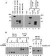
Figure 1
Figure 1
Phosphorylation of PTP-SL and STEP by PKA. (A) GST fusion proteins (1 μg) were phosphorylated in vitro by cPKA in the presence of γ-[32P]ATP as indicated. (B) COS-7 cells were mock-transfected (pRK5 vector alone) or transfected with the pRK5 PTP-SL wild type or S231A, as indicated, followed by 32P-labeling. Cells were left untreated (−) or were treated (+) with dibutyryl-cAMP, forskolin, or okadaic acid, as described in Materials and Methods. PTP-SL was precipitated from cell lysates with anti–PTP-SL antibody. All samples (A and B) were resolved by 10% SDS-PAGE under reducing conditions, and followed by autoradiography.
Figure 2
Figure 2
Effect of PTP-SL phosphorylation by PKA on the association with MAP kinases and their dephosphorylation. (A) GST-PTP-SL 147-288 wild type or S231A fusion proteins (1.5 μg) were left untreated (−) or were phosphorylated in vitro by cPKA (+) in the presence of cold ATP, as indicated. Rat-1 cell lysates (500 μg) were added, and the fusion proteins were precipitated with glutathione-Sepharose. The kinases were detected by immunoblot analysis with anti–ERK1/2 (top) or anti–p38α (bottom) antibodies. In lane 1, total lysate samples (20 μg) were loaded. Arrowheads indicate the migration of the kinases. (B) 293 cells were transfected with pRK5 GST-PTP-SL 147-549 (both panels); in the bottom panel, cells were cotransfected with pECE-HA-p38MAPK. After 48 h, cells were left untreated (−) or were treated with dibutyryl-cAMP, dibutyryl-cAMP plus H89, or forskolin, as indicated. The GST-PTP-SL fusion proteins were precipitated from the cell lysates with glutathione-Sepharose, and coprecipitated kinases were detected by immunoblot analysis with anti–ERK1/2 (top) or anti–HA (bottom) antibodies. (C) 293 cells were transfected with pRK5 GST (lane 2) or the pRK5 GST-PTP-SL 147-549 wild type or mutants, as indicated, and fusion proteins were precipitated as in B, followed by immunoblot with anti-ERK1/2 or anti-p38α antibodies. In lane 1, total lysate (20 μg) was loaded. All GST-PTP-SL proteins were equally expressed. (D) Tyrosine-phosphorylated HA-ERK2 or HA-p38α were precipitated with the anti–HA 12CA5 mAb from activated 293 cells, transfected with pCDNA3-HA-ERK2 (lanes 1–5) or pECE-HA-p38MAPK (lanes 6–10), and immune complexes were subjected to in vitro phosphatase assays during the indicated times (in minutes) in the presence of GST-PTP-SL 147-549 wild type (lanes 2, 3, 7, and 8) or S231E (lanes 4, 5, 9, and 10) (1 μg). In lanes 1 and 6, no fusion proteins were added, and samples were kept on ice. Tyrosine phosphorylation was detected by immunoblot with the anti-phosphotyrosine 4G10 mAb (top panels). Bottom panels show the equal presence of HA-ERK2 and HA-p38α in all lanes, after stripping of the filters and reprobing with the anti-HA 12CA5 mAb. Equal activities of GST-PTP-SL wild type and S231E towards pNPP were measured (not shown). All samples (A–D) were resolved by 10% SDS-PAGE under reducing conditions.
Figure 3
Figure 3
Transmembrane PTP-SL retains ERK2 and p38α outside of the nucleus. COS-7 cells were cotransfected with pcDNA3-HA-ERK2 or pECE-HA-p38MAPK, plus pRK5-PTP-SL 1-549 wild type or ΔKIM (Δ224-239) mutant, as indicated. 48 h after transfection, cells were costained and analyzed by immunofluorescence. HA-ERK2 and HA-p38α were stained with the mouse anti–HA mAb 12CA5 plus rhodamine-conjugated goat anti–mouse antibody (red, A, D, G, and J). PTP-SL was stained with rabbit polyclonal anti–PTP-SL antibody plus fluorescein isothiocyanate–conjugated goat anti–rabbit antibody (green, B, E, H, and K; subcellular localization of PTP-SL 1-549 corresponds to perinuclear areas in the cytoplasm). In C, F, I, and L, double color staining is shown; yellow areas correspond to colocalization of HA-ERK2 or HA-p38α, and PTP-SL.
Figure 4
Figure 4
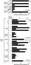
(A) PTP-SL retains HA-ERK2 in the cytoplasm independently of its PTP domain. COS-7 cells were cotransfected with pcDNA3-HA-ERK2 plus pRK5 (mock), or plus pRK5 PTP-SL 147-549, 1-549, or 1-288 wild type or mutants, as indicated. Cells were costained and analyzed by immunofluorescence as in Fig. 3. PTP-SL 147-549 is located in the cytoplasm. Subcellular localization of PTP-SL 1-549 and 1-288 is identical (see Fig. 3). (B, C) Effect of PKA on the nuclear localization of HA-ERK2 and HA-p38α in the presence of PTP-SL. COS-7 cells were cotransfected with pcDNA3-HA-ERK2 (B) or pECE-HA-p38MAPK (C) plus pRK5-PTP-SL 1-549 wild type or mutants, as indicated. Similar results were obtained with the double mutant C480S/S231A and the single mutant S231A (not shown). In some points, cells were additionally transfected with pCαEV (cPKAα) (+), and were induced as described in Materials and Methods. Cells were left untreated (−) or were treated (+) with dibutyryl-cAMP or dibutyryl-cAMP plus H89, as indicated, and then were costained and analyzed by immunofluorescence. HA-ERK2 or HA-p38α nuclear localization is presented as the percentage of cells coexpressing PTP-SL and HA-ERK2, or PTP-SL and HA-p38α that showed the MAP kinase located into the nucleus. For each point, at least 100 double positive cells were scored. Bars represent the mean ± SD of at least two separate experiments.
Figure 5
Figure 5
Model of MAP kinase regulation by PKA and PTP-SL. Pools of MAP kinases are maintained in the cytoplasm and into the nucleus by the balance between activation stimuli and the PTP-SL (or other KIM-containing PTPs) inhibitory effects. Upon PKA activation, the association of PTP-SL with the MAP kinase is impaired, and MAP kinase tyrosine phosphorylation and nuclear translocation is favored. The putative regulatory role for serine/threonine phosphatases (PP) in the dephosphorylation of PTP-SL, is indicated (see details in the text).

References

    1. Anderson N.G., Maller J.L., Tonks N.K., Sturgill T.W. Requirement for integration of signals from two distinct phosphorylation pathways for activation of MAP kinase. Nature. 1990;343:651–653. - PubMed
    1. Brunet A., Pouysségur J. Identification of MAP kinase domains by redirecting stress signals into growth factor responses. Science. 1996;272:1652–1655. - PubMed
    1. Burgering B.M.T., Bos J.L. Regulation of Ras-mediated signallingmore than one way to skin a cat. Trends Biochem. Sci. 1995;20:18–22. - PubMed
    1. Chajry N., Martin P.-M., Cochet C., Berthois Y. Regulation of p42 mitogen-activated-protein kinase activity by protein phosphatase 2A under conditions of growth inhibition by epidermal growth factor in A431 cells. Eur. J. Biochem. 1996;235:97–102. - PubMed
    1. Cobb M.H., Goldsmith E.J. How MAP kinases are regulated. J. Biol. Chem. 1995;270:14843–14846. - PubMed

Publication types

MeSH terms

Substances