. 1999 Dec 13;147(6):1145-52.
doi: 10.1083/jcb.147.6.1145.

Axo-glial interactions regulate the localization of axonal paranodal proteins

Affiliations

Axo-glial interactions regulate the localization of axonal paranodal proteins

J L Dupree et al. J Cell Biol. .

Abstract

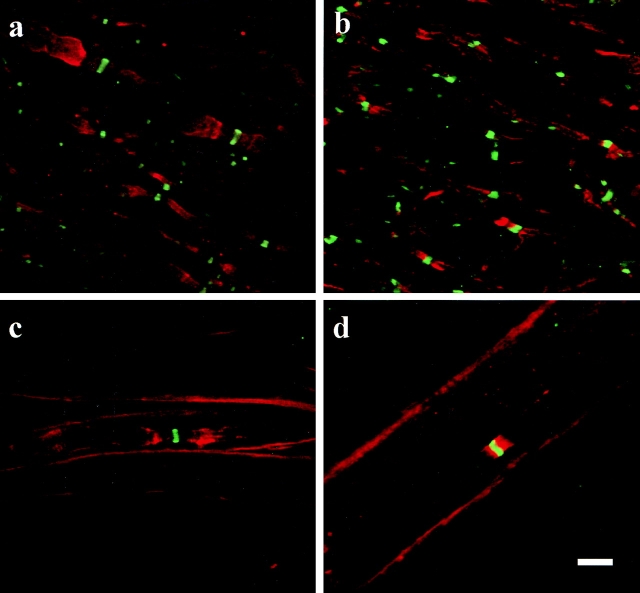
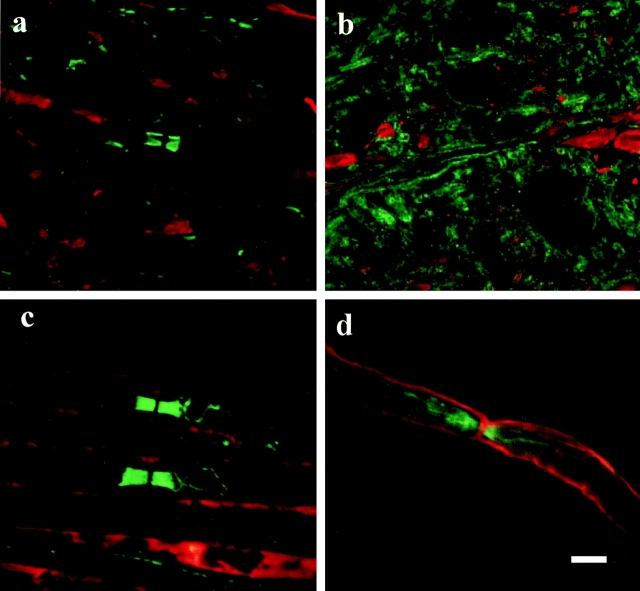
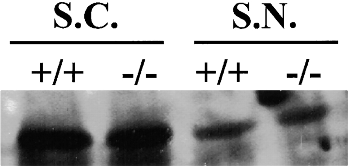
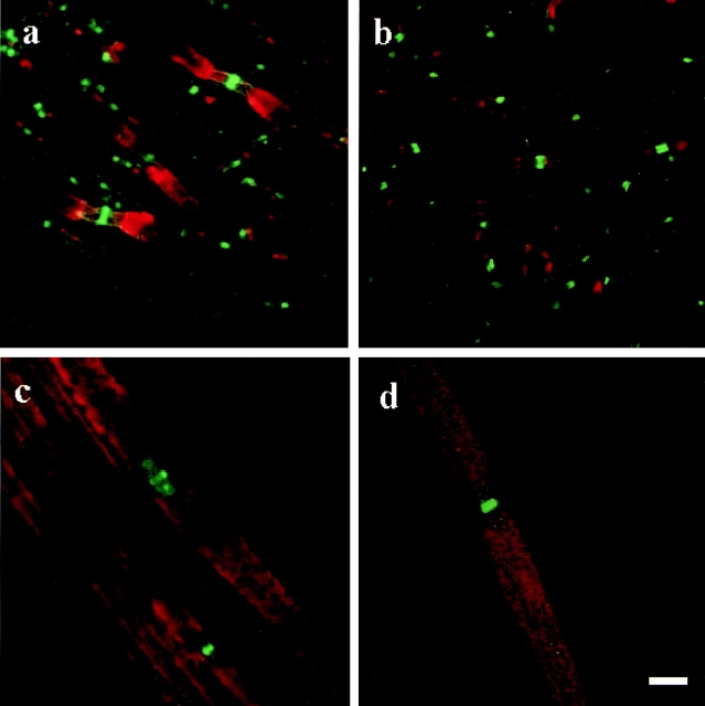
Mice incapable of synthesizing the abundant galactolipids of myelin exhibit disrupted paranodal axo-glial interactions in the central and peripheral nervous systems. Using these mutants, we have analyzed the role that axo-glial interactions play in the establishment of axonal protein distribution in the region of the node of Ranvier. Whereas the clustering of the nodal proteins, sodium channels, ankyrin(G), and neurofascin was only slightly affected, the distribution of potassium channels and paranodin, proteins that are normally concentrated in the regions juxtaposed to the node, was dramatically altered. The potassium channels, which are normally concentrated in the paranode/juxtaparanode, were not restricted to this region but were detected throughout the internode in the galactolipid-defi- cient mice. Paranodin/contactin-associated protein (Caspr), a paranodal protein that is a potential neuronal mediator of axon-myelin binding, was not concentrated in the paranodal regions but was diffusely distributed along the internodal regions. Collectively, these findings suggest that the myelin galactolipids are essential for the proper formation of axo-glial interactions and demonstrate that a disruption in these interactions results in profound abnormalities in the molecular organization of the paranodal axolemma.


Figures

Figure 1
Figure 1
(a) In the spinal cord of wild-type mice, potassium channels (red) are concentrated in the juxtaparanodes, and to a lesser extent, in the paranode, whereas the sodium channels (green) are restricted to the nodes of Ranvier. (b) In contrast, the potassium channels rarely accumulate in the juxtaparanodes of spinal cord tissue from the galactolipid-deficient mice. However, similar to the wild-type, the sodium channels cluster in the nodes but the sodium channel domains are slightly longer in the mutant as compared with the wild-type. The distribution of the potassium and sodium channels in the PNS is similar to the CNS for both the wild-type (c) and the mutant (d) mice. Note that in the CNS and the PNS of the galactolipid-deficient mice the channel domains occasionally overlap (yellow). a and b were generated as the compilation of eight consecutive images each 0.4 μm apart. c and d were generated as the compilation of 10 images each 0.26 μm apart. Bar, 5 μm.
Figure 2
Figure 2
In the wild-type mice paranodin (green) is highly concentrated in the paranodal regions of spinal cord (a) and sciatic nerve (c) axons. In contrast, the galactolipid-deficient mice exhibit a more diffuse labeling pattern. In the mutant spinal cord (b) paranodin is evenly distributed in the axolemma throughout the internode. In the sciatic nerve (d) of these mice, paranodin is concentrated in the paranode but the interface between the paranode and the juxtaparanode is not clearly demarcated. a and b, eight images 0.26 μm apart; double-labeled for paranodin in green and phosphorylated neurofilament in red. c and d, eight images 0.4 μm apart; double-labeled for paranodin in green and myelin basic protein in red. Bar, 5 μm.
Figure 3
Figure 3
Western blot analysis revealed no difference in the expression of paranodin between the wild-type (+/+) and the mutant (−/−) mice in either the spinal cord (S.C.) or the sciatic nerve (S.N.).
Figure 4
Figure 4
In the spinal cord, neurofascin (green) was restricted to the nodes of Ranvier in both the wild-type (a) and the mutant (b) mice. a and b are also labeled for the potassium channels (red) in an effort to assist with the recognition of nodal/paranodal regions. In the sciatic nerve (c and d), neurofascin also is concentrated in the node; however, some protein is also located in the paranodal regions of the wild-type (c) and mutant (d) mice. a and b, six images 0.31 μm apart; double-labeled for neurofascin in green and potassium in red. c and d, four images 0.26 μm apart; double-labeled for neurofascin in green and myelin basic protein in red. Bar, 5 μm.

References

    1. Black J.A., Kocsis J.D., Waxman S.G. Ion channel organization of the myelinated fiber. Trends Neurosci. 1990;13:48–54. - PubMed
    1. Bosio A., Bussow H., Adam J., Stoffel W. Galactosphingolipids and axono-glial interaction in myelin of the central nervous system. Cell Tissue Res. 1998;292:199–210. - PubMed
    1. Coetzee T., Fujita N., Dupree J., Shi R., Blight A., Suzuki K., Suzuki K., Popko B. Myelination in the absence of galactocerbroside and sulfatidenormal structure with abnormal function and regional instability. Cell. 1996;86:209–219. - PubMed
    1. Dugandzija-Novakovic S., Koszowski A.G., Levinson S.R., Shrager P. Clustering of Na+ channels and node of Ranvier formation in remyelinating axons. J. Neurosci. 1995;15:492–503. - PMC - PubMed
    1. Dupree J.L., Coetzee T., Blight A., Suzuki K., Popko B. Myelin galactolipids are essential for proper node of ranvier formation in the CNS. J. Neurosci. 1998;18:1642–1649. - PMC - PubMed

Publication types

MeSH terms