. 1999 Dec 13;147(6):1167-80.
doi: 10.1083/jcb.147.6.1167.

The A-kinase-anchoring protein AKAP95 is a multivalent protein with a key role in chromatin condensation at mitosis

Affiliations

The A-kinase-anchoring protein AKAP95 is a multivalent protein with a key role in chromatin condensation at mitosis

P Collas et al. J Cell Biol. .

Abstract

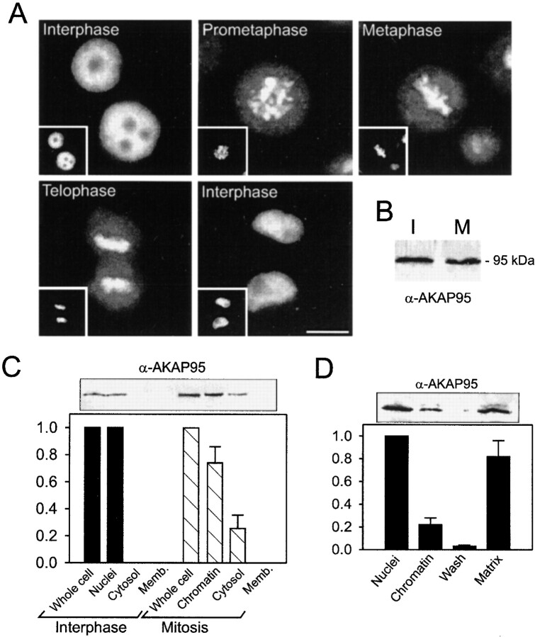
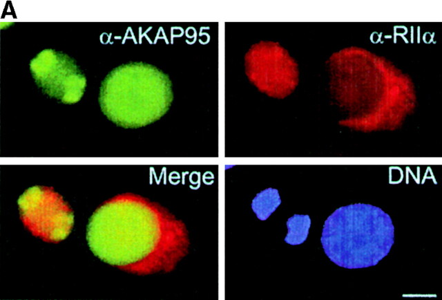
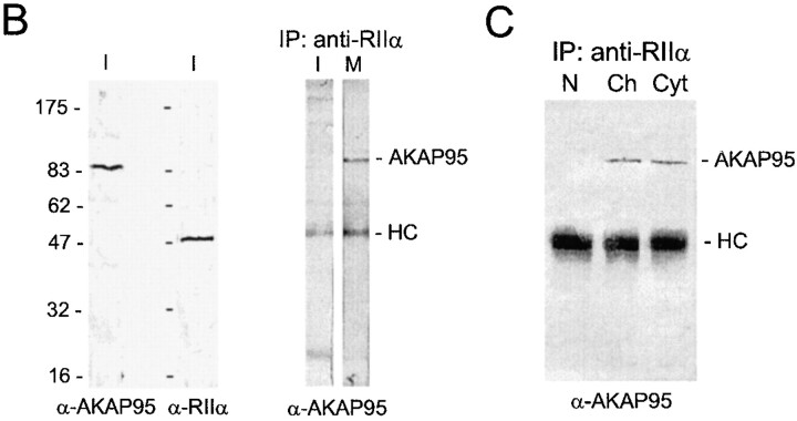
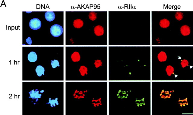
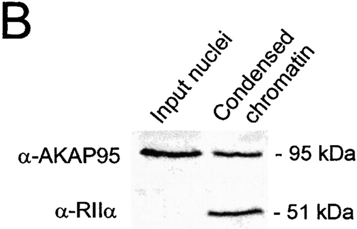
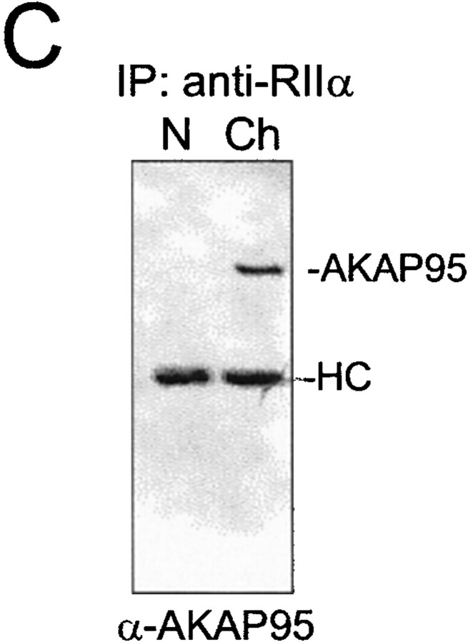
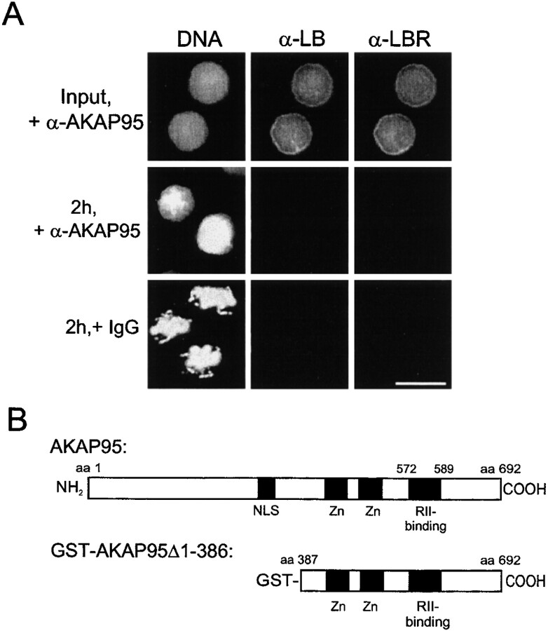
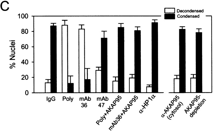
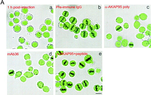
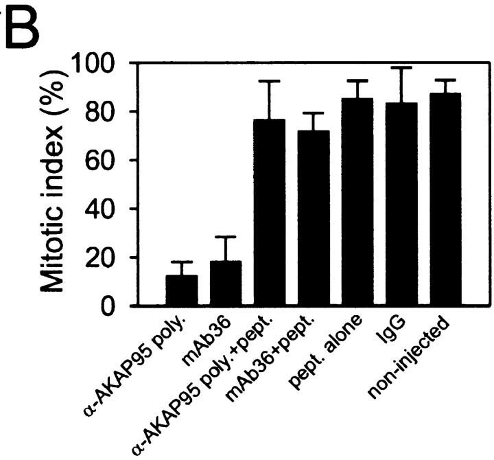
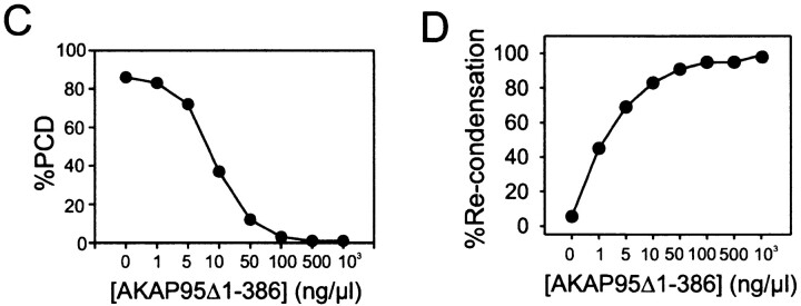
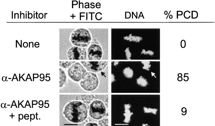
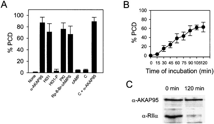
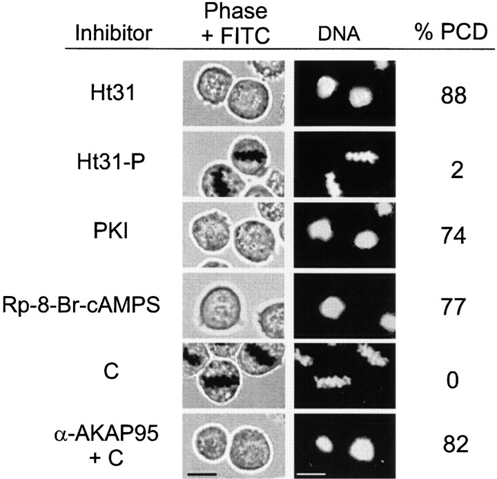
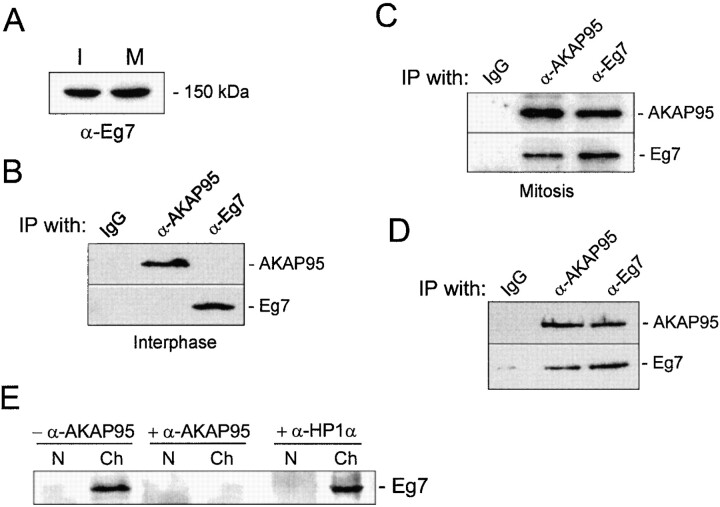
Protein kinase A (PKA) and the nuclear A-kinase-anchoring protein AKAP95 have previously been shown to localize in separate compartments in interphase but associate at mitosis. We demonstrate here a role for the mitotic AKAP95-PKA complex. In HeLa cells, AKAP95 is associated with the nuclear matrix in interphase and redistributes mostly into a chromatin fraction at mitosis. In a cytosolic extract derived from mitotic cells, AKAP95 recruits the RIIalpha regulatory subunit of PKA onto chromatin. Intranuclear immunoblocking of AKAP95 inhibits chromosome condensation at mitosis and in mitotic extract in a PKA-independent manner. Immunodepletion of AKAP95 from the extract or immunoblocking of AKAP95 at metaphase induces premature chromatin decondensation. Condensation is restored in vitro by a recombinant AKAP95 fragment comprising the 306-carboxy-terminal amino acids of the protein. Maintenance of condensed chromatin requires PKA binding to chromatin-associated AKAP95 and cAMP signaling through PKA. Chromatin-associated AKAP95 interacts with Eg7, the human homologue of Xenopus pEg7, a component of the 13S condensin complex. Moreover, immunoblocking nuclear AKAP95 inhibits the recruitment of Eg7 to chromatin in vitro. We propose that AKAP95 is a multivalent molecule that in addition to anchoring a cAMP/PKA-signaling complex onto chromosomes, plays a role in regulating chromosome structure at mitosis.


Figures

Figure 1
Figure 1
Subcellular and subnuclear localization of AKAP95 in HeLa cells. (A) The distribution of AKAP95 during the HeLa cell cycle was examined by immunofluorescence analysis of unsynchronized cells using an affinity-purified polyclonal anti–AKAP95 antibody. DNA (insets) was labeled with Hoechst 33342. Bar, 10 μm. (B) Interphase (I) and mitotic (M) HeLa cells (107 cells each) were dissolved in SDS-sample buffer, proteins separated by SDS-PAGE, and immunoblotted using the affinity-purified anti–AKAP95 antibody. (C) Interphase and mitotic HeLa cells (107 and 5 × 10⁷, respectively) were fractionated into nuclei or chromatin, cytosol, and cytoplasmic membranes, and proteins of each fraction were immunoblotted using anti–AKAP95 antibodies. Relative amounts of AKAP95 in each fraction were determined by densitometric analysis of duplicate blots. (D) Purified HeLa nuclei (108) were fractionated into chromatin and high salt–extracted nuclear matrices, proteins were immunoblotted using anti–AKAP95 antibodies, and duplicate blots analyzed by densitometry.
Figure 2
Figure 2
Dynamic association of AKAP95 with the RIIα regulatory subunit of PKA in HeLa cells. (A) Double immunofluorescence analysis of AKAP95 and RIIα using affinity-purified anti–AKAP95 antibodies and an mAb against human RIIα. DNA was labeled with Hoechst 33342. In each panel, the left cell is in mitosis, whereas the right cell is in interphase. Bar, 10 μm. (B, left two panels) Immunoblotting of AKAP95 and RIIα in interphase (I) cells using antibodies described in A. (right) Coimmunoprecipitation of AKAP95 with RIIα from mitotic (M) but not interphase (I) cells. Cells were lysed by sonication, the lysate was cleared at 15,000 g, and RIIα was immunoprecipitated (IP) from the supernatant using anti–RIIα mAbs. Precipitates were immunoblotted with anti–AKAP95 antibodies. (C) Anti-RIIα immunoprecipitation from interphase nuclei (N), mitotic condensed chromatin (Ch), or mitotic extract (Cyt). Precipitates were immunoblotted with anti–AKAP95 antibodies. HC, IgG heavy chain.
Figure 2
Figure 2
Dynamic association of AKAP95 with the RIIα regulatory subunit of PKA in HeLa cells. (A) Double immunofluorescence analysis of AKAP95 and RIIα using affinity-purified anti–AKAP95 antibodies and an mAb against human RIIα. DNA was labeled with Hoechst 33342. In each panel, the left cell is in mitosis, whereas the right cell is in interphase. Bar, 10 μm. (B, left two panels) Immunoblotting of AKAP95 and RIIα in interphase (I) cells using antibodies described in A. (right) Coimmunoprecipitation of AKAP95 with RIIα from mitotic (M) but not interphase (I) cells. Cells were lysed by sonication, the lysate was cleared at 15,000 g, and RIIα was immunoprecipitated (IP) from the supernatant using anti–RIIα mAbs. Precipitates were immunoblotted with anti–AKAP95 antibodies. (C) Anti-RIIα immunoprecipitation from interphase nuclei (N), mitotic condensed chromatin (Ch), or mitotic extract (Cyt). Precipitates were immunoblotted with anti–AKAP95 antibodies. HC, IgG heavy chain.
Figure 3
Figure 3
Association of the RIIα regulatory subunit of PKA with AKAP95 during chromatin condensation. (A) Purified HeLa nuclei were disassembled in mitotic extract and double-labeled with anti–AKAP95 and anti–RIIα antibodies at indicated time points. Arrows in merged images point to initial sites of RIIα accumulation on the chromatin. Bar, 10 μm. (B) Input nuclei and condensed chromatin (after 2 h in the extract as in A) were immunoblotted using anti–AKAP95 and anti–RIIα antibodies. (C) Immunoprecipitation of RIIα from lysates of input nuclei (N) or condensed chromatin (Ch). Immune complexes were immunoblotted using anti-AKAP95 antibodies. HC, IgG heavy IgG chain.
Figure 3
Figure 3
Association of the RIIα regulatory subunit of PKA with AKAP95 during chromatin condensation. (A) Purified HeLa nuclei were disassembled in mitotic extract and double-labeled with anti–AKAP95 and anti–RIIα antibodies at indicated time points. Arrows in merged images point to initial sites of RIIα accumulation on the chromatin. Bar, 10 μm. (B) Input nuclei and condensed chromatin (after 2 h in the extract as in A) were immunoblotted using anti–AKAP95 and anti–RIIα antibodies. (C) Immunoprecipitation of RIIα from lysates of input nuclei (N) or condensed chromatin (Ch). Immune complexes were immunoblotted using anti-AKAP95 antibodies. HC, IgG heavy IgG chain.
Figure 4
Figure 4
Anti–AKAP95 antibodies introduced into nuclei inhibit chromatin condensation in mitotic extract. (A) HeLa nuclei were loaded with either affinity-purified anti–AKAP95 antibodies (+α-AKAP95) or preimmune IgGs (+IgGs) and incubated for 2 h in a cytosolic extract prepared from mitotic HeLa cells. Aliquots of extracts were immunofluorescently labeled with human anti–lamin B (α-LB) and rabbit anti–LBR (α-LBR) antibodies. DNA was stained with Hoechst 33342. Note the lack of chromatin condensation with anti–AKAP95 antibodies (middle). (B) Schematic representation of full-length human AKAP95 (top; Eide et al. 1998) and of the GST-AKAP95Δ1-386 peptide (bottom). (C) HeLa nuclei were loaded with either preimmune IgGs (IgG), affinity-purified anti–AKAP95 antibodies (Poly) alone, or together with a competitor GST-AKAP95Δ1-386 peptide (Poly + AKAP95), mAb36 alone or with GST-AKAPΔ951-386 (mAb36 + AKAP95), mAb47 or affinity-purified anti–HP1α antibodies (α-HP1α). Nuclei were incubated in mitotic extract for 2 h as in A, the DNA was stained, and proportions (percent ± SD) of decondensed (open bars) and condensed (filled bars) chromatin masses determined. Alternatively, untreated nuclei were incubated in mitotic extract containing affinity-purified anti–AKAP95 antibodies or immunodepleted of soluble AKAP95, and proportions of decondensed and condensed chromatin masses were determined (right two data sets).
Figure 4
Figure 4
Anti–AKAP95 antibodies introduced into nuclei inhibit chromatin condensation in mitotic extract. (A) HeLa nuclei were loaded with either affinity-purified anti–AKAP95 antibodies (+α-AKAP95) or preimmune IgGs (+IgGs) and incubated for 2 h in a cytosolic extract prepared from mitotic HeLa cells. Aliquots of extracts were immunofluorescently labeled with human anti–lamin B (α-LB) and rabbit anti–LBR (α-LBR) antibodies. DNA was stained with Hoechst 33342. Note the lack of chromatin condensation with anti–AKAP95 antibodies (middle). (B) Schematic representation of full-length human AKAP95 (top; Eide et al. 1998) and of the GST-AKAP95Δ1-386 peptide (bottom). (C) HeLa nuclei were loaded with either preimmune IgGs (IgG), affinity-purified anti–AKAP95 antibodies (Poly) alone, or together with a competitor GST-AKAP95Δ1-386 peptide (Poly + AKAP95), mAb36 alone or with GST-AKAPΔ951-386 (mAb36 + AKAP95), mAb47 or affinity-purified anti–HP1α antibodies (α-HP1α). Nuclei were incubated in mitotic extract for 2 h as in A, the DNA was stained, and proportions (percent ± SD) of decondensed (open bars) and condensed (filled bars) chromatin masses determined. Alternatively, untreated nuclei were incubated in mitotic extract containing affinity-purified anti–AKAP95 antibodies or immunodepleted of soluble AKAP95, and proportions of decondensed and condensed chromatin masses were determined (right two data sets).
Figure 5
Figure 5
Injection of anti–AKAP95 antibodies into HeLa cell nuclei prevents mitotic chromosome condensation but not nuclear membrane disassembly. (A) Cells were synchronized in S phase by double thymidine block (a, input cells), and nuclei were microinjected with ∼2 pg of either preimmune IgGs (b), affinity-purified anti–AKAP95 polyclonal antibodies (c), mAb 36 (d), or anti–AKAP95 polyclonal antibodies with ∼250 pg competitor GST-AKAP95Δ1-386 peptide (e). In each case, injection solution contained 10 μg/ml of a 150-kD FITC-dextran (green) to verify injections into nuclei (a). Subsequently, injected cells were synchronized in metaphase with nocodazole for 18 h and examined by bright field and fluorescence microscopy (b–e). (B) Mitotic index of cells injected with indicated reagents or control noninjected cells was determined after labeling DNA with Hoechst 33342. (C) Immunofluorescence detection of LBR in noninjected interphase cells, or cells injected with indicated reagents as in A. DNA was labeled with Hoechst 33342. Bars, 10 μm.
Figure 5
Figure 5
Injection of anti–AKAP95 antibodies into HeLa cell nuclei prevents mitotic chromosome condensation but not nuclear membrane disassembly. (A) Cells were synchronized in S phase by double thymidine block (a, input cells), and nuclei were microinjected with ∼2 pg of either preimmune IgGs (b), affinity-purified anti–AKAP95 polyclonal antibodies (c), mAb 36 (d), or anti–AKAP95 polyclonal antibodies with ∼250 pg competitor GST-AKAP95Δ1-386 peptide (e). In each case, injection solution contained 10 μg/ml of a 150-kD FITC-dextran (green) to verify injections into nuclei (a). Subsequently, injected cells were synchronized in metaphase with nocodazole for 18 h and examined by bright field and fluorescence microscopy (b–e). (B) Mitotic index of cells injected with indicated reagents or control noninjected cells was determined after labeling DNA with Hoechst 33342. (C) Immunofluorescence detection of LBR in noninjected interphase cells, or cells injected with indicated reagents as in A. DNA was labeled with Hoechst 33342. Bars, 10 μm.
Figure 6
Figure 6
AKAP95 is required for the maintenance of condensed chromatin in mitotic extract. (A) Immunoblocking or immunodepletion of AKAP95 promotes PCD. Morphology of in vitro–condensed chromatin exposed for 2 h to mitotic extract containing either affinity-purified anti–AKAP95 antibodies (+α-AKAP95), preimmune IgGs (+IgG), or anti–AKAP95 antibodies with 0.1 mg/ml GST-AKAP95Δ1-386 competitor peptide (+α-AKAP95 + GST-AKAP95Δ1-386). Alternatively, chromatin was also exposed to mitotic extract immuno- or mock-depleted of AKAP95 (AKAP95-depl. and Mock-depl., respectively). Chromatin was also incubated in interphase extract for 1 h (Interphase). Bar, 10 μm. (B) Histone H1 kinase activity of either mitotic extracts after 2 h of incubation at 30°C without (M) or with anti–AKAP95 polyclonal antibodies (M, + α-AKAP95), mitotic extract immunodepleted of AKAP95 (M, AKAP95-depl.), interphase cytosolic extract to assess baseline H1 kinase activity (I), or cell lysis buffer used to prepare the mitotic extracts (Buffer). (C) Inhibition of PCD with GST-AKAP95Δ1-386. Purified condensed chromatin was incubated for 2 h in mitotic extract immunodepleted of AKAP95 and containing increasing concentrations of GST-AKAP95Δ1-386. Proportions of PCD were determined after DNA staining with Hoechst. (D) Restoration of condensation of prematurely decondensed chromatin with GST-AKAP95Δ1-386. Purified condensed chromatin was allowed to undergo PCD for 2 h in mitotic extract immunodepleted of AKAP95. Increasing concentrations of GST-AKAP95Δ1-386 were added, and proportions of recondensed chromatin units determined after another hour of incubation.
Figure 6
Figure 6
AKAP95 is required for the maintenance of condensed chromatin in mitotic extract. (A) Immunoblocking or immunodepletion of AKAP95 promotes PCD. Morphology of in vitro–condensed chromatin exposed for 2 h to mitotic extract containing either affinity-purified anti–AKAP95 antibodies (+α-AKAP95), preimmune IgGs (+IgG), or anti–AKAP95 antibodies with 0.1 mg/ml GST-AKAP95Δ1-386 competitor peptide (+α-AKAP95 + GST-AKAP95Δ1-386). Alternatively, chromatin was also exposed to mitotic extract immuno- or mock-depleted of AKAP95 (AKAP95-depl. and Mock-depl., respectively). Chromatin was also incubated in interphase extract for 1 h (Interphase). Bar, 10 μm. (B) Histone H1 kinase activity of either mitotic extracts after 2 h of incubation at 30°C without (M) or with anti–AKAP95 polyclonal antibodies (M, + α-AKAP95), mitotic extract immunodepleted of AKAP95 (M, AKAP95-depl.), interphase cytosolic extract to assess baseline H1 kinase activity (I), or cell lysis buffer used to prepare the mitotic extracts (Buffer). (C) Inhibition of PCD with GST-AKAP95Δ1-386. Purified condensed chromatin was incubated for 2 h in mitotic extract immunodepleted of AKAP95 and containing increasing concentrations of GST-AKAP95Δ1-386. Proportions of PCD were determined after DNA staining with Hoechst. (D) Restoration of condensation of prematurely decondensed chromatin with GST-AKAP95Δ1-386. Purified condensed chromatin was allowed to undergo PCD for 2 h in mitotic extract immunodepleted of AKAP95. Increasing concentrations of GST-AKAP95Δ1-386 were added, and proportions of recondensed chromatin units determined after another hour of incubation.
Figure 7
Figure 7
Induction of PCD in mitotic HeLa cells by immunoblocking of AKAP95. HeLa cells were synchronized in metaphase by thymidine block and nocodazole treatment. Mitotic cells were injected with either 2–5 pg affinity-purified polyclonal anti–AKAP95 antibodies or anti–AKAP95 antibodies together with 250 pg competitor GST-AKAP95Δ1-386 peptide. Control cells were injected with FITC-dextran only (top). Cells remained in nocodazole after injection and were examined after 1 h by phase-contrast and fluorescence (FITC) microscopy. DNA was labeled with Hoechst 33342. Percentage of PCD was calculated from 30–40 cells injected per treatment. Arrow points to a noninjected cell that did not undergo PCD. Bars, 10 μm.
Figure 8
Figure 8
AKAP95–RIIα interaction, cAMP signaling, and PKA activity are required for maintenance of condensed chromatin in mitotic extract. (A) Chromatin condensed in mitotic extract was purified and exposed to fresh mitotic extract containing either anti–AKAP95 antibodies (1:50 dilution), 500 nM Ht31, 500 nM Ht31-P, 1 μM PKI, 100 μM Rp-8-Br-cAMPS, 1 μM cAMP, 15 ng/μl recombinant catalytic subunit of PKA (C), or C plus anti–AKAP95 antibodies. Proportions (percent ± SD) of PCD were determined by DNA labeling after 90 min. (B) Chromatin condensed in mitotic extract was purified and exposed to fresh mitotic extract immunodepleted of RIIα. Proportions (percent ± SD) of PCD were determined by DNA staining of sample aliquots at regular intervals. (C) Chromatin fractions at the start (Input) and at the end (120 min) of incubation in RIIα-depleted mitotic extract were sedimented and proteins were immunoblotted using anti–AKAP95 and anti–RIIα antibodies.
Figure 9
Figure 9
Disruption of AKAP95–RII interaction and downregulation of cAMP/PKA signaling promote PCD in mitotic cells. Mitotic HeLa cells were injected as in Fig. 7 with either 50 nM RII-anchoring inhibitor peptide Ht31, 50 nM control Ht31-P, 10 nM PKI, 10 μM cAMP antagonist Rp-8-Br-cAMPS, ∼1 ng C, or ∼1 ng C together with 2–5 pg anti–AKAP95 antibodies. Cells remained in nocodazole after injection and were examined as in Fig. 7. Percentage of PCD was calculated from 30–40 injected cells. Bars, 10 μm.
Figure 10
Figure 10
Immunoblocking of AKAP95 inhibits the association of Eg7 to condensed chromatin in mitotic extract. (A) Immunoblot of interphase (I) and mitotic (M) HeLa cell lysates with antibodies against Eg7, a component of the human condensin complex. (B) AKAP95 and Eg7 were immunoprecipitated from interphase HeLa cell lysates and precipitates were immunoblotted with each precipitating antibody. (C) AKAP95 and Eg7 were immunoprecipitated from mitotic HeLa cell lysates and precipitates immunoblotted as in B. (D) AKAP95 and Eg7 were immunoprecipitated from mitotic chromatin after solubilization with micrococcal nuclease and digestion with DNase I. Precipitates were immunoblotted as in B. (B–D) Control immunoprecipitations were carried out with preimmune rabbit IgGs (IgG). (E) Interphase nuclei (N) loaded with preimmune IgGs (−α-AKAP95), anti–AKAP95 antibodies (+α-AKAP95) or anti–HP1α antibodies (+α-HP1α) were allowed to condense in mitotic extract. After 2 h, chromatin masses (Ch) were sedimented and immunoblotted using anti–Eg7 antibodies.

References

    1. Adachi Y., Luke M., Laemmli U.K. Chromosome assembly in vitrotopoisomerase II is required for condensation. Cell. 1991;64:137–148. - PubMed
    1. Buendia B., Courvalin J.-C. Domain-specific disassembly and reassembly of nuclear membranes during mitosis. Exp. Cell Res. 1997;230:133–144. - PubMed
    1. Carr D.W., Stofko-Hahn R.E., Fraser I.D., Bishop S.M., Acott T.S., Brennan R.G., Scott J.D. Interaction of the regulatory subunit (RII) of cAMP-dependent protein kinase with RII-anchoring proteins occurs through an amphipathic helix binding motif. J. Biol. Chem. 1991;266:14188–14192. - PubMed
    1. Chaudhary N., Courvalin J.-C. Stepwise reassembly of the nuclear envelope at the end of mitosis. J. Cell Biol. 1993;122:295–306. - PMC - PubMed
    1. Coghlan V.M., Langeberg L.K., Fernandez A., Lamb N.J., Scott J.D. Cloning and characterization of AKAP 95, a nuclear protein that associates with the regulatory subunit of type II cAMP-dependent protein kinase. J. Biol. Chem. 1994;269:7658–7665. - PubMed

Publication types

MeSH terms