. 1999 Dec 13;147(6):1205-22.
doi: 10.1083/jcb.147.6.1205.

ER to Golgi transport: Requirement for p115 at a pre-Golgi VTC stage

Affiliations

ER to Golgi transport: Requirement for p115 at a pre-Golgi VTC stage

C Alvarez et al. J Cell Biol. .

Abstract

The membrane transport factor p115 functions in the secretory pathway of mammalian cells. Using biochemical and morphological approaches, we show that p115 participates in the assembly and maintenance of normal Golgi structure and is required for ER to Golgi traffic at a pre-Golgi stage. Injection of antibodies against p115 into intact WIF-B cells caused Golgi disruption and inhibited Golgi complex reassembly after BFA treatment and wash-out. Addition of anti-p115 antibodies or depletion of p115 from a VSVtsO45 based semi-intact cell transport assay inhibited transport. The inhibition occurred after VSV glycoprotein (VSV-G) exit from the ER but before its delivery to the Golgi complex, and resulted in VSV-G protein accumulating in peripheral vesicular tubular clusters (VTCs). The p115-requiring step of transport followed the rab1-requiring step and preceded the Ca(2+)-requiring step. Unexpectedly, mannosidase I redistributed from the Golgi complex to colocalize with VSV-G protein arrested in pre-Golgi VTCs by p115 depletion. Redistribution of mannosidase I was also observed in cells incubated at 15 degrees C. Our data show that p115 is essential for the translocation of pre-Golgi VTCs from peripheral sites to the Golgi stack. This defines a previously uncharacterized function for p115 at the VTC stage of ER to Golgi traffic.


Figures
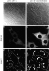
Figure 1
Figure 1
Antibodies against p115 cause Golgi complex disassembly. mAbs against p115 mixed with TR-dextran (A–C), or mAbs against 5′-nucleotidase mixed with TR-dextran (D–F) were microinjected into the cytoplasm of WIF-B cells. Phase images of cells are shown (A and D). Injected cells are identified by TR-dextran (B and E) and are traced in outline in C and F. Cells were fixed 2 h after injection and processed for immunofluorescence using antibodies against Mann II (C and F). Cells injected with anti–p115 antibodies, but not uninjected cells or cells injected with control antibodies show disassembly of Golgi complexes. Bar, 10 μm.
Figure 2
Figure 2
Antibodies against p115 do not block BFA-induced retrograde Golgi to ER traffic. Mouse mAbs against p115 were microinjected into the cytoplasm of WIF-B cells. Cells were fixed immediately after injection (A and B) or after a 30-min BFA treatment (C and D). Cell were processed for double label immunofluorescence using antibodies against Mann II (A and C) or against mouse IgG (B and D). Relocation of Mann II from the Golgi to the ER appears indistinguishable in injected and uninjected cells. Golgi remnants were detected in some cells after the 30-min BFA treatment (C, arrowheads).
Figure 3
Figure 3
Antibodies against p115 block Golgi complex reassembly during BFA wash-out. WIF-B cells were treated with BFA for 30 min, and then injected with mAbs against p115 mixed with TR-dextran. Cells were fixed after 10 min (A and B), 30 min (C and D), or 120 min (E and F) of BFA wash-out, and processed for immunofluorescence using antibodies against Mann II (A, C, and E). Injected cells were identified by their content of TR-dextran (B, D, and F), and are traced in outline in A, C, and E. After 10 min of BFA wash-out, Mann II relocation to punctate structures is indistinguishable in injected and uninjected cells. After 30 and 120 min of BFA wash-out, Mann II appears in morphologically normal Golgi structures in uninjected cells, but remains in punctate structures in injected cells. Arrowheads mark Golgi in injected cells, arrows point to Golgi in uninjected cells. Asterisk denotes a more compact Golgi complex in injected cells.
Figure 4
Figure 4
p115 is associated with VTCs moving cargo VSV-G protein from the ER to the Golgi. NRK cells were infected with VSVtsO45 at 42°C for 3 h. The cells were either fixed (A–C) or incubated at 15°C for 3 h (D–F) or at 32°C for 1 h (G–I) before fixation. Cell were processed for double label immunofluorescence using antibodies against p115 (A, D, and G) and antibodies against VSV-G protein (B, E, and H). At 42°C, VSV-G protein is present in the ER (B), whereas p115 is predominantly localized to the Golgi (A). p115 and VSV-G protein colocalize in peripheral VTCs after 15°C incubation (D–F), and in the Golgi after 32°C incubation (G–I). Bar 10 μm.
Figure 5
Figure 5
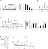
p115 is essential for ER to Golgi transport. ER to Golgi transport was performed in semi-intact NRK cells. Transport is measured as the percentage of VSV-G protein processed from the endo-H–sensitive (S) to the endo-H–resistant (R) form. (A) Transport reactions contained complete transport cocktail (lanes 2–6), or cocktail with ATP-depleting system (lane 1). Reactions were supplemented with increasing amounts of affinity-purified anti–p115 antibodies (lanes 3–6). Transport of VSV-G protein was proportionally inhibited in the presence of antibodies against p115. Analogous gels (n = 3) were quantitated by densitometry and the averages are presented in the bar graph. Transport in lane 1 is set as 0% and in lane 2 as 100%. (B) Transport reactions contained complete transport cocktail (lane 2), cocktail with ATP-depleting system (lane 1), or cocktail supplemented with anti–p115 antibodies preincubated with GST-p115 (lane 3), or GST (lane 4). Preincubation of anti–p115 antibodies with GST-p115 neutralized their inhibitory effect on transport. (C) Transport reactions contained complete transport cocktail (lanes 2–7), or cocktail with ATP-depleting system (lane 1). In lane 3, transport cocktail contained cytosol preincubated with control IgGs. In lane 4, transport cocktail contained cytosol preincubated with anti–p115 antibodies. Reduced level of p115 is visible in lane 4 and leads to inhibition of VSV-G protein transport. Increasing amounts of purified p115 were added to the cytosol shown in lane 4. Addition of purified p115 overcame the inhibitory effect of p115 removal and supported VSV-G protein transport (lanes 5–7). Analogous gels (n = 3) were quantitated by densitometry, and the averages are presented in the bar graph. Transport in lane 1 is set as 0% and in lane 2 as 100%. An aliquot of each transport reaction was probed by immunoblotting with anti–p115 antibodies and the immunoblot is shown in panel p115. The same amount of p115 was used in lane 1 as in lane 2 and only reaction in lane 2 was analyzed. (D) Rat liver cytosol was incubated with anti–p115 antibodies or control IgGs cross-linked to protein A–Sepharose. Bound material was eluted and analyzed by SDS-PAGE. An ∼110-kD band was visible after Coomassie blue staining in material eluted from anti-p115 column (lane 1) but not in control eluate (lane 2). The ∼110-kD band was excised, digested with trypsin, and the resulting peptides analyzed by MALDI mass spectrometry. The peptide mass map of the 10 most abundant peptides (marked by asterisks) matched the sequence of p115.
Figure 7
Figure 7
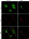
Antibodies against p115 block ER to Golgi transport of VSV-G protein at a pre-Golgi stage. NRK cells were infected with VSVtsO45 for 3 h at 42°C. Cells were permeabilized and supplemented with complete transport cocktail supplemented either with anti–p115 antibodies (A–C) or control antibodies (D–F). After transport at 32°C for 90 min, cells were processed by double label immunofluorescence using anti-p115 (A and D) and anti–VSV-G protein (B and E) antibodies. Addition of anti–p115 antibodies to the transport assay has no effect on VSV-G protein exit from the ER, but prevents VSV-G protein transport to the Golgi (A–C) and causes accumulation of VSV-G protein in scattered VTCs. Arrowheads point to peripheral structures containing VSV-G protein and anti–p115 antibodies. Arrows indicate Golgi elements labeled with anti–p115 antibodies but lacking VSV-G protein. Addition of control antibodies to the transport assay had no effect on VSV-G protein transport, and VSV-G protein was efficiently delivered to the Golgi (D–F). Bar 10 μm.
Figure 8
Figure 8
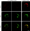
p115 depletion blocks ER to Golgi transport of VSV-G protein at a pre-Golgi stage. NRK cells were infected with VSVtsO45 for 3 h at 42°C. Cells were permeabilized and supplemented with transport cocktails containing p115-depleted cytosol (A–C) or complete cytosol (D–F). After transport at 32°C for 90 min, cells were processed by double label immunofluorescence using anti–VSV-G protein (A and D) and anti–Mann II (B and E) antibodies. Depletion of p115 from the transport assay had no effect on VSV-G protein exit from the ER, but prevented VSV-G protein transport to the Golgi (A–C) and caused accumulation of VSV-G protein in peripheral VTCs lacking Mann II. In the presence of complete cytosol, VSV-G protein was efficiently delivered to the Golgi (D–F). Bar 10 μm.
Figure 10
Figure 10
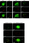
Mann I relocates from the Golgi to arrested pre-Golgi VTCs. NRK cells grown at 37°C (A–C) or incubated for 3 h at 15°C (D–F) were processed for double label immunofluorescence using anti–Mann I (A and D) and anti–Mann II (B and E) antibodies. In cells grown at 37°C, Mann I colocalized with Mann II in the Golgi region (C), but after 15°C incubation, Mann I relocated to peripheral punctate structures and did not colocalize with the Golgi localized Mann II (F). NRK cells infected with VSVtsO45 were incubated for 2 h at 42°C and for an additional 3 h at 15°C (G–L). Cells were processed for double label immunofluorescence using anti–Mann I (G) and anti–VSV-G protein (I) antibodies, or anti–Mann II (J) and anti–VSV-G protein antibodies (K). Mann I is present in dispersed punctate structures, some of which contain VSV-G protein (I, arrowheads). Mann II remains within the Golgi and does not relocate to peripheral structures containing VSV-G protein (L). NRK cells infected with VSVtsO45 were incubated for 3 h at 42°C, permeabilized, and supplemented with transport cocktails containing complete cytosol (M–O) or p115-depleted cytosol (P–R). After transport at 32°C for 90 min, cells were processed for double label immunofluorescence using anti–Mann I (M and P) and anti–VSV-G protein (N and Q) antibodies. In reactions containing complete cytosol, VSV-G protein is delivered to the Golgi where it colocalizes with Mann I (O). In reactions containing p115-depleted cytosol, Mann I relocates to pre-Golgi VTCs containing arrested VSV-G protein (R, arrowheads). Bar, 10 μm.
Figure 9
Figure 9
VSV-G protein is differentially endo-D resistant/sensitive in LEC-1 and NRK cells. (A and B) ER to Golgi transport was performed in semi-intact LEC-1 cells. After transport, the sensitivity/resistance of VSV-G protein to endo-D was analyzed. (A) VSV-G protein is endo-D resistant when transport cocktail lacking ATP is used (−ATP), and this is set as 0% relative processing. A proportion of VSV-G protein is endo-D sensitive when complete transport cocktail is used (+ATP), and this is set as 100% relative processing. A similar proportion of VSV-G protein is endo-D sensitive when complete transport cocktails containing control antibodies (control) or anti–p115 antibodies (anti-p115) are used. (B) VSV-G protein is endo-D resistant when transport cocktail lacking ATP is used (lane 1), and this is set as 0% relative processing. A proportion of VSV-G protein is endo-D sensitive when complete transport cocktail is used (lane 2), and this is set as 100% relative processing. A similar proportion of VSV-G protein is endo-D sensitive when complete transport cocktails containing cytosol immunodepleted with control antibodies (lane 3), preimmune antibodies (lane 4), or anti–p115 antibodies (lanes 5 and 6) are used. Analogous gels (n = 3) were quantitated by densitometry and averages are presented as a bar graph. An aliquot of each transport reaction was probed by immunoblotting with anti–p115 antibodies and the immunoblot is shown in panel p115. The relative amounts of p115 in each transport reaction are presented in the bar graph. The amount of p115 in lane 1 is set as 100%. (C) LEC-1 cells were infected with VSVtsO45 for 3 h at 42°C, and either analyzed directly or incubated at 32°C for additional 1 h or at 15°C for additional 3 h before analysis. Cells were collected and analyzed directly or after endo-D digestion. Processing of VSV-G protein from the endo-D–resistant (R) to the endo-D–sensitive (S) form is shown. A single VSV-G band is seen in untreated samples regardless of incubation temperature (lanes 4–6). VSV-G is resistant to endo-D when retained in the ER during the 42°C incubation (lane 1), but becomes endo-D sensitive when transported to the Golgi during a 32°C incubation (lane 2) or when arrested in peripheral VTCs during a 15°C incubation (lane 3). (D, lanes 1–3 and 5–7) ER to Golgi transport was performed in semi-intact NRK cells. After transport, the sensitivity/resistance of VSV-G protein to endo-H and endo-D was analyzed in parallel. VSV-G protein is endo-H sensitive (lane 1) and endo-D resistant (lane 5) when transport cocktail lacking ATP is used. VSV-G protein is endo-H resistant (lane 2) and endo-D resistant (lane 6) when complete transport cocktail is used. VSV-G protein is endo-H sensitive (lane 3) and endo-D resistant (lane 7) when complete transport cocktails containing anti–p115 antibodies are used. (D, lanes 4 and 8) NRK cells were infected with VSVtsO45 (3 h at 32°C), pulsed-labeled at the restrictive temperature (10 min at 42°C) and chased in complete medium for 3 h at 15°C. Cells were collected and digested with either endo-H (lane 4) or endo-D (lane 8). VSV-G protein is endo-H sensitive and endo-D resistant when arrested in peripheral VTCs at 15°C.

References

    1. Aridor M., Bannykh S.I., Rowe T., Balch W.E. Sequential coupling between COPII and COPI vesicle coats in endoplasmic reticulum to Golgi transport. J. Cell Biol. 1995;131:875–893. - PMC - PubMed
    1. Aridor M., Weissman J., Bannykh S., Nuoffer C., Balch W.E. Cargo selection by the COPII budding machinery during export from the ER. J. Cell Biol. 1998;141:61–70. - PMC - PubMed
    1. Balch W.E., Elliott M.M., Keller D.S. ATP-coupled transport of vesicular stomatitis virus G protein between the endoplasmic reticulum and the Golgi. J. Biol. Chem. 1986;261:14681–14689. - PubMed
    1. Balch W.E., McCaffery J.M., Plutner H., Farquhar M.G. Vesicular stomatitis virus glycoprotein is sorted and concentrated during export from the endoplasmic reticulum. Cell. 1994;76:841–852. - PubMed
    1. Bannykh I., Balch W. Membrane dynamics at the endoplasmic reticulum–Golgi interface. J. Cell Biol. 1997;138:1–4. - PMC - PubMed

MeSH terms