. 1999 Dec 13;147(6):1223-36.
doi: 10.1083/jcb.147.6.1223.

Role for Drs2p, a P-type ATPase and potential aminophospholipid translocase, in yeast late Golgi function

Affiliations

Role for Drs2p, a P-type ATPase and potential aminophospholipid translocase, in yeast late Golgi function

C Y Chen et al. J Cell Biol. .

Abstract

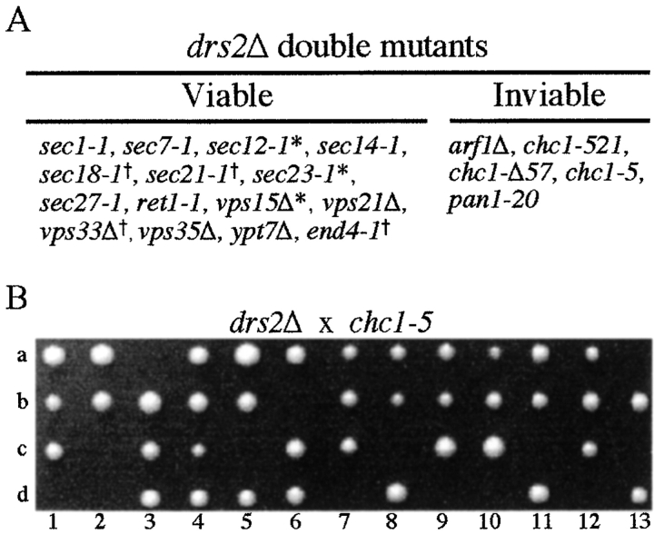
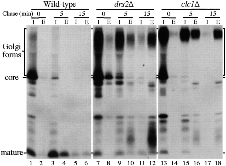
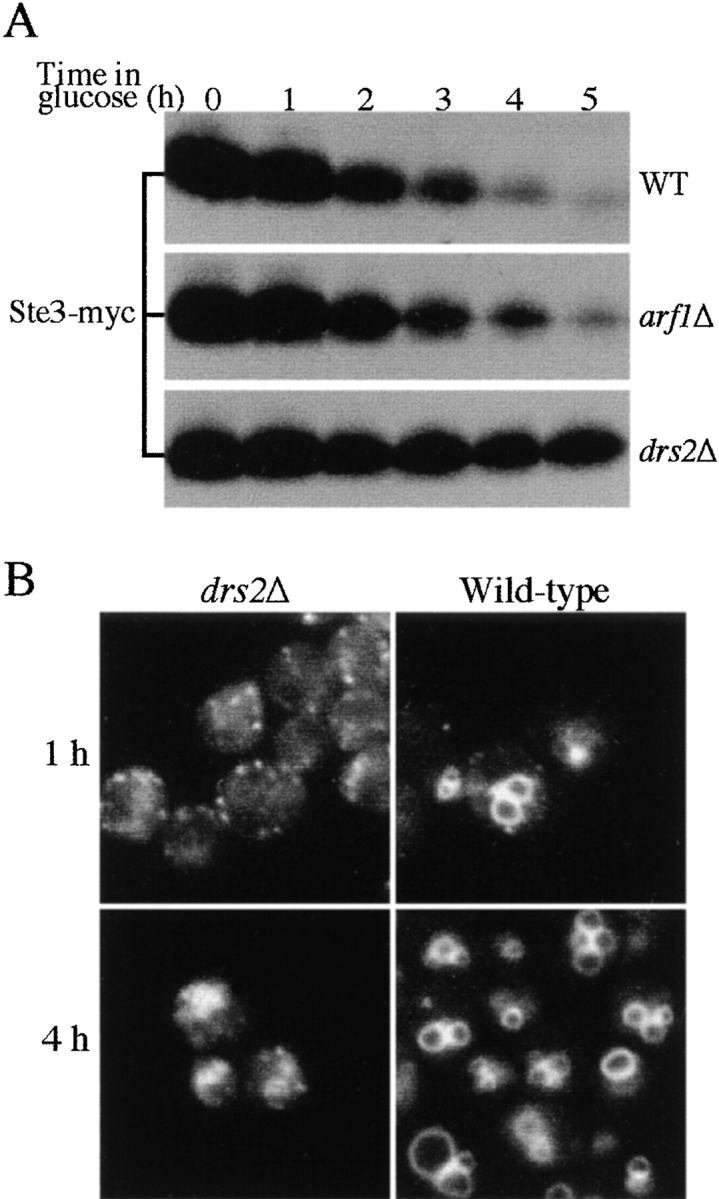
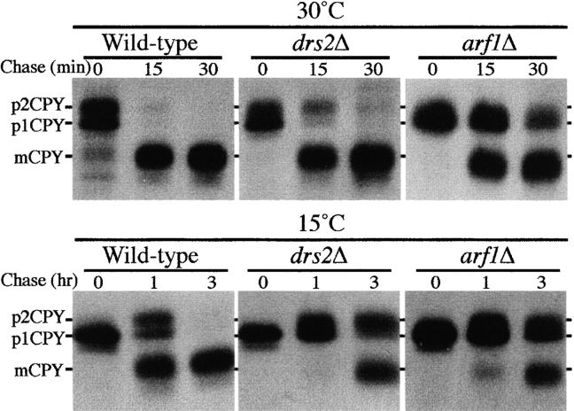
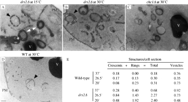
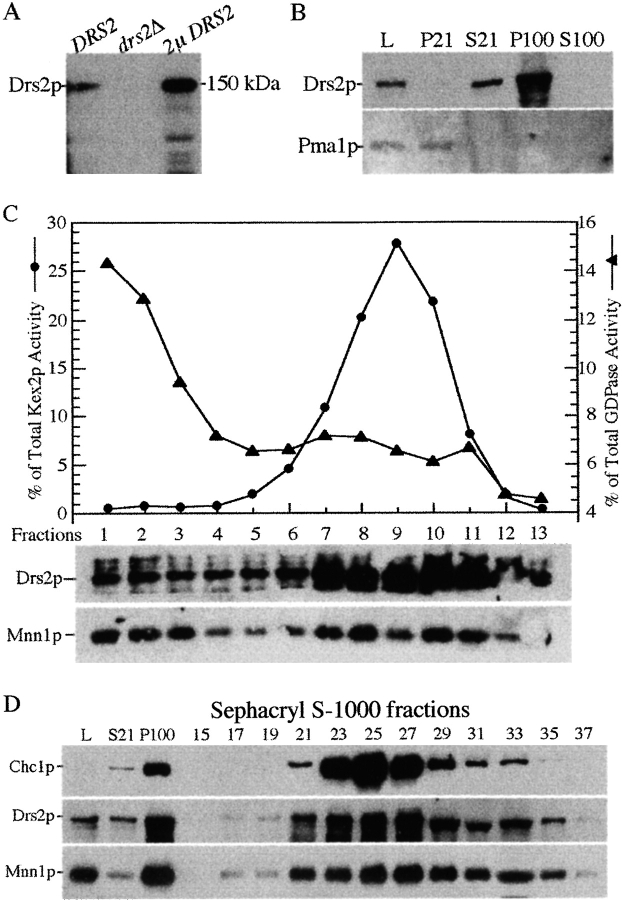
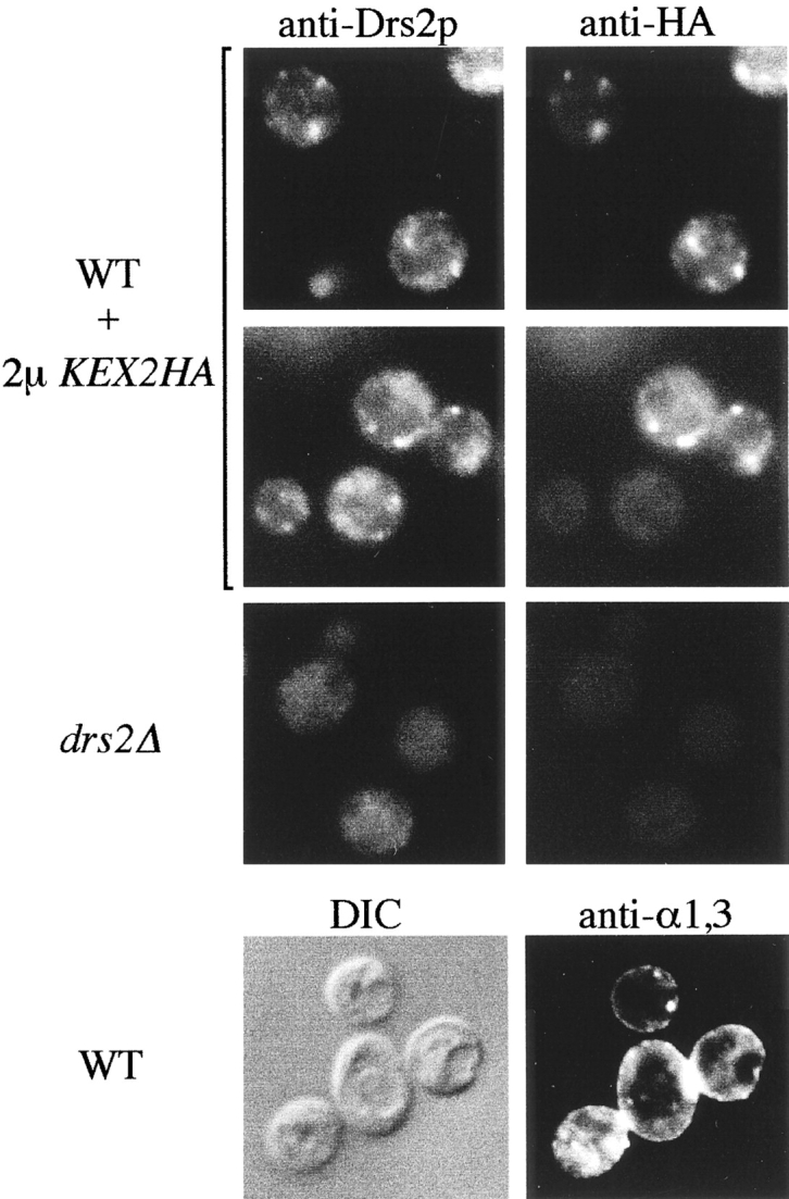
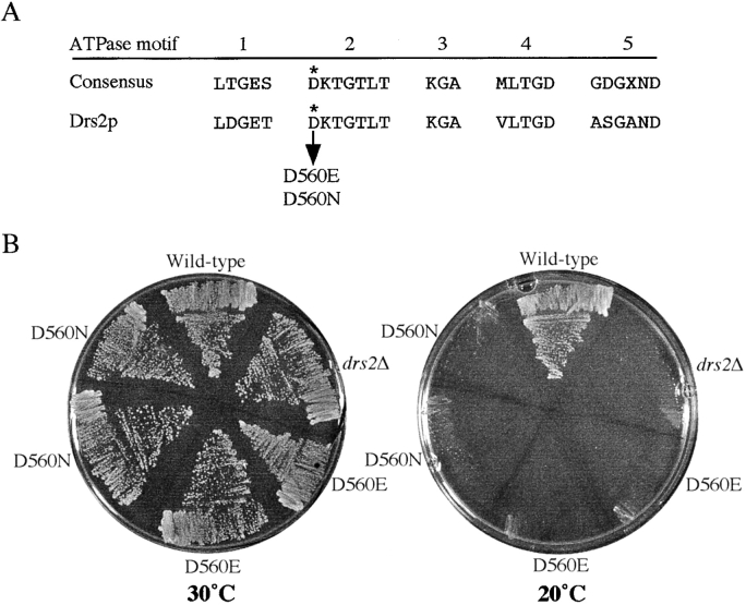
ADP-ribosylation factor appears to regulate the budding of both COPI and clathrin-coated transport vesicles from Golgi membranes. An arf1Delta synthetic lethal screen identified SWA3/DRS2, which encodes an integral membrane P-type ATPase and potential aminophospholipid translocase (or flippase). The drs2 null allele is also synthetically lethal with clathrin heavy chain (chc1) temperature-sensitive alleles, but not with mutations in COPI subunits or other SEC genes tested. Consistent with these genetic analyses, we found that the drs2Delta mutant exhibits late Golgi defects that may result from a loss of clathrin function at this compartment. These include a defect in the Kex2-dependent processing of pro-alpha-factor and the accumulation of abnormal Golgi cisternae. Moreover, we observed a marked reduction in clathrin-coated vesicles that can be isolated from the drs2Delta cells. Subcellular fractionation and immunofluorescence analysis indicate that Drs2p localizes to late Golgi membranes containing Kex2p. These observations indicate a novel role for a P-type ATPase in late Golgi function and suggest a possible link between membrane asymmetry and clathrin function at the Golgi complex.


Figures

Figure 1
Figure 1
drs2Δ is synthetically lethal with arf1Δ, chc1, and pan1 alleles. (A) Genetic analyses between drs2Δ and mutations that perturb the secretory pathway. Strains carrying the indicated mutations (see Materials and Methods) were crossed with a drs2Δ mutant (6210 drs2Δ or PRY6222) to generate diploids. After tetrad analyses of the progeny, the viable double mutants were streaked at 20°, 26.5°, and 37°C to compare the growth relative to parental strains carrying single mutations. *Double mutants of the alleles and drs2 that were able to grow at 20°C, where single drs2 mutants could not. Double mutants that grew more slowly than either single mutant at 26.5°C. (B) Tetrad analysis of progeny derived from crossing PRY6222 (drs2Δ) with 6210 chc1-5 (chc1-5). Spores that failed to grow were predicted to be drs2Δ chc1-5 double mutants.
Figure 2
Figure 2
The drs2Δ mutant exhibits a cold-sensitive defect in pro–α-factor processing. Wild-type, drs2Δ (hereafter used to represent SEY6210 and the isogenic strain 6210 drs2Δ, respectively), and clc1Δ (LSY93.1-10A) strains were labeled for 10 min at 15°C, chased for the times indicated, and converted to spheroplasts. Intracellular (I) and extracellular (E) portions were separated by centrifugation and subjected to immunoprecipitation with antiserum to α-factor.
Figure 3
Figure 3
The drs2Δ mutant exhibits a cold-sensitive defect in the endocytic pathway. (A) Wild-type, drs2Δ, and arf1Δ strains were transformed with a plasmid carrying the Ste3-myc construct (pSL2624; Givan and Sprague 1997) and grown to early log phase in galactose to induce expression of the STE3-myc protein. Glucose was added to 3% final concentration to repress new synthesis of Ste3-myc and the cultures were shifted to 15°C. Lysates were prepared at the times indicated and immunoblotted for Ste3-myc using the 9E10 c-myc antibody. (B) Wild-type and drs2Δ cells were stained with FM4-64 on ice and shifted to 15°C to initiate endocytosis of the dye. Images were captured at 1 and 4 h after shift to 15°C.
Figure 4
Figure 4
The drs2Δ mutant exhibits a cold-sensitive kinetic defect in CPY transport to the vacuole. Wild-type, drs2Δ, and arf1Δ (all isogenic to SEY6210) strains were labeled for 10 min at 15° or 30°C, and then chased for the times indicated. CPY was recovered from each sample by immunoprecipitation and subjected to SDS-PAGE.
Figure 5
Figure 5
The drs2Δ mutant accumulates abnormal membrane structures that are similar to Berkeley bodies. Electron micrographs of drs2Δ cells incubated for 2 h at 15°C (A) or kept at 30°C (B) showing numerous double-membrane ring and crescent-shaped structures. Plasma membrane (PM) and vacuoles (V) are labeled. The black arrowhead in A denotes a modestly fenestrated ring structure. The arrowhead in B shows a ring structure with a stacked crescent membrane. For comparison, an electron micrograph of similar structures observed in a chc1Δ cell (C, GPY1103) is shown. Narrower, more fenestrated, and typically incomplete ring structures were also found in wild-type (WT) cells grown at 30°C (D, arrowhead). These wild-type ring structures did not stain as darkly as rings found in the drs2Δ mutant (compare to double arrowhead in A). Bars, 0.2 μm. E, drs2Δ and wild-type cells grown at the indicated temperatures were visualized by transmission electron microscopy. Membrane bound structures from 23–25 randomly selected cells were counted and expressed as an average number of structures per cell section.
Figure 6
Figure 6
The drs2Δ mutant accumulates aberrant Golgi membranes and exhibits a deficiency of clathrin-coated vesicles. (A) Wild-type and drs2Δ cells were grown at 30°C and shifted to 15°C for 1 h, and then lysed and subjected to centrifugation at 21,000 g for 30 min. The 21,000-g supernatant was centrifuged at 100,000 g for 80 min to generate a 100,000-g pellet. This pellet was resuspended and further resolved on a Sephacryl S-1000 column. Samples of every other fraction from fraction 15 to 35 were assayed by immunoblotting for the clathrin heavy chain (Chc1p), a late Golgi protein Mnn1p, and an endosomal t-SNARE Pep12p. (B–D) Membranes in fraction 16 from the drs2Δ sample where Mnn1p was found (B), and fraction 26 from wild-type (C) and drs2Δ (D) samples where Chc1p was enriched were fixed, pelleted, and examined by EM. Bars, 50 nm.
Figure 7
Figure 7
Drs2p cofractionates with high-density membranes containing the late Golgi markers, Kex2p and Mnn1p. (A) Specificity of the Drs2p antibodies. Wild-type, drs2Δ, and SEY6210 pRS425-DRS2 (2μ DRS2) cells were grown at 30°C before lysing with glass beads in SDS-urea sample buffer. Total cellular proteins (0.1 OD per lane) were subjected to SDS-PAGE and immunoblotted with affinity-purified antibodies against Drs2p. (B) Wild-type cells grown at 30°C were lysed (lysate, L) and subjected to centrifugation at 21,000 g for 30 min to generate pellet (P21) and supernatant (S21) fractions. The S21 fraction was further centrifuged at 100,000 g for 80 min to generate pellet (P100) and supernatant (S100) fractions. 20 μg of total protein per fraction was immunoblotted for Drs2p and the plasma membrane ATPase, Pma1p. (C) Wild-type cells grown at 30°C were lysed and subjected to differential centrifugation and sucrose gradient fractionation of the P100 fraction. These gradient fractions have been previously used to examine Mnn1p distribution in Figure 6 a of Reynolds et al. 1998. Equal volumes per gradient fraction were assayed by enzyme activity for Kex2p or GDPase and by immunoblotting for Drs2p, Mnn1p, and Pep12p. (D) Wild-type cells were grown at 30°C, lysed, and subjected to differential centrifugation to produce a P100 fraction, which was then subjected to gel filtration chromatography as described in Fig. 6. Samples of every other fraction from fraction 15 to 35 were assayed by immunoblotting for the clathrin heavy chain (Chc1p), the late Golgi protein Mnn1p and Drs2p.
Figure 8
Figure 8
Drs2p colocalizes with Kex2p in internal, punctate membranes. The wild-type strain carrying the HA-tagged KEX2 on a 2-μm plasmid (WT + 2μ KEX2HA), and the drs2Δ strain were grown overnight at 30°C in minimal medium. After dilution in rich medium to 0.2 OD600, the wild-type strain was shifted to 20°C and cultured for 7 h while the drs2Δ strain was cultured at 30°C for 5 h, and then shifted to 20°C for 2 h. Cells were then fixed and prepared for immunofluorescence as previously described (Graham et al. 1994). Affinity-purified anti–Drs2p and monoclonal anti–HA antibodies were used in the top six panels to visualize Drs2p (left) and the HA-tagged Kex2p (right) in the same cells. The bottom two panels are DIC and immunofluorescence images of wild-type cells grown at 20°C and labeled with an antibody specific for α1,3-linked mannose. Notice how the punctate and rim staining pattern (plasma membrane) of α1,3-mannose epitopes is different from the punctate staining of Drs2p in wild-type cells.
Figure 9
Figure 9
Mutation of a conserved ATPase motif causes loss of Drs2p function in vivo. (A) Comparison of five conserved consensus motifs of yeast P-type ATPases and the corresponding sequence in Drs2p (adapted from Catty et al. 1997). The arrow indicates the aspartic acid residue (D) at position 560, which was mutated to glutamic acid (E) or asparagine (N). (B) The drs2Δ strain containing plasmids pDRS2(D560E), pDRS2(D560N), pRS315-DRS2 (wild-type), and the pRS315 empty vector (drs2Δ) were grown at 30° or 20°C for 3 d.

References

    1. Allen G., Green N.M. A 31-residue tryptic peptide from the active site of the [Ca++]-transporting ATPase of rabbit sarcoplasmic reticulum. FEBS Lett. 1976;63:188–192. - PubMed
    1. Auland M.E., Roufogalis B.D., Devaux P.F., Zachowski A. Reconstitution of ATP-dependent aminophospholipid translocation in proteoliposomes. Proc. Natl. Acad. Sci. USA. 1994;91:10938–10942. - PMC - PubMed
    1. Barik S., Galinski M.S. Megaprimer method of PCRincreased template concentration improves yield. Biotechnology. 1991;10:489–490. - PubMed
    1. Becherer K.A., Rieder S.E., Emr S.D., Jones E.W. Novel syntaxin homologue, Pep12p, required for the sorting of lumenal hydrolases to the lysosome-like vacuole in yeast. Mol. Biol. Cell. 1996;7:579–594. - PMC - PubMed
    1. Catty P., de Kerchove d'Exaerde A., Goffeau A. The complete inventory of the yeast Saccharomyces cerevisiae P-type transport ATPases. FEBS Lett. 1997;409:325–332. - PubMed

Publication types

MeSH terms

Substances