. 1999 Dec 13;147(6):1299-312.
doi: 10.1083/jcb.147.6.1299.

Blood platelets are assembled principally at the ends of proplatelet processes produced by differentiated megakaryocytes

Affiliations

Blood platelets are assembled principally at the ends of proplatelet processes produced by differentiated megakaryocytes

J E Italiano Jr et al. J Cell Biol. .

Abstract

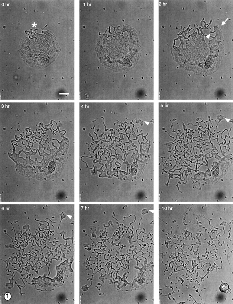
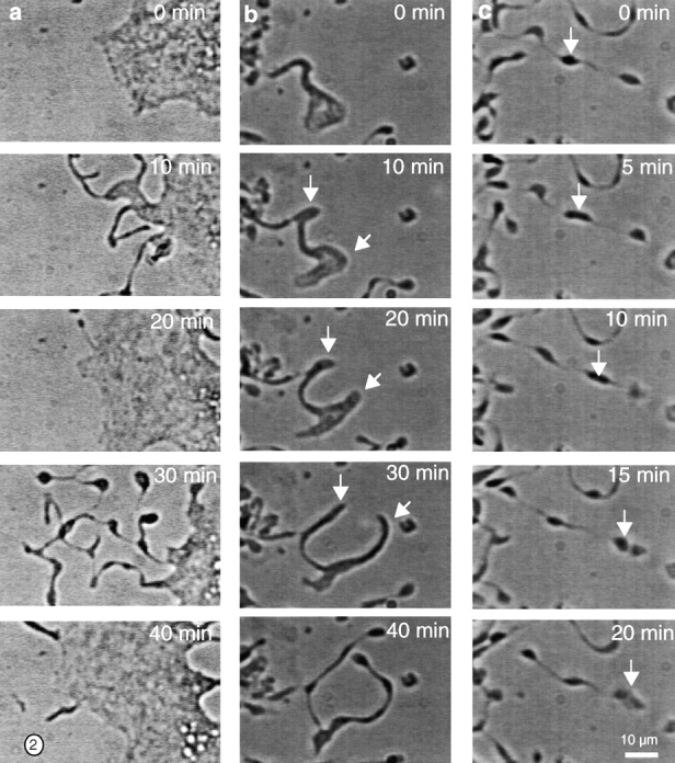
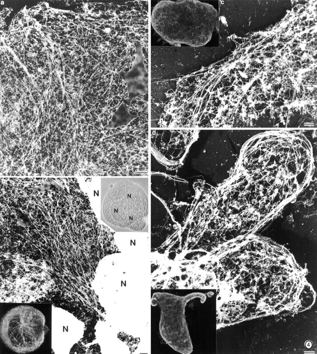
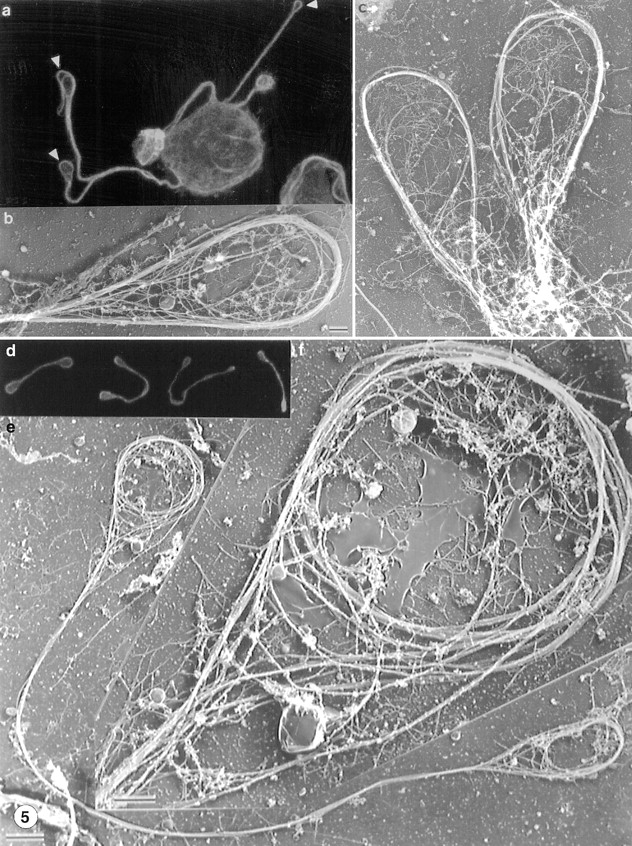
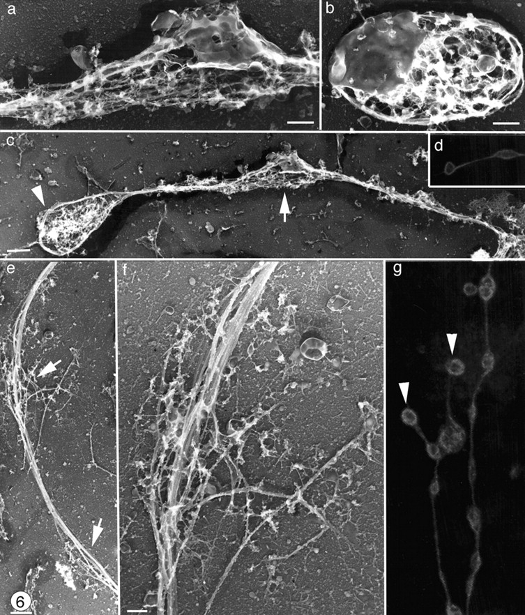
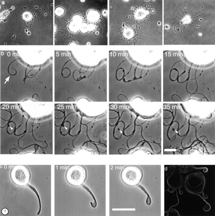
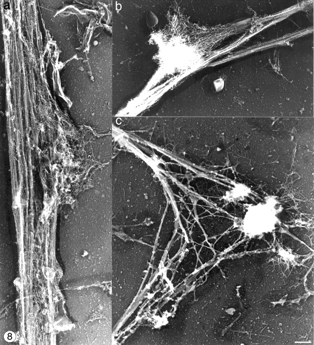
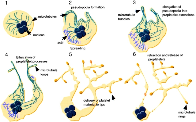
Megakaryocytes release mature platelets in a complex process. Platelets are known to be released from intermediate structures, designated proplatelets, which are long, tubelike extensions of the megakaryocyte cytoplasm. We have resolved the ultrastructure of the megakaryocyte cytoskeleton at specific stages of proplatelet morphogenesis and correlated these structures with cytoplasmic remodeling events defined by video microscopy. Platelet production begins with the extension of large pseudopodia that use unique cortical bundles of microtubules to elongate and form thin proplatelet processes with bulbous ends; these contain a peripheral bundle of microtubules that loops upon itself and forms a teardrop-shaped structure. Contrary to prior observations and assumptions, time-lapse microscopy reveals proplatelet processes to be extremely dynamic structures that interconvert reversibly between spread and tubular forms. Microtubule coils similar to those observed in blood platelets are detected only at the ends of proplatelets and not within the platelet-sized beads found along the length of proplatelet extensions. Growth and extension of proplatelet processes is associated with repeated bending and bifurcation, which results in considerable amplification of free ends. These aspects are inhibited by cytochalasin B and, therefore, are dependent on actin. We propose that mature platelets are assembled de novo and released only at the ends of proplatelets, and that the complex bending and branching observed during proplatelet morphogenesis represents an elegant mechanism to increase the numbers of proplatelet ends.


Figures

Figure 1
Figure 1
Video-enhanced light microscopy of a terminally differentiated mouse megakaryocyte forming proplatelets in vitro. During the initial stages of proplatelet formation, the megakaryocyte spreads and its cortical cytoplasm begins to unravel at one pole (this zone of erosion is labeled with an asterisk in the first panel). As the cell spreads, the cytoplasm at the erosion site is remodeled into large pseudopodia (white arrow at 2 h) that elongate and become thinner over time, forming narrow tubes of 1–4 μm diam. The proplatelet extensions frequently bend (white arrowhead at 2 h), and it is at sites of bending that the tube bifurcates to generate a new process. In this manner, the entire cytoplasmic space of the megakaryocyte is converted into anastomosing proplatelet extensions. Proplatelets also develop segmented constrictions along their length that impart a beaded appearance. The process of proplatelet elaboration culminates in a rapid retraction that separates the many strands of proplatelets from a residual nuclear mass (asterisk in last panel). Note the crawling of small cytoplasmic fragments (5–10 μm) at the ends of some of the proplatelet extensions (arrowheads at 4–7 h). Crawling begins when the end of the proplatelet adheres and flattens, forms a leading lamellipodia, and migrates away from the cell center, dragging a trail of proplatelets behind. Bar, 20 μm.
Figure 2
Figure 2
The dynamic behavior of proplatelets. (a) Interconversion between spread lamellar and condensed tubular forms. Phase-contrast images taken 10 min apart showing the dynamic interconversion between proplatelet morphologies. Proplatelets were observed to reversibly flatten, and then convert to thin tubular proplatelet processes. (b) Bifurcation of proplatelets. Phase-contrast images taken 10 min apart showing the bending and branching of a proplatelet extensions. Bends are converted into loops that become compressed and elongate, resulting in a bifurcation of the original tube. White arrows denote the branch points. (c) Platelet-sized particle movement along proplatelets. The white arrow denotes a platelet-sized nodule translocating along a proplatelet process, while the end of the proplatelet is stationary. In the last panel, this particle fuses with a stationary particle. The images are at 5-min intervals. Bar, 10 μm.
Figure 4
Figure 4
Structure of the megakaryocyte cytoskeleton before and during early proplatelet formation. (a and b) Structure of a megakaryocyte cytoskeleton lacking extensions. (a) Electron micrograph showing the structure of the cortical cytoskeleton in a megakarytocyte. A meshwork of actin filaments and microtubules is present in the cell cortex. Individual microtubules radiate outward from the cell center. (b) Electron micrograph showing the structure of the megakaryocyte cytoskeleton near its nuclear mass (N). Microtubules gather into arrays in the cell center. (right inset) Morphology of megakaryocyte in the light microscope photographed with phase contrast optics. (left inset) Confocal photograph of an early stage megakaryocyte stained with antitubulin antibody. Arrays of microtubules originate near the cell center and radiate out into the cell cortex. (c) Electron micrograph of a more mature megakaryocyte cytoskeleton before pseudopodia formation. Microtubules become densely packed into cortical bundles situated parallel to and just beneath the plasma membrane. (d) Organization of microtubules within pseudopodia. These structures contain dense rims composed of large bundles of microtubules. (Inset) Antitubulin immunofluorescent staining reveals the concentration of microtubules at the margins of the pseudopodia. Bars, 1 μm.
Figure 5
Figure 5
Organization of microtubules at the ends of proplatelet extensions (a–c) and in released proplatelet forms (d–g). (a) Antitubulin staining of a megakaryocyte and its early proplatelet extension. Microtubules concentrate along the edges of the megakaryocyte and enter into the proplatelet extensions. Bundles enter the projections from both sides and separate periodically within the extensions. The distal end of each proplatelet has a teardrop-shaped enlargement that contains a microtubule loop (arrowheads). (b and c) Representative electron micrographs showing the organization of microtubules within the tips of proplatelet termini. The end of each proplatelet contains a microtubule bundle that loops beneath the plasma membrane and reenters the shaft to form a teardrop-shaped structure. (d) Gallery of released platelet forms stained for tubulin by immunofluorescence confocal microscopy. Released platelet-sized particles are connected by thin cytoplasmic bridges, the most abundant being barbell shapes composed of two platelet-like particles connected by a single cytoplasmic strand. Microtubules line the shaft and rim the bulbous ends. (e) Low magnification electron micrograph showing the microtubule-based cytoskeleton of a representative released proplatelet form. A microtubule bundle lines the shaft of the proplatelet. Bar, 2 μm. (f) Higher magnification electron micrograph of one end of the proplatelet shown in e. Ends have microtubule bundles arranged into teardrop-shaped loops, similar to those at the ends of proplatelets extending from megakaryocytes. Within these loops are microtubule coils structurally similar to those observed in mature platelets isolated from the blood. Bar, 0.5 μm.
Figure 6
Figure 6
Organization of microtubules along the shaft of proplatelets. Swellings along the proplatelet (a) contain detergent-insoluble membrane and vesicles that are similar to those found in released platelets (b) in these samples. (c) Low magnification electron micrograph showing the microtubules bundles lining the shaft of a typical proplatelet. Although proplatelet ends (arrowhead) have distinct microtubule coils, microtubules observed in swellings along the shaft (arrow) are not coiled. (d and g) Proplatelets stained for tubulin by immunofluorescence and photographed on a confocal microscope. Proplatelets show periodic segments connected by thin cytoplasmic bridges, but only the ends (arrowheads) have microtubule bundles arranged into teardrop-shaped loops. (e and f) Higher magnification electron micrograph of a swelling along a proplatelet shaft showing that these are points where the microtubule bundle separates for a short distance (arrows) but does not form a loop. Bars: (b and f) 0.5 μm; (c and e) 1 μm.
Figure 7
Figure 7
Effect of cytoskeletal disrupting agents on proplatelet growth. (a) Gallery of phase-contrast micrographs of megakaryocytes treated with 2 μM cytochalasin B. Extensions from the megakaryocyte surface show reduced beading and absence of branching. (b) Images showing a time series at a higher magnification of processes extending from the megakaryocyte surface in the presence of 2 μM cytochalasin B. The white arrowheads indicate the tip of one extension, which remains almost stationary while the diameter of the loop formed by the extension increases with time. (c) Effect of taxol on proplatelet formation. Phase-contrast images showing a time series of a tubelike extension growing from a taxol-treated megakaryocyte. In the presence of 10 μM taxol, the megakaryocyte forms a single pseudopodia. As this tube elongates, its distal tip bends and curves back on itself (0–1 min) until its tip contacts its shaft and appears to fuse (2 min). This forms a teardrop-shaped structure on the end of the projection. (d) Antitubulin immunofluorescence staining of a megakaryocyte cultured in the presence of 10 μM taxol. Taxol treatment reduces the number of extensions made by cells. Microtubule staining concentrates at the cell edge and in the proplatelet-like extensions grown from the cell surface. The microtubule bundles in the shafts are thickened compared with those extended by untreated cells, and are observed to curl within the bulbous ends. Bars: (b) 10 μm; (c) 25 μm.
Figure 8
Figure 8
Cytoskeletal organization at proplatelet bends. (a) Representative micrograph showing the cytoskeleton of a proplatelet shaft. Parallel bundles of microtubules line the tube and prospective branch points are defined by outpouchings of 10-nm filaments. (b) Micrograph showing a platelet-sized swelling along the length of a proplatelet that has spread on the surface. This spread region contains a dense meshwork of F-actin, with ends that are collapsed and bound to the sides of the microtubule bundles. Microtubule bundles are bent and appear to have separated. A meshwork of spread actin filaments is observed extending from the platelet-sized segment. (c) Micrograph showing a pronounced bend in a proplatelet. The elbow of the bend contains aggregates of filamentous material, which contact the substratum in the elbow region. Bar, 1 μm.
Figure 9
Figure 9
Model of platelet production suggested by these experiments and previous studies. After commitment to the megakaryocyte lineage, cells undergo polyploidization and cytoplasmic maturation (Stage 1). During the initial stages of proplatelet formation, megakaryocytes remodel their cytoplasm into thick pseudopodia (Stage 2), which contain bundles of microtubules situated just beneath the protruding membrane. Blunt pseudopodia elongate into proplatelet processes, which harbor thick bundles of microtubules in their core and contain a teardrop-shaped loop at their distal tip (Stage 3). Proplatelets frequently bend and form a branched structure from which new processes extend (Stage 4). These proplatelet processes form constrictions along their length giving the beaded appearance to proplatelets (Stage 5). The swellings along proplatelets are unstable structures presumed to contain packets of platelet material in the process of being delivered to the ends. Proplatelets are released from the megakaryocyte body after a retraction (Stage 6), and may undergo further fragmentation to yield individual platelets.

References

    1. An E., Ogata K., Kuriya S., Nomura T. Interleukin-6 and erythropoietin act as direct potentiators and inducers of in vitro cytoplasmic process formation on purified mouse megakaryocytes. Exp. Hematol. 1994;22:149–156. - PubMed
    1. Andrews R.K., Fox J.B. Interaction of purified actin-binding protein with glycoprotein Ib-IX complex. J. Biol. Chem. 1991;266:7144–7147. - PubMed
    1. Becker R.P., DeBruyn P.P. The transmural passage of blood cells into myeloid sinusoids and the entry of platelets into the sinusoidal circulationa scanning electron microscopic investigation. Am. J. Anat. 1976;145:1046–1052. - PubMed
    1. Behnke O. An electron microscope study of megakaryocytes of rat bone marrow. I. The development of the demarcation membrane system and the platelet surface coat. J. Ultrastruct. Res. 1968;24:412–428. - PubMed
    1. Behnke O. An electron microscope study of the rat megakaryocyte. II. Some aspects of platelet release and microtubules. J. Ultrastruct. Res. 1969;26:111–129. - PubMed

Publication types

MeSH terms