

Cell elongation induces laminin alpha2 chain expression in mouse embryonic mesenchymal cells: role in visceral myogenesis
- PMID: 10601345
- PMCID: PMC2168094
- DOI: 10.1083/jcb.147.6.1341
Cell elongation induces laminin alpha2 chain expression in mouse embryonic mesenchymal cells: role in visceral myogenesis
Abstract
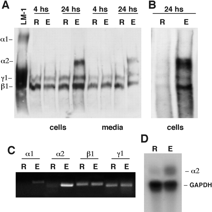
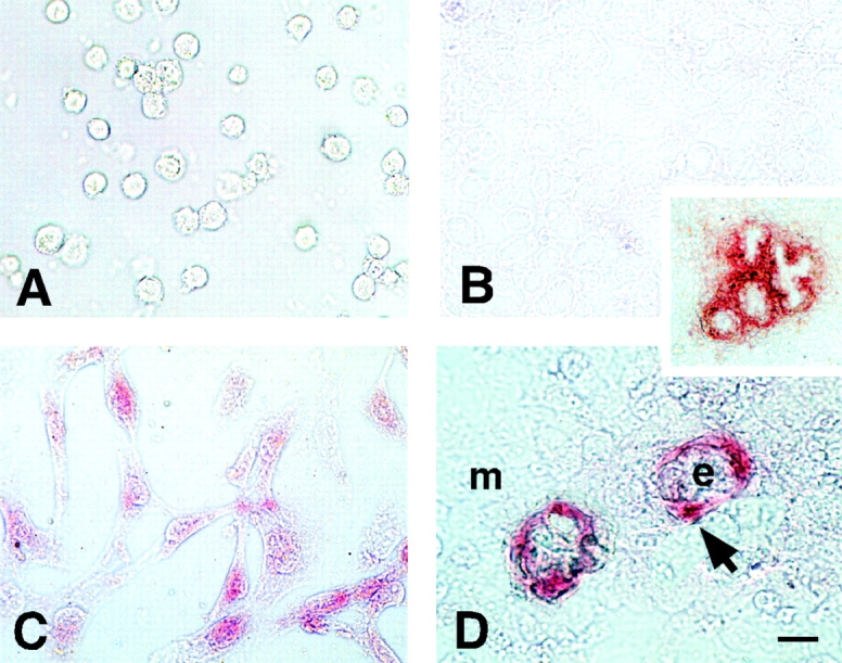
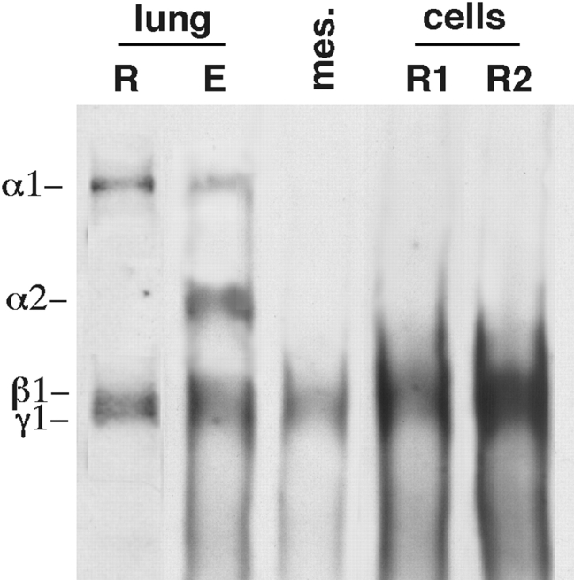
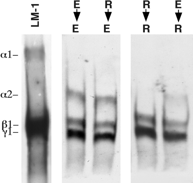
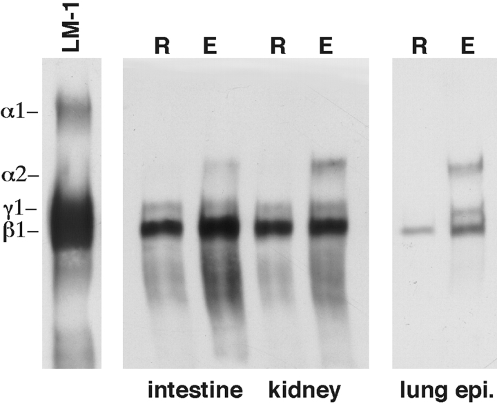
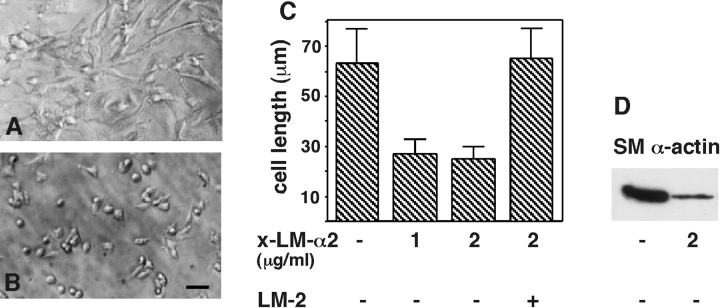
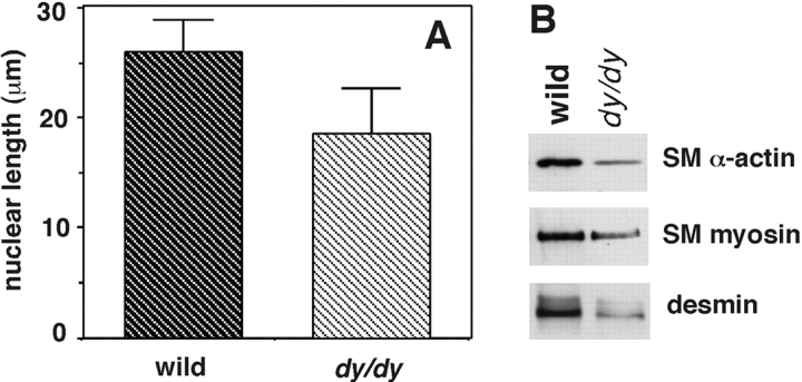
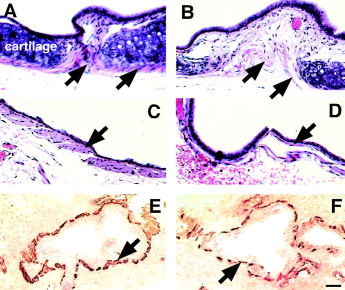
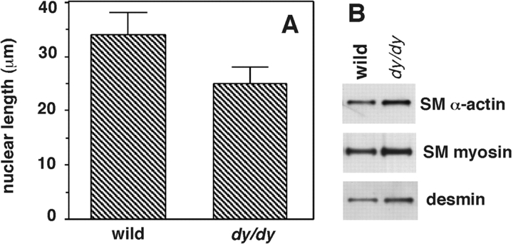
Bronchial smooth muscle (SM) mesenchymal cell precursors change their shape from round to spread/elongated while undergoing differentiation. Here we show that this change in cell shape induces the expression of laminin (LM) alpha2 chain not present in round mesenchymal cells. LM alpha2 expression is reversible and switched on and off by altering the cell's shape in culture. In comparison, the expression of LM beta1 and gamma1 remains unchanged. Functional studies showed that mesenchymal cell spreading and further differentiation into SM are inhibited by an antibody against LM alpha2. Dy/dy mice express very low levels of LM alpha2 and exhibit congenital muscular dystrophy. Lung SM cells isolated from adult dy/dy mice spread defectively and synthesized less SM alpha-actin, desmin, and SM-myosin than controls. These deficiencies were completely corrected by exogenous LM-2. On histological examination, dy/dy mouse airways and gastrointestinal tract had shorter SM cells, and lungs from dy/dy mice contained less SM-specific protein. The intestine, however, showed compensatory hyperplasia, perhaps related to its higher contractile activity. This study therefore demonstrated a novel role for the LM alpha2 chain in SM myogenesis and showed that its decrease in dy/dy mice results in abnormal SM.
Figures


Similar articles
-
High RhoA activity maintains the undifferentiated mesenchymal cell phenotype, whereas RhoA down-regulation by laminin-2 induces smooth muscle myogenesis.J Cell Biol. 2002 Mar 4;156(5):893-903. doi: 10.1083/jcb.200107049. Epub 2002 Mar 4. J Cell Biol. 2002. PMID: 11877460 Free PMC article.
-
Bronchial smooth muscle hypoplasia in mouse embryonic lungs exposed to a laminin beta1 chain antisense oligonucleotide.Mech Dev. 1999 Dec;89(1-2):15-23. doi: 10.1016/s0925-4773(99)00198-7. Mech Dev. 1999. PMID: 10559476
-
Embryonic mesenchymal cells share the potential for smooth muscle differentiation: myogenesis is controlled by the cell's shape.Development. 1999 Jul;126(13):3027-33. doi: 10.1242/dev.126.13.3027. Development. 1999. PMID: 10357945
-
Embryological origin of airway smooth muscle.Proc Am Thorac Soc. 2008 Jan 1;5(1):4-10. doi: 10.1513/pats.200704-049VS. Proc Am Thorac Soc. 2008. PMID: 18094078 Free PMC article. Review.
-
Laminin isoforms and lung development: all isoforms are not equal.Dev Biol. 2006 Jun 15;294(2):271-9. doi: 10.1016/j.ydbio.2006.03.032. Epub 2006 Apr 3. Dev Biol. 2006. PMID: 16643883 Review.
Cited by
-
P311 induces a TGF-beta1-independent, nonfibrogenic myofibroblast phenotype.J Clin Invest. 2002 Nov;110(9):1349-58. doi: 10.1172/JCI15614. J Clin Invest. 2002. PMID: 12417574 Free PMC article.
-
Laminin-2 stimulates the proliferation of epithelial cells in a conjunctival epithelial cell line.Cell Prolif. 2004 Apr;37(2):161-75. doi: 10.1111/j.1365-2184.2004.00292.x. Cell Prolif. 2004. PMID: 15030550 Free PMC article.
-
Synthetic nanostructures inducing differentiation of human mesenchymal stem cells into neuronal lineage.Exp Cell Res. 2007 May 15;313(9):1820-9. doi: 10.1016/j.yexcr.2007.02.031. Epub 2007 Mar 12. Exp Cell Res. 2007. PMID: 17428465 Free PMC article.
-
Knockdown of Laminin gamma-3 (Lamc3) impairs motoneuron guidance in the zebrafish embryo.Wellcome Open Res. 2017 Nov 16;2:111. doi: 10.12688/wellcomeopenres.12394.1. eCollection 2017. Wellcome Open Res. 2017. PMID: 29417095 Free PMC article.
-
Clonogenic multipotent stem cells in human adipose tissue differentiate into functional smooth muscle cells.Proc Natl Acad Sci U S A. 2006 Aug 8;103(32):12167-72. doi: 10.1073/pnas.0604850103. Epub 2006 Jul 31. Proc Natl Acad Sci U S A. 2006. PMID: 16880387 Free PMC article.
References
-
- Anastasi S., Giordano S., Sthandier O., Gambarotta G., Maione R., Comoglio P., Amati P. A natural hepatocyte growth factor/scatter factor autocrine loop in myoblast cells and the effect of the constitutive Met kinase activation on myogenic differentiation. J. Cell Biol. 1997;137:1057–1068. - PMC - PubMed
-
- Bernier S.M., Utani A., Sugiyama S., Doi T., Polistina C., Yamada Y. Cloning and expression of laminin α2 chain (m-chain) in the mouse. Matrix Biol. 1995;14:447–455. - PubMed
-
- Bidwell J.P., Alvarex M., Feister H., Onyia J., Hock J. Nuclear matrix proteins and osteoblast gene expression. J. Bone Miner. Res. 1998;2:155–167. - PubMed
-
- Burgeson R.E., Chiquet M., Deutzmann R., Ekblom P., Engel J., Kleinman H., Martin G.R., Meneguzzi M., Sanes J. A new nomenclature for the laminins. Matrix Biol. 1994;263:16536–16544. - PubMed
-
- Burry P.H. Structural aspects of prenatal and postnatal development and growth of the lung. In: McDonald J.A., editor. In Lung Growth and Development. Marcel Dekker, Inc.; N.Y: 1997. pp. 1–36.
Publication types
MeSH terms
Substances
Grants and funding
LinkOut - more resources
Full Text Sources
Other Literature Sources
Molecular Biology Databases
