. 1999 Dec 13;147(6):1351-63.
doi: 10.1083/jcb.147.6.1351.

Direct binding of three tight junction-associated MAGUKs, ZO-1, ZO-2, and ZO-3, with the COOH termini of claudins

Affiliations

Direct binding of three tight junction-associated MAGUKs, ZO-1, ZO-2, and ZO-3, with the COOH termini of claudins

M Itoh et al. J Cell Biol. .

Abstract

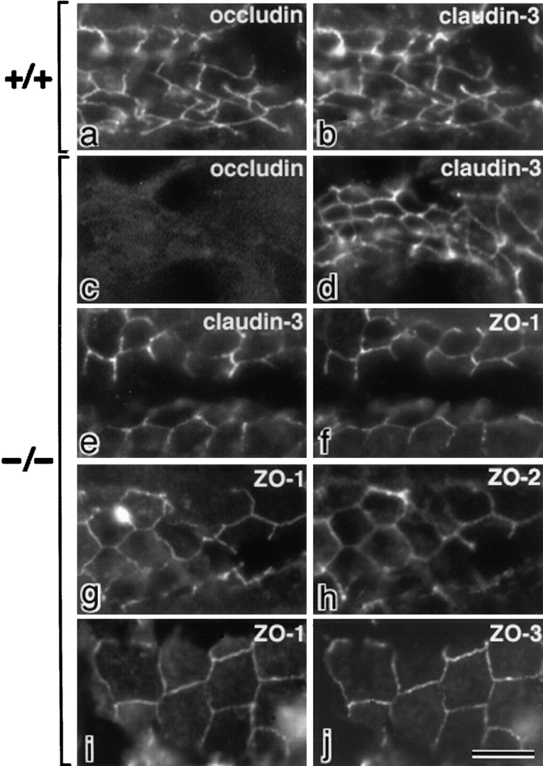
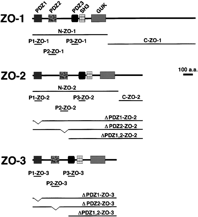
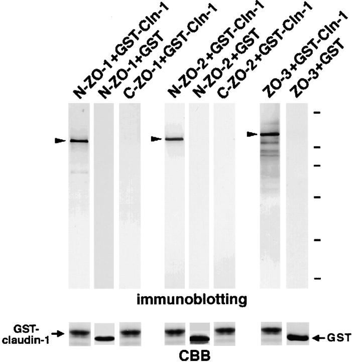
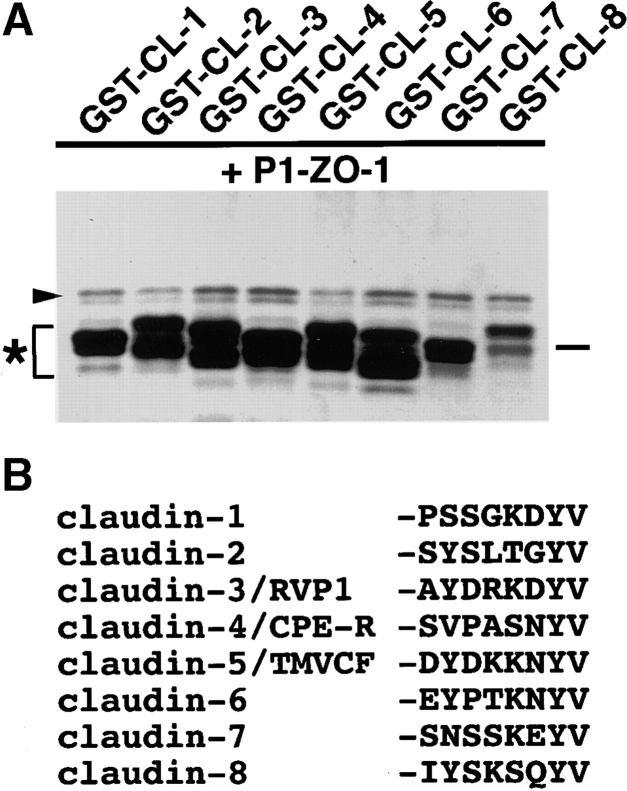
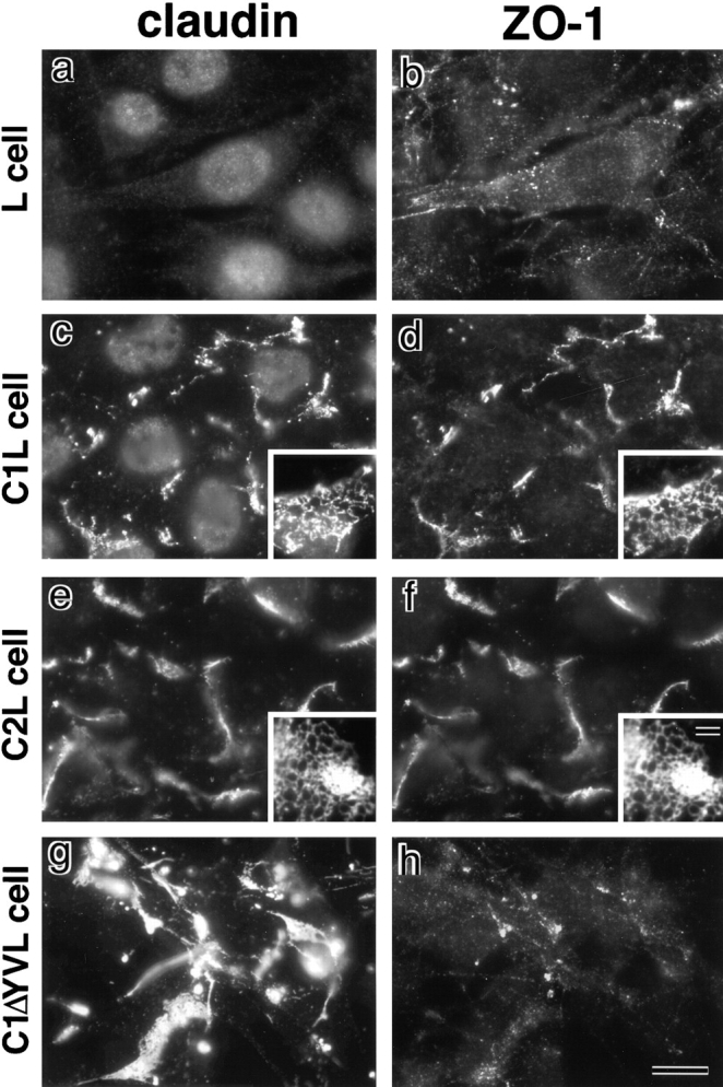
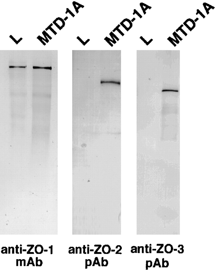
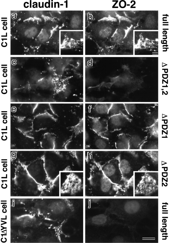
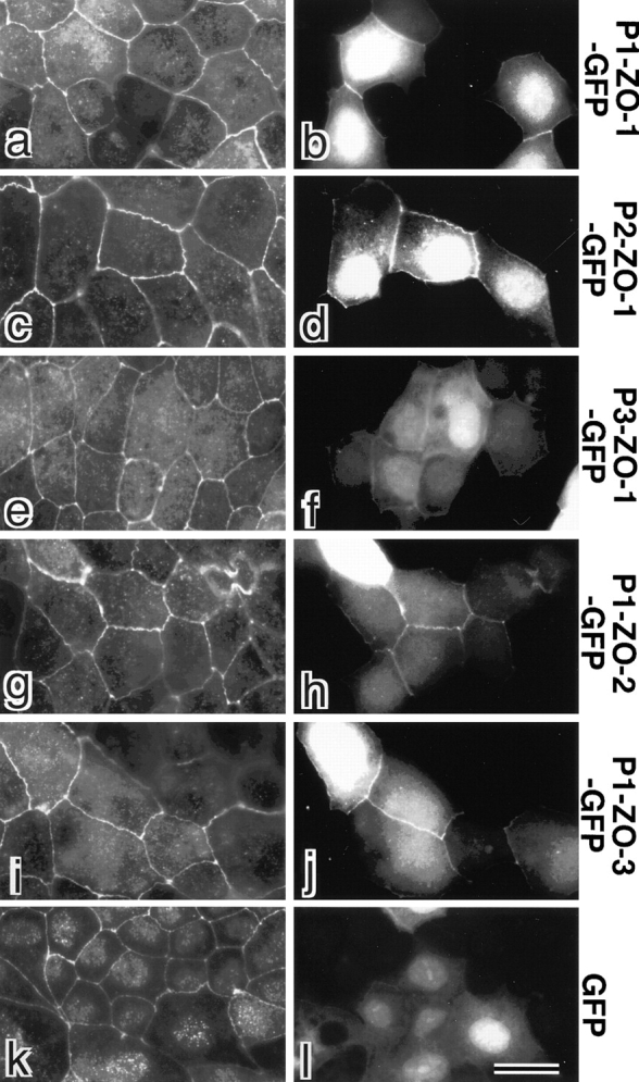
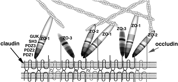
ZO-1, ZO-2, and ZO-3, which contain three PDZ domains (PDZ1 to -3), are concentrated at tight junctions (TJs) in epithelial cells. TJ strands are mainly composed of two distinct types of four-transmembrane proteins, occludin, and claudins, between which occludin was reported to directly bind to ZO-1/ZO-2/ZO-3. However, in occludin-deficient intestinal epithelial cells, ZO-1/ZO-2/ZO-3 were still recruited to TJs. We then examined the possible interactions between ZO-1/ZO-2/ZO-3 and claudins. ZO-1, ZO-2, and ZO-3 bound to the COOH-terminal YV sequence of claudin-1 to -8 through their PDZ1 domains in vitro. Then, claudin-1 or -2 was transfected into L fibroblasts, which express ZO-1 but not ZO-2 or ZO-3. Claudin-1 and -2 were concentrated at cell-cell borders in an elaborate network pattern, to which endogenous ZO-1 was recruited. When ZO-2 or ZO-3 were further transfected, both were recruited to the claudin-based networks together with endogenous ZO-1. Detailed analyses showed that ZO-2 and ZO-3 are recruited to the claudin-based networks through PDZ2 (ZO-2 or ZO-3)/PDZ2 (endogenous ZO-1) and PDZ1 (ZO-2 or ZO-3)/COOH-terminal YV (claudins) interactions. In good agreement, PDZ1 and PDZ2 domains of ZO-1/ZO-2/ZO-3 were also recruited to claudin-based TJs, when introduced into cultured epithelial cells. The possible molecular architecture of TJ plaque structures is discussed.


Figures

Figure 1
Figure 1
Recruitment of ZO-1/ZO-2/ZO-3 to TJs of intestinal epithelial cells in occludin-deficient mice. Frozen sections of intestinal epithelial cells in wild-type (a and b) and occludin-deficient mice (c–j) were double stained with rat anti–occludin mAb (a and c)/rabbit anti–claudin-3 pAb (b and d), rabbit anti–claudin-3 pAb (e)/mouse anti–ZO-1 mAb (f), mouse anti–ZO-1 mAb (g)/rabbit anti–ZO-2 pAb (h), or mouse anti–ZO-1 mAb (i)/rabbit anti–ZO-3 pAb (j). In occludin-deficient intestinal epithelial cells, not only ZO-1 but also ZO-2 and ZO-3 were concentrated at the claudin-3–positive TJs. Bar, 10 μm.
Figure 2
Figure 2
Structures of ZO-1, ZO-2, and ZO-3 and their deletion mutants. ZO-1, ZO-2, and ZO-3 contain three PDZ domains (PDZ1, PDZ2, PDZ3), one SH3 domain (SH3) and one guanylate kinase-like domain (GUK) in this order from their NH2 termini. ZO-1 and ZO-2 were divided into NH2-terminal (N-ZO-1, N-ZO-2) and COOH-terminal regions (C-ZO-1, C-ZO-2). When these constructs were expressed in Sf9 cells by baculovirus infection, N- and C-ZO-2, but not N- or C-ZO-1, were tagged with 6xHis. When the other constructs were expressed in E. coli or mammalian cells, they were tagged with 6xHis or c-myc, respectively.
Figure 3
Figure 3
Association of N-ZO-1, N-ZO-2, and full-length ZO-3 with the cytoplasmic domain of claudin-1 in vitro. The GST fusion protein with the cytoplasmic domain of claudin-1 (GST-Cln-1) or GST protein (GST), which was bound to glutathione–Sepharose beads, was incubated with the lysate of Sf9 cells expressing N-ZO-1, C-ZO-1, 6xHis-N-ZO-2, or 6xHis-C-ZO-2, or with the lysate of E. coli expressing 6xHis-tagged full-length ZO-3. In each lysate, the amount of recombinant protein was adjusted to be the same. After washing, the proteins associated with GST fusion protein or GST were eluted from the beads with a buffer containing glutathione. The eluates from N-ZO-1–, C-ZO-1–, 6xHis-N-ZO-2–, 6xHis-C-ZO-2–, or 6xHis-ZO-3–incubated beads were separated by SDS-PAGE followed by immunoblotting with anti–ZO-1 mAb T8754, anti–ZO-1 mAb T7713, anti-His tag mAb, anti-His tag mAb, or anti-His tag mAb (immunoblotting). The amount of GST–claudin-1 (GST-claudin-1) and GST (GST) in each eluate was determined by Coomassie brilliant blue staining (CBB). As indicated by arrowheads, N-ZO-1, N-ZO-2, full-length ZO-3, but not C-ZO-1 or C-ZO-2, showed binding affinity to the cytoplasmic domain of claudin-1. Bars indicate molecular masses of 200, 116, 97, 66, 45, and 31 kD, respectively, from the top.
Figure 4
Figure 4
Association of PDZ1 domains of ZO-1, ZO-2, and ZO-3 with the COOH-terminal YV sequence of claudin-1. All SDS-PAGE gels were stained with Coomassie brilliant blue. (A) The GST fusion protein with the cytoplasmic domain of claudin-1 (GST-CL-1), which was bound to glutathione–Sepharose beads, was incubated with the lysate of E. coli expressing 6xHis-PDZ1 domains (P1-ZO-1, P1-ZO-2, P1-ZO-3), 6xHis-PDZ2 domains (P2-ZO-1, P2-ZO-2, P2-ZO-3), and 6xHis-PDZ3 domains (P3-ZO-1, P3-ZO-2, P3-ZO-3) of ZO-1, ZO-2, and ZO-3. After washing, the proteins associated with GST fusion proteins were eluted from the beads with a buffer containing glutathione. The eluates from PDZ domain-incubated beads were separated by SDS-PAGE followed by Coomassie brilliant blue staining or by immunoblotting with anti-His tag mAb (anti-His tag mAb). Among the 9 PDZ domains, only P1-ZO-1, P1-ZO-2, and P1-ZO-3 were associated with the cytoplasmic domain of claudin-1 (arrowheads). (B) Similar binding assay was performed between P1-ZO-1/P1-ZO-2/P1-ZO-3 and GST fusion protein with the COOH-terminal YV-deleted cytoplasmic domain of claudin-1 (GST-CL-1ΔYV). P1-ZO-1, P1-ZO-2, and P1-ZO-3 bound to GST-CL-1 (arrowheads), but not to GST-CL-1ΔYV. Bars indicate a molecular mass of 31 kD. (C) Quantitative analysis of the binding between PDZ1 domains of ZO-1/ZO-2/ZO-3 and the cytoplasmic domain of claudin-1. Glutathione–Sepharose bead slurry containing GST-claudin-1 was incubated with E. coli lysate containing 0.01–0.5 μg of the PDZ1 domain of ZO-1, ZO-2, or ZO-3 (from the top). The amounts of the PDZ1 domain of ZO-1, ZO-2, or ZO-3 in the E. coli lysate and in each eluate (inset) were estimated as described in Materials and Methods. Each point represents the mean value of triplicate determinations. The binding was saturable, and Scatchard analysis (inset) indicated that the K d values for the PDZ1 domains of ZO-1 (P1-ZO-1), ZO-2 (P1-ZO-2), and ZO-3 (P1-ZO-3) were ∼13, 11, and 18 nM, respectively.
Figure 5
Figure 5
Association of PDZ1 domains of ZO-1 (P1-ZO-1) with the COOH-terminal YV sequence of claudin-1 to -8 (GST-CL-1 to -8). (A) In vitro binding analyses similar to Fig. 4 were performed. P1-ZO-1 bound to all of GST-CL-1 to -8 (arrowhead). Some of the GST fusion proteins (asterisk) were partly degraded. Bar indicates a molecular mass of 31 kD. (B) COOH-terminal amino acid sequences of claudin-1 to -8. All end in YV.
Figure 7
Figure 7
Recruitment of endogenous ZO-1 to claudin-based networks in L transfectants expressing claudin-1 or -2. Parental L cells (L cell; a and b) and L transfectants expressing claudin-1 (C1L cell; c and d) were double stained with rat anti–claudin-1 mAb (claudin; a and c) and mouse anti–ZO-1 mAb (ZO-1; b and d). L transfectants expressing claudin-2 (C2L cell; e and f) were double stained with anti–claudin-2 mAb (e) and anti–ZO-1 mAb (f). L transfectants expressing claudin-1 mutant lacking the COOH-terminal YV sequence (C1ΔYVL cell; g and h) were double stained with anti–claudin-1 pAb (g) and anti–ZO-1 mAb (h). ZO-1 showed no characteristic concentration in L cells (b), whereas in C1L and C2L cells ZO-1 was coconcentrated with claudin-1 (c and d) and claudin-2 (e and f) as planes at cell–cell borders. Close inspection revealed that in these cells both claudins and ZO-1 were concentrated in elaborate network patterns, and that their network patterns were mostly overlapped (insets in c–f). In C1ΔYVL cells, mutant claudin-1 was concentrated at cell–cell borders (g), whereas ZO-1 showed no concentration (h). Bar: (a–h) 10 μm; (insets) 15 μm.
Figure 6
Figure 6
Expression of ZO-1, ZO-2, and ZO-3 in mouse L fibroblasts. The total cell lysate of cultured mouse L fibroblasts and MTD-1A epithelial cells were subjected to immunoblotting with anti–ZO-1 mAb (anti–ZO-1 mAb), anti–ZO-2 pAb (anti–ZO-2 pAb), or anti–ZO-3 pAb (anti–ZO-3 pAb). ZO-1, ZO-2, and ZO-3 were expressed in MTD-1A cells, whereas only ZO-1 was detected in L fibroblasts. Bars indicate molecular masses of 200, 116, 97, 66, 45, and 31 kD, respectively, from the top.
Figure 8
Figure 8
Recruitment of exogenous ZO-2 to claudin-based networks in L transfectants expressing claudin-1. Full-length ZO-2 (full-length; a and b), deletion mutant of ZO-2 lacking both PDZ1 and -2 domains (ΔPDZ1,2; c and d), deletion mutant of ZO-2 lacking PDZ1 domain (ΔPDZ1; e and f), and deletion mutant of ZO-2 lacking PDZ2 domain (ΔPDZ2; g and h) were transfected into L transfectants expressing claudin-1 (C1L cells), and stable transfectants were obtained. Furthermore, full-length ZO-2 (full-length; i and j) was transfected into L transfectants expressing claudin-1 mutant lacking its COOH-terminal YV sequence (C1ΔYVL cell), and stable transfectants were obtained. These introduced proteins were tagged with c-myc epitope. These stable transfectants were double stained with anti–claudin-1 mAb (a, c, e, and g) or pAb (i) and anti–c-myc mAb (b, d, f, h, and j). In C1L cells where claudin-1 was concentrated at cell–cell borders in an elaborate network pattern, full-length ZO-2 (b), ΔPDZ1-ZO-2 (f), and ΔPDZ2-ZO-2 (h), but not ΔPDZ1,2-ZO-2 (d), were recruited to the claudin-1–based networks. Insets represent the network patterns of concentrated claudin-1 (a and g), full-length ZO-2 (b), and ΔPDZ2-ZO-2 (h). No concentration of full-length ZO-2 was observed in C1ΔYVL cells (j). Bar: (a–j) 10 μm; (insets) 15 μm.
Figure 10
Figure 10
Behavior of GFP-fusion proteins with PDZ1, -2, and -3 domains of ZO-1/ZO-2/ZO-3 in cultured epithelial cells (MDCK cells). GFP (GFP) or GFP-fusion proteins with PDZ1 domain of ZO-1 (P1-ZO-1-GFP), PDZ2 domain of ZO-1 (P2-ZO-1-GFP), PDZ3 domain of ZO-1 (P3-ZO-1-GFP), PDZ1 domain of ZO-2 (P1-ZO-2-GFP) or PDZ1 domain of ZO-3 (P1-ZO-3-GFP) were exogenously and transiently expressed in MDCK cells. These cells were fixed and stained with anti–claudin-1 pAb (a, c, e, g, i, and k) in red. Expressed GFP or GFP-fusion proteins were visualized by green fluorescence (b, d, f, h, j, and l). PDZ1 domains of ZO-1 (b, P1-ZO-1-GFP), ZO-2 (h, P1-ZO-2-GFP), ZO-3 (j, P1-ZO-3-GFP), and PDZ2 domain of ZO-1 (d, P2-ZO-1-GFP) were recruited to claudin-1–positive TJs. PDZ3 domain of ZO-1 (f, P3-ZO-1-GFP) also appeared to be concentrated at TJs, although very faintly as compared with b, d, h, and j. No concentration of GFP at cell–cell borders was observed (l). Bar, 10 μm.
Figure 11
Figure 11
Schematic drawing of the molecular architecture of the TJ plaque. TJ strands are represented as linear co-polymers of claudins and occludin with short and long COOH-terminal tails, respectively. ZO-1, ZO-2, and ZO-3 are directly associated with the COOH termini of claudins at their PDZ1 domains. These molecules are depicted here to interact with occludin at their GUK domains. ZO-1/ZO-2 and ZO-1/ZO-3 heterodimers are depicted to be formed through direct PDZ2/PDZ2 interaction, but the possibility cannot be completely excluded that these interaction requires some linker protein. It remains unclear whether ZO-1, ZO-2, and ZO-3 exist as monomers and/or homodimers (not depicted here), except that ZO-1/ZO-1 homodimers were reported to be undetectable. The COOH-terminal region of ZO-1 and ZO-2 has a binding affinity to actin filaments to function as cross-linkers between TJ strands and actin filaments. The interaction between ZO-3 and actin filaments has not yet been examined.

Similar articles

Cited by

References

    1. Anderson J.M. Cell signallingMAGUK magic. Curr. Biol. 1996;6:382–384. - PubMed
    1. Ando-Akatsuka Y., Saitou M., Hirase T., Kishi M., Sakakibara A., Itoh M., Yonemura S., Furuse M., Tsukita Sh. Interspecies diversity of the occludin sequencecDNA cloning of human, mouse, dog, and rat-kangaroo homologues. J. Cell Biol. 1996;133:43–47. - PMC - PubMed
    1. Balda M.S., Anderson J.M., Matter K. The SH3 domain of the tight junction protein ZO-1 binds to a serine protein kinase that phosphorylates a region C-terminal to this domain. FEBS Lett. 1996;399:326–332. - PubMed
    1. Balda M.S., González-Mariscal L., Matter K., Cereijido M., Anderson J.M. Assembly of the tight junctionThe role of diacylglycerol. J. Cell Biol. 1993;123:293–302. - PMC - PubMed
    1. Citi S., Sabanay H., Jakes R., Geiger B., Kendrick-Jones J. Cingulin, a new peripheral component of tight junctions. Nature. 1988;333:272–276. - PubMed

Publication types

MeSH terms

Substances

Associated data