. 1999 Dec 20;190(12):1769-82.
doi: 10.1084/jem.190.12.1769.

Targeted gene disruption demonstrates that P-selectin glycoprotein ligand 1 (PSGL-1) is required for P-selectin-mediated but not E-selectin-mediated neutrophil rolling and migration

Affiliations

Targeted gene disruption demonstrates that P-selectin glycoprotein ligand 1 (PSGL-1) is required for P-selectin-mediated but not E-selectin-mediated neutrophil rolling and migration

J Yang et al. J Exp Med. .

Abstract

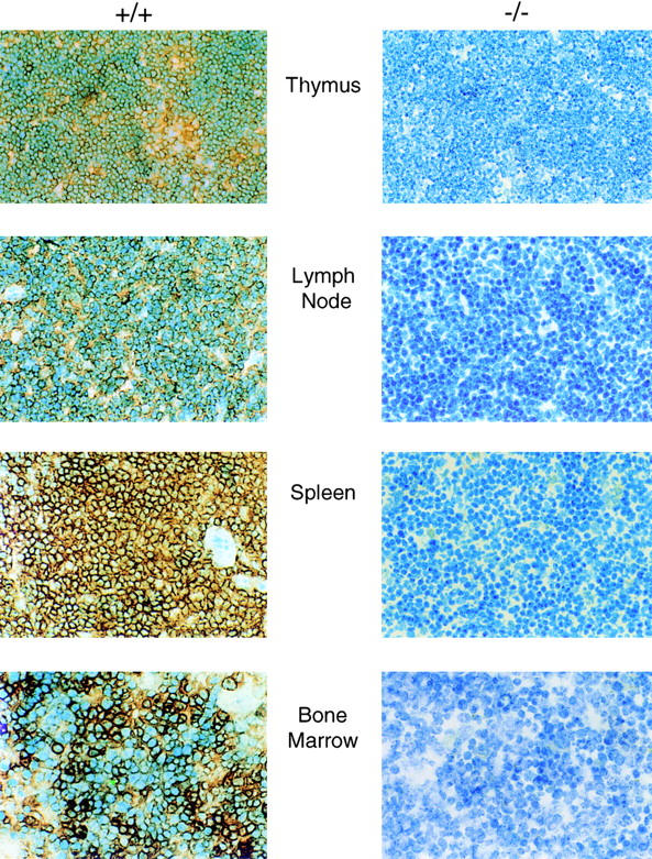
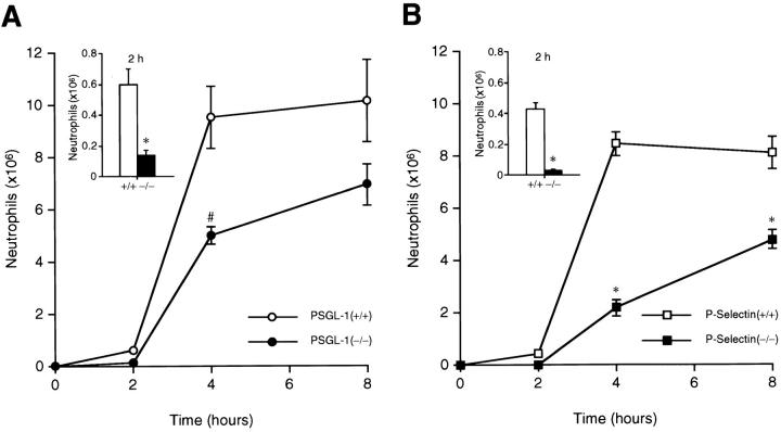
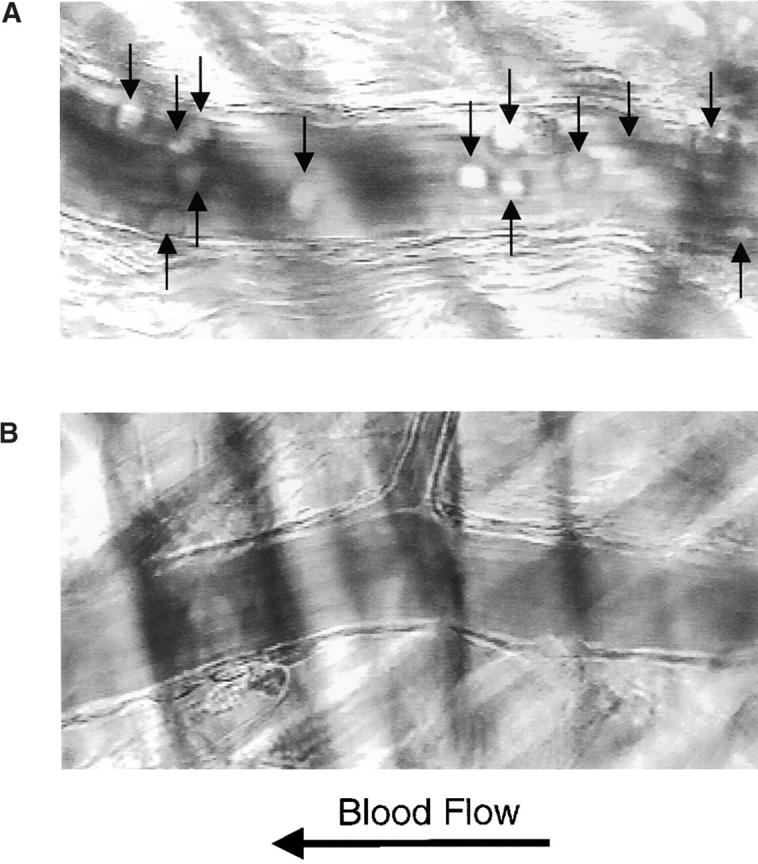
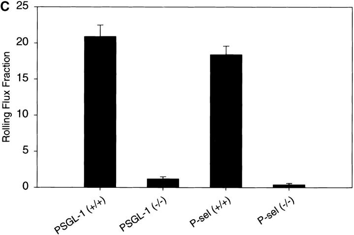
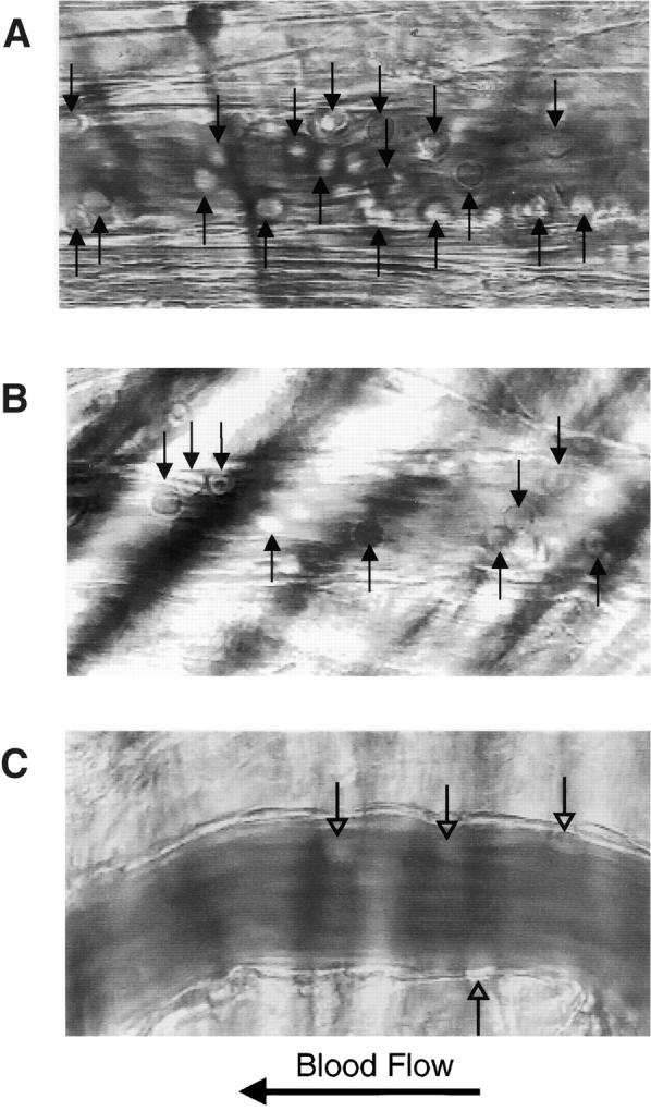
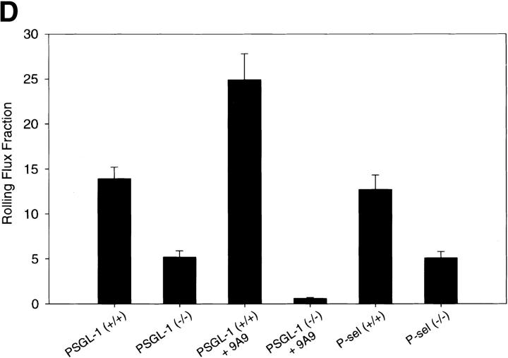
P-selectin glycoprotein ligand 1 (PSGL-1) is a mucin-like selectin counterreceptor that binds to P-selectin, E-selectin, and L-selectin. To determine its physiological role in cell adhesion as a mediator of leukocyte rolling and migration during inflammation, we prepared mice genetically deficient in PSGL-1 by targeted disruption of the PSGL-1 gene. The homozygous PSGL-1-deficient mouse was viable and fertile. The blood neutrophil count was modestly elevated. There was no evidence of spontaneous development of skin ulcerations or infections. Leukocyte infiltration in the chemical peritonitis model was significantly delayed. Leukocyte rolling in vivo, studied by intravital microscopy in postcapillary venules of the cremaster muscle, was markedly decreased 30 min after trauma in the PSGL-1-deficient mouse. In contrast, leukocyte rolling 2 h after tumor necrosis factor alpha stimulation was only modestly reduced, but blocking antibodies to E-selectin infused into the PSGL-1-deficient mouse almost completely eliminated leukocyte rolling. These results indicate that PSGL-1 is required for the early inflammatory responses but not for E-selectin-mediated responses. These kinetics are consistent with a model in which PSGL-1 is the predominant neutrophil P-selectin ligand but is not a required counterreceptor for E-selectin under in vivo physiological conditions.


Figures

Figure 1
Figure 1
Disruption of the PSGL-1 gene in mice: targeting vector and Southern blot analysis. (A) Partial restriction maps of the PSGL-1 gene and the targeting vector KO.pNT. Restriction enzyme sites are marked as follows: E, EcoRI; H, HindIII; B, BamHI; S, SmaI; N, NotI. Probes used for Southern analyses (A–D, Neo), primers used for generating the vector (P1–P4), and the repeat elements are indicated. (B) Southern blot analyses of ES clones. Genomic DNA from two clones, W19 and W34, were digested with HindIII. After gel electrophoresis, DNA was transferred to the membrane and hybridized with probe C. The blot was stripped and reprobed with probe Neo. The targeted band (KO) and the wild-type band (WT) are indicated. (C) Southern blot analyses of DNA isolated from F1 and F2 generations. Genomic DNA from tail biopsies was digested with EcoRI. The fragments were separated by gel electrophoresis, transferred to a membrane, and hybridized with probe A. The bands corresponding to the targeted allele (KO) and the wild-type allele (WT) are indicated.
Figure 1
Figure 1
Disruption of the PSGL-1 gene in mice: targeting vector and Southern blot analysis. (A) Partial restriction maps of the PSGL-1 gene and the targeting vector KO.pNT. Restriction enzyme sites are marked as follows: E, EcoRI; H, HindIII; B, BamHI; S, SmaI; N, NotI. Probes used for Southern analyses (A–D, Neo), primers used for generating the vector (P1–P4), and the repeat elements are indicated. (B) Southern blot analyses of ES clones. Genomic DNA from two clones, W19 and W34, were digested with HindIII. After gel electrophoresis, DNA was transferred to the membrane and hybridized with probe C. The blot was stripped and reprobed with probe Neo. The targeted band (KO) and the wild-type band (WT) are indicated. (C) Southern blot analyses of DNA isolated from F1 and F2 generations. Genomic DNA from tail biopsies was digested with EcoRI. The fragments were separated by gel electrophoresis, transferred to a membrane, and hybridized with probe A. The bands corresponding to the targeted allele (KO) and the wild-type allele (WT) are indicated.
Figure 2
Figure 2
Northern blot analysis of tissue for PSGL-1 mRNA. Total RNA isolated from the thymus and spleen of PSGL-1 (−/−) and PSGL-1 (+/+) mice was subjected to electrophoresis, and the RNA was transferred to a Duralon-UV™ membrane. The PSGL-1 messenger RNA was detected with a probe representing most of the coding region. The membrane was stripped and probed with a cDNA probe for β-actin.
Figure 3
Figure 3
Flow cytometric analysis of PSGL-1 expression on peripheral blood neutrophils. Cells from wild-type (+/+; top), PSGL-1+/− (middle), and PSGL-1−/− (bottom) mice were stained with anti–PSGL-1 antibodies (solid line) or polyclonal rabbit nonimmune IgG (dotted line) followed by FITC-labeled goat anti–rabbit IgG. Fluorescence intensity is shown on the x-axis; cell number is presented on the y-axis.
Figure 4
Figure 4
Immunohistochemical analysis of spleen, thymus, lymph node, and bone marrow. Tissue from homozygous null (−/−) and wild-type (+/+) mice is indicated. Fixed tissue sections were treated with normal goat serum followed by rabbit anti–mouse PSGL-1 antibody. Bound anti–mouse PSGL-1 antibody was visualized with biotinylated goat anti–rabbit antibody reacted with peroxidase-conjugated streptavidin. The samples were developed with 3,3′-diaminobenzidine and counterstained with methylene blue.
Figure 5
Figure 5
Neutrophil migration in thioglycollate-induced peritonitis. PSGL-1–deficient mice with matched controls and P-selectin–deficient mice with matched controls were studied in parallel. Absolute neutrophil counts in the peritoneal exudate were determined at the indicated times after induction of experimental peritonitis with thioglycollate. An expanded comparison at 2 h is presented in the inset. (A) PSGL-1−/− mice (•, and [inset] black bars) and matched wild-type control mice (○, and [inset] white bars). (B) P-selectin−/− mice (▪, and [inset] black bars) and matched wild-type control mice (□, and [inset] white bars). Each data point represents the average of the results of 8–10 mice. Data are presented as mean ± SEM. Differences were statistically significant (*P < 0.001; # P = 0.002) compared with respective wild-type controls.
Figure 6
Figure 6
Leukocyte rolling after mild trauma. Intravital microscopy of cremaster muscle venules within 30 min of the initiation of surgery revealed the presence of a large number of rolling leukocytes (arrows) associated with the vascular wall in wild-type mice (A) and no leukocytes associated with the vascular wall in PSGL-1–deficient mice (B). Videos available at http://www.jem.org/cgi/content/full/190/12/1769/F6/DC1 correspond to A and B. The leukocyte rolling flux fraction, measured as the number of rolling leukocytes compared with the total leukocytes in flowing blood per unit time, is compared for PSGL-1+/+, PSGL-1−/−, P-selectin (P-sel)+/+, and P-selectin−/− mice in this assay system (C). Both PSGL-1−/− and P-selectin−/− mice have low but measurable leukocyte rolling flux fractions.
Figure 6
Figure 6
Leukocyte rolling after mild trauma. Intravital microscopy of cremaster muscle venules within 30 min of the initiation of surgery revealed the presence of a large number of rolling leukocytes (arrows) associated with the vascular wall in wild-type mice (A) and no leukocytes associated with the vascular wall in PSGL-1–deficient mice (B). Videos available at http://www.jem.org/cgi/content/full/190/12/1769/F6/DC1 correspond to A and B. The leukocyte rolling flux fraction, measured as the number of rolling leukocytes compared with the total leukocytes in flowing blood per unit time, is compared for PSGL-1+/+, PSGL-1−/−, P-selectin (P-sel)+/+, and P-selectin−/− mice in this assay system (C). Both PSGL-1−/− and P-selectin−/− mice have low but measurable leukocyte rolling flux fractions.
Figure 7
Figure 7
Leukocyte rolling after TNF-α stimulation. Intravital microscopy of cremaster muscle venules 2 h after injection of TNF-α into the scrotal sac revealed the presence of rolling leukocytes (arrows) associated with the vascular wall in wild-type mice (A) and in PSGL-1–deficient mice (B). Infusion of the blocking E-selectin antibody 9A9 into PSGL-1–deficient mice greatly reduced the number of rolling leukocytes; leukocytes visible in C are stationary. Videos available at http://www.jem.org/cgi/content/full/190/12/1769/F7/DC1 correspond to A and B, and to two time points in C, showing leukocyte rolling before infusion of anti–E-selectin antibody, and leukocyte rolling during and after infusion of anti–E-selectin antibody, respectively. The leukocyte rolling flux fraction, measured as the number of rolling leukocytes compared with the total leukocytes in flowing blood per unit time, is compared for PSGL-1+/+ mice, PSGL-1−/− mice, PSGL-1−/− mice treated with anti–E-selectin antibody 9A9, PSGL-1+/+ mice treated with anti–E-selectin antibody 9A9, P-selectin (P-sel)+/+ mice, and P-selectin−/− mice in this assay system (D).
Figure 7
Figure 7
Leukocyte rolling after TNF-α stimulation. Intravital microscopy of cremaster muscle venules 2 h after injection of TNF-α into the scrotal sac revealed the presence of rolling leukocytes (arrows) associated with the vascular wall in wild-type mice (A) and in PSGL-1–deficient mice (B). Infusion of the blocking E-selectin antibody 9A9 into PSGL-1–deficient mice greatly reduced the number of rolling leukocytes; leukocytes visible in C are stationary. Videos available at http://www.jem.org/cgi/content/full/190/12/1769/F7/DC1 correspond to A and B, and to two time points in C, showing leukocyte rolling before infusion of anti–E-selectin antibody, and leukocyte rolling during and after infusion of anti–E-selectin antibody, respectively. The leukocyte rolling flux fraction, measured as the number of rolling leukocytes compared with the total leukocytes in flowing blood per unit time, is compared for PSGL-1+/+ mice, PSGL-1−/− mice, PSGL-1−/− mice treated with anti–E-selectin antibody 9A9, PSGL-1+/+ mice treated with anti–E-selectin antibody 9A9, P-selectin (P-sel)+/+ mice, and P-selectin−/− mice in this assay system (D).

Similar articles

Cited by

References

    1. Springer T.A. Traffic signals for lymphocyte recirculation and leukocyte emigrationthe multistep paradigm. Cell. 1994;76:301–314. - PubMed
    1. Tedder T.F., Steeber D.A., Chen A., Engel P. The selectinsvascular adhesion molecules. FASEB J. 1995;9:866–873. - PubMed
    1. McEver R.P., Moore K.L., Cummings R.D. Leukocyte trafficking mediated by selectin-carbohydrate interactions. J. Biol. Chem. 1995;270:11025–11028. - PubMed
    1. Stenberg P.E., McEver R.P., Shuman M.A., Jacques Y.V., Bainton D.F. A platelet alpha-granule membrane protein GMP140 is expressed on the plasma membrane after activation. J. Cell Biol. 1985;101:880–886. - PMC - PubMed
    1. Berman C.L., Yeo E., Wencel-Drake J.D., Furie B.C., Ginsberg M.H., Furie B. A platelet alpha granule membrane protein that is incorporated into the plasma membrane during activation. Characterization and subcellular localization of PADGEM protein. J. Clin. Invest. 1986;78:130–137. - PMC - PubMed

Publication types