. 1999 Dec 20;190(12):1837-48.
doi: 10.1084/jem.190.12.1837.

Lineage-specific modulation of interleukin 4 signaling by interferon regulatory factor 4

Affiliations

Lineage-specific modulation of interleukin 4 signaling by interferon regulatory factor 4

S Gupta et al. J Exp Med. .

Abstract

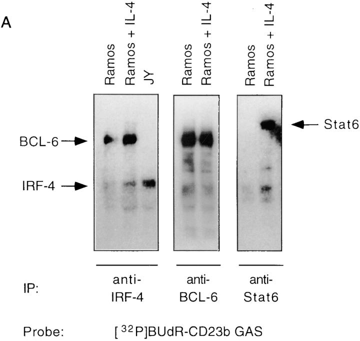
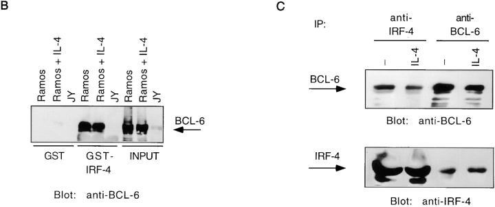
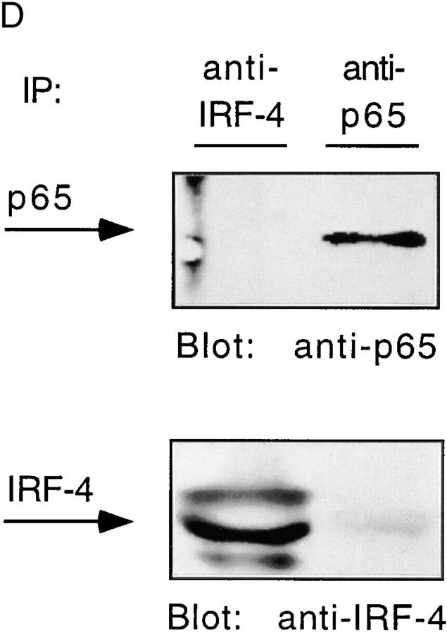
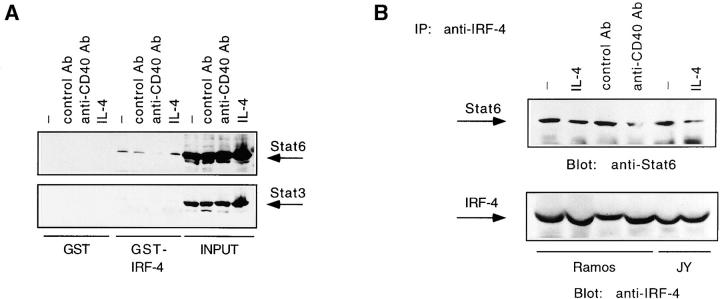
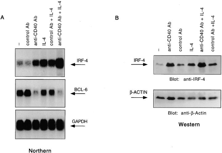
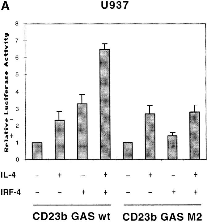
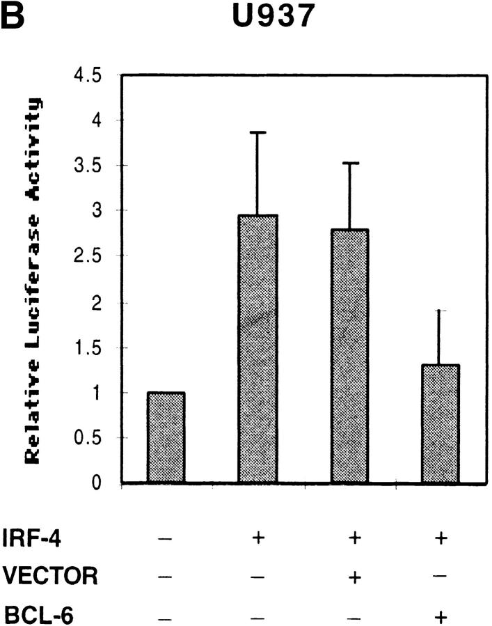
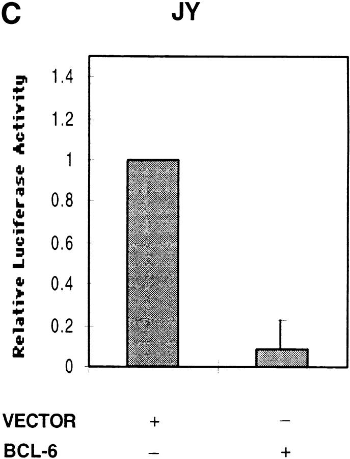
Interleukin (IL)-4 is an immunoregulatory cytokine that exerts distinct biological activities on different cell types. Our studies indicate that interferon regulatory factor (IRF)-4 is both a target and a modulator of the IL-4 signaling cascade. IRF-4 expression is strongly upregulated upon costimulation of B cells with CD40 and IL-4. Furthermore, we find that IRF-4 can interact with signal transducer and activator of transcription (Stat)6 and drive the expression of IL-4-inducible genes. The transactivating ability of IRF-4 is blocked by the repressor factor BCL-6. Since expression of IRF-4 is mostly confined to lymphoid cells, these data provide a potential mechanism by which IL-4-inducible genes can be regulated in a lineage-specific manner.


Figures

Figure 1
Figure 1
The synergistic upregulation of CD23 in response to CD40 and IL-4 reflects an induction of both CD23a and CD23b isoforms. Ramos cells were cultured for 24 h either in the absence of any stimulus or in the presence of an anti-CD40 Ab (0.1 μg/ml), an isotype-matched control Ab (0.1 μg/ml), human IL-4 (100 U/ml), a combination of anti-CD40 Ab plus IL-4, or a control Ab plus IL-4. U937 cells were either unstimulated or stimulated with IL-4. JY cells were left unstimulated. After harvesting the cells, total RNA was extracted and subjected to RNase protection analysis. The antisense riboprobe corresponds to a 600-bp 5′ EcoRI-HindIII fragment of the CD23a cDNA. The protected fragments of 588 and 488 nucleotides correspond to two isoforms of CD23a transcript which differ by one base change at position 96 in the 5′ untranslated region, while the 381-nucleotide protected fragment corresponds to the CD23b transcript (reference 44).
Figure 2
Figure 2
The CD23b GAS is targeted by Stat6, BCL-6, and IRF-4. (A) Ramos cells were either unstimulated or stimulated with anti-CD40 Ab (0.1 μg/ml), an isotype-matched control Ab (0.1 μg/ml), or human IL-4 (100 U/ml) for 24 h. JY cells were left unstimulated. Nuclear extracts were then prepared and analyzed by EMSA using a 32P-labeled CD23b GAS wt probe. (B) Ramos cells were cultured and assayed as described in A. Oligonucleotide competition assays were performed either in the absence or presence of a 100-fold molar excess of cold GAS oligonucleotides (see Table ) added to the shift reaction as indicated. (C) Ramos and JY cells were cultured as described in A. Nuclear extracts were then prepared and analyzed by EMSA using either a 32P-labeled CD23b GAS wt probe or a 32P-labeled CD23b GAS M1 probe (Table ) as indicated. Ab interference mobility shift assays were carried out by addition of antisera against Stat6, BCL-6, IRF-4, or control. All antisera were added at a final dilution of 1:20 for 30 min at 4°C before incubation with the probe for 20 min at 25°C. (D) JY cells were unstimulated. Nuclear extracts were then prepared and analyzed by EMSA using a 32P-labeled CD23b GAS wt probe (left). Ab interference mobility shift assays were carried out by addition of antisera against IRF-2, IRF-4, ICSBP, or control as indicated. All antisera were added as outlined above. As a control for the IRF-2 and ICSBP antisera, Ab interference analysis using a 32P-labeled GBP-ISRE probe (reference 32) was simultaneously performed on Ramos extracts (right).
Figure 2
Figure 2
The CD23b GAS is targeted by Stat6, BCL-6, and IRF-4. (A) Ramos cells were either unstimulated or stimulated with anti-CD40 Ab (0.1 μg/ml), an isotype-matched control Ab (0.1 μg/ml), or human IL-4 (100 U/ml) for 24 h. JY cells were left unstimulated. Nuclear extracts were then prepared and analyzed by EMSA using a 32P-labeled CD23b GAS wt probe. (B) Ramos cells were cultured and assayed as described in A. Oligonucleotide competition assays were performed either in the absence or presence of a 100-fold molar excess of cold GAS oligonucleotides (see Table ) added to the shift reaction as indicated. (C) Ramos and JY cells were cultured as described in A. Nuclear extracts were then prepared and analyzed by EMSA using either a 32P-labeled CD23b GAS wt probe or a 32P-labeled CD23b GAS M1 probe (Table ) as indicated. Ab interference mobility shift assays were carried out by addition of antisera against Stat6, BCL-6, IRF-4, or control. All antisera were added at a final dilution of 1:20 for 30 min at 4°C before incubation with the probe for 20 min at 25°C. (D) JY cells were unstimulated. Nuclear extracts were then prepared and analyzed by EMSA using a 32P-labeled CD23b GAS wt probe (left). Ab interference mobility shift assays were carried out by addition of antisera against IRF-2, IRF-4, ICSBP, or control as indicated. All antisera were added as outlined above. As a control for the IRF-2 and ICSBP antisera, Ab interference analysis using a 32P-labeled GBP-ISRE probe (reference 32) was simultaneously performed on Ramos extracts (right).
Figure 5
Figure 5
IRF-4 interacts with BCL-6. (A) Ramos cells were either unstimulated or stimulated with IL-4 (100 U/ml). JY cells were unstimulated. Nuclear extracts were then prepared as described in the legend to Fig. 2, and incubated with a radiolabeled 5-bromodeoxyuridine–substituted CD23b GAS wt probe (BUdR-CD23b GAS wt) in a standard shift reaction. DNA–protein complexes were irradiated twice with 1,000 mJ of UV, and then immunoprecipitated (IP) with either an IRF-4, a BCL-6, or a Stat6 antiserum. The immunoprecipitates were resolved by 7% SDS-PAGE, transferred to a nitrocellulose membrane, and then exposed to an x-ray film. No cross-linking was detected in the absence of UV irradiation. (B) Ramos and JY cells were cultured as described in A. Nuclear extracts were then prepared and incubated with immobilized GST–IRF-4 fusion protein. Bound proteins were eluted, fractionated by 7% SDS-PAGE, transferred to a nitrocellulose membrane, and then probed with a BCL-6 antiserum. Binding to immobilized GST alone is shown as a control. (C) Ramos cells were either unstimulated or stimulated with IL-4 (100 U/ml). Nuclear extracts were prepared and immunoprecipitated with either an anti–IRF-4 or an anti–BCL-6 antiserum. The immune complexes were resolved by 7% SDS-PAGE, and then analyzed by Western blotting using an anti–BCL-6 antiserum (top). The blot was later stripped and reprobed with an anti–IRF-4 antiserum (bottom), as described in the legend to Fig. 3. (D) JY cells were unstimulated. Nuclear extracts were prepared and immunoprecipitated with either an anti–IRF-4 or an anti-p65 antiserum. The immune complexes were resolved by 7% SDS-PAGE, and then analyzed by Western blotting using an anti-p65 antiserum (top). The blot was later stripped and reprobed with an anti–IRF-4 antiserum (bottom), as described above.
Figure 5
Figure 5
IRF-4 interacts with BCL-6. (A) Ramos cells were either unstimulated or stimulated with IL-4 (100 U/ml). JY cells were unstimulated. Nuclear extracts were then prepared as described in the legend to Fig. 2, and incubated with a radiolabeled 5-bromodeoxyuridine–substituted CD23b GAS wt probe (BUdR-CD23b GAS wt) in a standard shift reaction. DNA–protein complexes were irradiated twice with 1,000 mJ of UV, and then immunoprecipitated (IP) with either an IRF-4, a BCL-6, or a Stat6 antiserum. The immunoprecipitates were resolved by 7% SDS-PAGE, transferred to a nitrocellulose membrane, and then exposed to an x-ray film. No cross-linking was detected in the absence of UV irradiation. (B) Ramos and JY cells were cultured as described in A. Nuclear extracts were then prepared and incubated with immobilized GST–IRF-4 fusion protein. Bound proteins were eluted, fractionated by 7% SDS-PAGE, transferred to a nitrocellulose membrane, and then probed with a BCL-6 antiserum. Binding to immobilized GST alone is shown as a control. (C) Ramos cells were either unstimulated or stimulated with IL-4 (100 U/ml). Nuclear extracts were prepared and immunoprecipitated with either an anti–IRF-4 or an anti–BCL-6 antiserum. The immune complexes were resolved by 7% SDS-PAGE, and then analyzed by Western blotting using an anti–BCL-6 antiserum (top). The blot was later stripped and reprobed with an anti–IRF-4 antiserum (bottom), as described in the legend to Fig. 3. (D) JY cells were unstimulated. Nuclear extracts were prepared and immunoprecipitated with either an anti–IRF-4 or an anti-p65 antiserum. The immune complexes were resolved by 7% SDS-PAGE, and then analyzed by Western blotting using an anti-p65 antiserum (top). The blot was later stripped and reprobed with an anti–IRF-4 antiserum (bottom), as described above.
Figure 3
Figure 3
IRF-4 interacts with Stat6. (A) Ramos cells were stimulated as described in the legend to Fig. 2. WCEs were then prepared and incubated with immobilized GST–IRF-4 fusion protein. Bound proteins were eluted, resolved by 7% SDS-PAGE, blotted onto a nitrocellulose membrane, and then probed with a Stat6 (top) or Stat3 Ab (bottom). Binding to immobilized GST alone is shown as a control. (B) Ramos cells were stimulated as described in the legend to Fig. 2. JY cells were either unstimulated or stimulated with IL-4 (100 U/ml). WCEs were then prepared and immunoprecipitated with an IRF-4 antiserum. The immunoprecipitated proteins were resolved by 7% SDS-PAGE and then analyzed by Western blotting using a Stat6 Ab (top). The blot was later stripped and reprobed with an anti–IRF-4 antiserum (bottom) using protein A–horseradish peroxidase conjugate as a secondary detection reagent to probe the primary Ab binding.
Figure 4
Figure 4
IRF-4 expression is upregulated in response to CD40 and/or IL-4. (A) Ramos cells were stimulated as described in the legend to Fig. 1. Total RNA was then extracted, and 10 μg of the RNA was assayed by Northern blotting as per standard protocols. The blot was then probed with either an IRF-4 cDNA (top), a BCL-6 cDNA (middle), or a GAPDH cDNA (bottom) radiolabeled by random hexamer priming. (B) Tonsillar mononuclear cells were stimulated as described in the legend to Fig. 1. WCEs were prepared, electrophoresed on a 7% SDS-polyacrylamide gel, and then analyzed by Western blotting using an anti–IRF-4 Ab (top). The blot was later stripped and reprobed with a β-actin Ab to ensure for equal loading.
Figure 6
Figure 6
Effects of IRF-4 on the transactivation of CD23b. (A) U937 cells were cotransfected with a luciferase reporter construct driven by either an oligomerized CD23b GAS wt or an oligomerized CD23b GAS M2 element, and either an IRF-4 expression plasmid (pCEP4-IRF4) or equivalent amounts of empty vector. The transfected cells were equally split into two 2-ml aliquots and then incubated for 24 h in the presence or absence of IL-4 (10 ng/ml). The data are presented relative to the activity of the reporter construct in control U937 cells, which was set to 1.0, as indicated, in each experiment. Results show the mean ± SE of four independent experiments. (B) U937 cells were cotransfected with a luciferase reporter construct driven by an oligomerized CD23b GAS wt element, and an IRF-4 expression plasmid (pCEP4-IRF4) in the presence of either an BCL-6 expression vector (pMT2T-BCL6) or equivalent amounts of empty pMT2T vector, as indicated. The transfected cells were left unstimulated for 16 h. The data are presented relative to the activity of the reporter construct in control U937 cells, which was set to 1.0, as indicated, in each experiment. Results show the mean ± SE of three independent experiments. (C) JY cells were cotransfected with a CD23b promoter luciferase reporter construct in the presence of an expression plasmid for BCL-6 (pMT2T-BCL6) or of equivalent amounts of empty pMT2T vector. The transfected cells were left unstimulated for 16 h. The data are presented relative to the activity of the reporter construct in control JY cells, which was set to 1.0, as indicated, in each experiment. Results show the mean ± SE of three independent experiments.
Figure 6
Figure 6
Effects of IRF-4 on the transactivation of CD23b. (A) U937 cells were cotransfected with a luciferase reporter construct driven by either an oligomerized CD23b GAS wt or an oligomerized CD23b GAS M2 element, and either an IRF-4 expression plasmid (pCEP4-IRF4) or equivalent amounts of empty vector. The transfected cells were equally split into two 2-ml aliquots and then incubated for 24 h in the presence or absence of IL-4 (10 ng/ml). The data are presented relative to the activity of the reporter construct in control U937 cells, which was set to 1.0, as indicated, in each experiment. Results show the mean ± SE of four independent experiments. (B) U937 cells were cotransfected with a luciferase reporter construct driven by an oligomerized CD23b GAS wt element, and an IRF-4 expression plasmid (pCEP4-IRF4) in the presence of either an BCL-6 expression vector (pMT2T-BCL6) or equivalent amounts of empty pMT2T vector, as indicated. The transfected cells were left unstimulated for 16 h. The data are presented relative to the activity of the reporter construct in control U937 cells, which was set to 1.0, as indicated, in each experiment. Results show the mean ± SE of three independent experiments. (C) JY cells were cotransfected with a CD23b promoter luciferase reporter construct in the presence of an expression plasmid for BCL-6 (pMT2T-BCL6) or of equivalent amounts of empty pMT2T vector. The transfected cells were left unstimulated for 16 h. The data are presented relative to the activity of the reporter construct in control JY cells, which was set to 1.0, as indicated, in each experiment. Results show the mean ± SE of three independent experiments.

Similar articles

Cited by

References

    1. Brown M., Hurai J. Functions of IL-4 and control of its expression. Crit. Rev. Immunol. 1997;17:1–32. - PubMed
    1. Chomarat P., Banchereau J. An update on interleukin-4 and its receptor. Eur. Cytokine Netw. 1997;8:333–344. - PubMed
    1. Paul W.E. Interleukin-4a prototypic immunoregulatory lymphokine. Blood. 1991;77:1859–1870. - PubMed
    1. Laman J., Claassen E., Noelle R. Functions of CD40 and its ligand, gp39 (CD40L) Crit. Rev. Immunol. 1996;16:59–108. - PubMed
    1. Darnell J., Kerr I., Stark G. Jak-STAT pathways and transcriptional activation in response to IFNs and other extracellular signaling proteins. Science. 1994;264:1415–1420. - PubMed

Publication types

MeSH terms