

Impaired response of human motoneurones to corticospinal stimulation after voluntary exercise
- PMID: 10601504
- PMCID: PMC2269689
- DOI: 10.1111/j.1469-7793.1999.00749.x
Impaired response of human motoneurones to corticospinal stimulation after voluntary exercise
Abstract
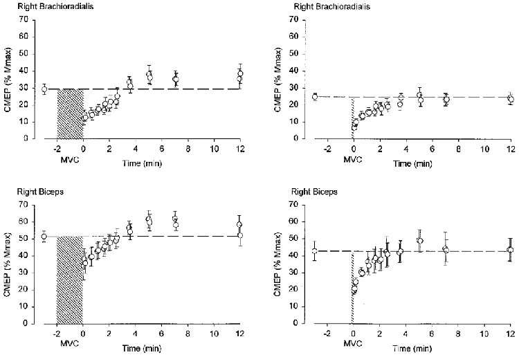
1. Activation of descending corticospinal tracts with transmastoid electrical stimuli has been used to assess changes in the behaviour of motoneurones after voluntary contractions. Stimuli were delivered before and after maximal voluntary isometric contractions (MVCs) of the elbow flexor muscles. 2. Following a sustained MVC of the elbow flexors lasting 5-120 s there was an immediate reduction of the response to transmastoid stimulation to about half of the control value. The response recovered to control levels after about 2 min. This was evident even when the size of the responses was adjusted to accommodate changes in the maximal muscle action potential (assessed with supramaximal stimuli at the brachial plexus). 3. To determine whether the post-contraction depression required activity in descending motor paths, motoneurones were activated by supramaximal tetanic stimulation of the musculocutaneous nerve for 10 s. This did not depress the response to transmastoid stimulation. 4. Following a sustained MVC of 120 s duration, the response to transcranial magnetic stimulation of the motor cortex gradually declined to a minimal level by about 2 min and remained depressed for more than 10 min. 5. Additional studies were performed to check that the activation of descending tracts by transmastoid stimulation was likely to involve excitation of direct corticospinal paths. When magnetic cortical stimuli and transmastoid stimuli were timed appropriately, the response to magnetic cortical stimulation could be largely occluded. 6. This study describes a novel depression of effectiveness of corticospinal actions on human motoneurones. This depression may involve the corticomotoneuronal synapse.
Figures


Comment in
-
Experiments using transcranial magnetic brain stimulation in man could reveal important new mechanisms in motor control.J Physiol. 1999 Dec 15;521 Pt 3(Pt 3):565. doi: 10.1111/j.1469-7793.1999.00565.x. J Physiol. 1999. PMID: 10601488 Free PMC article. No abstract available.
Similar articles
-
Ischaemia after exercise does not reduce responses of human motoneurones to cortical or corticospinal tract stimulation.J Physiol. 2000 Jun 15;525 Pt 3(Pt 3):793-801. doi: 10.1111/j.1469-7793.2000.00793.x. J Physiol. 2000. PMID: 10856130 Free PMC article.
-
Maximal force, voluntary activation and muscle soreness after eccentric damage to human elbow flexor muscles.J Physiol. 2005 Aug 15;567(Pt 1):337-48. doi: 10.1113/jphysiol.2005.087767. Epub 2005 Jun 9. J Physiol. 2005. PMID: 15946963 Free PMC article.
-
Unexpected factors affecting the excitability of human motoneurones in voluntary and stimulated contractions.J Physiol. 2016 May 15;594(10):2707-17. doi: 10.1113/JP272164. Epub 2016 Apr 24. J Physiol. 2016. PMID: 26940402 Free PMC article.
-
Corticospinal transmission after voluntary contractions.Adv Exp Med Biol. 2002;508:435-41. doi: 10.1007/978-1-4615-0713-0_49. Adv Exp Med Biol. 2002. PMID: 12171140 Review.
-
Magnetic brain stimulation: a tool to explore the action of the motor cortex on single human spinal motoneurones.Trends Neurosci. 1991 Sep;14(9):401-5. doi: 10.1016/0166-2236(91)90029-t. Trends Neurosci. 1991. PMID: 1720580 Review.
Cited by
-
Visual feedback alters the variations in corticospinal excitability that arise from rhythmic movements of the opposite limb.Exp Brain Res. 2005 Mar;161(3):325-34. doi: 10.1007/s00221-004-2076-x. Epub 2004 Oct 23. Exp Brain Res. 2005. PMID: 15517219
-
Corticospinal output and loss of force during motor fatigue.Exp Brain Res. 2009 Aug;197(2):111-23. doi: 10.1007/s00221-009-1897-z. Epub 2009 Jul 2. Exp Brain Res. 2009. PMID: 19572125
-
Human motoneurone excitability is depressed by activation of serotonin 1A receptors with buspirone.J Physiol. 2017 Mar 1;595(5):1763-1773. doi: 10.1113/JP273200. Epub 2016 Dec 17. J Physiol. 2017. PMID: 27859267 Free PMC article. Clinical Trial.
-
Critical considerations of the contribution of the corticomotoneuronal pathway to central fatigue.J Physiol. 2022 Dec;600(24):5203-5214. doi: 10.1113/JP282564. Epub 2022 Nov 23. J Physiol. 2022. PMID: 36326193 Free PMC article. Review.
-
The effects of forearm position and contraction intensity on cortical and spinal excitability during a submaximal force steadiness task of the elbow flexors.J Neurophysiol. 2020 Feb 1;123(2):522-528. doi: 10.1152/jn.00349.2019. Epub 2019 Nov 27. J Neurophysiol. 2020. PMID: 31774348 Free PMC article.
References
-
- Allen GM, Gandevia SC, Neering IR, Hickie I, Jones R, Middleton J. Muscle performance, voluntary activation and perceived effort in normal subjects and patients with prior poliomyelitis. Brain. 1994;117:661–670. - PubMed
-
- Bellemare F, Woods JJ, Johansson R, Bigland-Ritchie B. Motor-unit discharge rates in maximal voluntary contractions of three human muscles. Journal of Neurophysiology. 1983;50:1380–1392. - PubMed
-
- Bergmans J. The Physiology of Single Human Nerve Fibres. Louvain: Vander; 1970.
-
- Brasil-Neto JP, Pascual-Leone A, Valls-Sole J, Cammarota A, Cohen LG, Hallett M. Postexercise depression of motor evoked potentials: a measure of central nervous system fatigue. Experimental Brain Research. 1993;93:181–184. - PubMed
Publication types
MeSH terms
LinkOut - more resources
Full Text Sources
Medical