. 1999 Dec;128(7):1616-22.
doi: 10.1038/sj.bjp.0702957.

Modulation of mPer1 gene expression by anxiolytic drugs in mouse cerebellum

Affiliations

Modulation of mPer1 gene expression by anxiolytic drugs in mouse cerebellum

M Akiyama et al. Br J Pharmacol. 1999 Dec.

Abstract

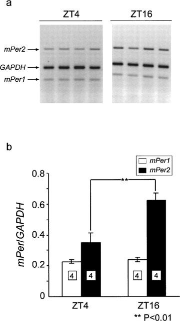
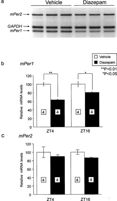
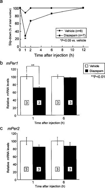
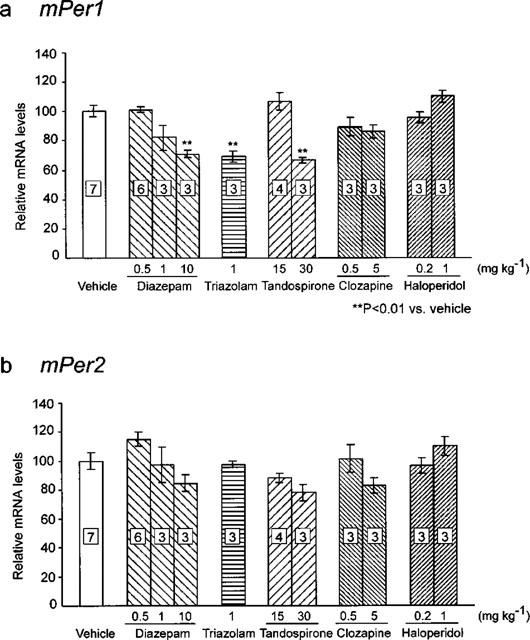
1. The mPer1 and mPer2 genes are putative mouse clock genes that regulate circadian oscillator present in the suprachiasmatic nucleus (SCN) neuron. While they are also expressed in the granular cell layer in the cerebellum, their function is unknown. In a first step to verify the physiological roles of mPer1 and mPer2 genes in the cerebellum, we examined the effects of benzodiazepines on the expression of the mPer1 and mPer2 genes. 2. mPer2 mRNA expression was higher at ZT16 than ZT4 in the mouse cerebellum. 3. High-dose administration of diazepam (10 mg kg-1) or triazolam (1 mg kg-1) reduced mPer1 mRNA level 1 h after treatment in the cerebellum. 4. Reduced expression of mPer1 by diazepam treatment was transient. No difference of mPer1 mRNA level between diazepam (10 mg kg-1)- and vehicle-treated group was observed 6 h after treatment. 5. Administration of high doses of tandospirone (30 mg kg-1), a non-benzodiazepine anxiolytic also reduced mPer1 mRNA expression 1 h after treatment. 6. Administration of high doses of clozapine (5 mg kg-1) or haloperidol (1 mg kg-1) impaired the rota-rod performance without affecting on mPer1 mRNA level. 7. Diazepam and tandospirone inhibited the expression of mPer1 mRNA in the primary cultured cerebellum granule cells. 8. Transient reductions of mPer1 mRNA levels by various benzodiazepines and tandospirone is associated with impairment of coordinated movement, such as rota-rod performance and equilibrium.
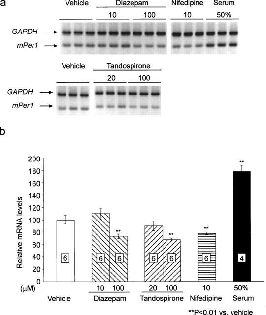

Figures

Figure 1
Figure 1
Expression of the mPer1 and mPer2 gene in the mouse cerebellum at ZT4 and ZT16. (a) Representative electrophoretic image of RT–PCR products of mPer1, mPer2 and GAPDH mRNA from ZT4 and ZT16. (b) Relative mRNA levels of mPer1 and mPer2 at ZT4 and ZT16 were shown by ratio to GAPDH mRNA levels. Numbers in boxes in the columns indicate the number of experiments. mPer2 mRNA is highly expressed at ZT16 than at ZT4 (**P>0.01, Student's t-test).
Figure 2
Figure 2
Effect of diazepam treatment on the expression of mPer1 and mPer2 at ZT4 and ZT16. (a) Representative example of electrophoretic image of RT–PCR products of mPer1, mPer2 and GAPDH mRNA from vehicle- or diazepam-treated mice at ZT16. mRNA levels of mPer1 (b) and mPer2 (c) of vehicle- or diazepam-treated group 1 h after injection at ZT4 and ZT16 were normalized to GAPDH mRNA. Vehicle-treated group was set to 100 in both ZT for avoiding the effects of daily change of mPer mRNA. Numbers in boxes in the columns indicate the number of experiments. mPer1 mRNA level was reduced by diazepam treatment at both ZT (**P<0.01 at ZT4, *P<0.05 at ZT16, Student's t-test).
Figure 3
Figure 3
Temporal effect of diazepam administration on the expression of mPer1 and mPer2. (a) Temporal inhibition of cooperative motor activity by diazepam administration was examined by rota-rod test. Most severe inhibition of locomotor activity was observed at 1 h after injection. (*P<0.05 vs vehicle, Fisher's test) mRNA levels of mPer1 (b) and mPer2 (c) of vehicle- or diazepam-treated group 1 and 6 h after injection of vehicle or diazepam (10 mg kg−1) were normalized by GAPDH mRNA. Vehicle-treated group was set to 100 at both experiments. Numbers in boxes in the columns indicate the number of experiments. Reduction of mPer1 mRNA by diazepam (*P<0.01, Student's t-test) restored after 6 h after treatment.
Figure 4
Figure 4
Effects of various drugs on mPer1 (a) and mPer2 (b) expression 1 h after administration. mRNA levels of mPer1 and mPer2 of vehicle-, diazepam-, triazolam-, tandospirone-, clozapine- and haloperidol-treated group 1 h after injection were normalized to GAPDH mRNA levels. The number in the Figure indicates the amounts (mg kg−1) of administered drugs. Numbers in boxes indicate the number of experiments. Vehicle-treated group was set to 100. Numbers in boxes in the columns indicate the number of experiments. High dose administration of diazepam (10 mg kg−1), triazolam (1 mg kg−1) and tandospirone (30 mg kg−1) reduced mPer1 expression (**P<0.01 vs vehicle, Student's t-test).
Figure 5
Figure 5
Effects of diazepam, tandospirone, nifedipine and 50% serum on mPer1 expression in cerebellar granule cell culture. (a) Representative example of electrophoretic image of RT–PCR products of mPer1, mPer2 and GAPDH mRNA from vehicle- diazepam- nifedipine- or high concentration of serum-treated granule cell culture. (b) mRNA levels of mPer1 1 h after treatment were normalized to GAPDH mRNA. Vehicle-treated group was set to 100. Numbers in boxes in the columns indicate the number of experiments. High dose treatment of diazepam (100 μM), tandospirone (100 μM) or nifedipine (10 μM) reduced mPer1 expression (**P<0.01 vs vehicle, Student's t-test).

References

    1. AIBA A., KANO M., CHEN C., STANTON M.E., FOX G.D., HERRUP K., ZWINGMAN T.A., TONEGAWA S. Deficient cerebellar long-term depression and impaired motor learning in mGluR1 mutant mice. Cell. 1994;79:377–388. - PubMed
    1. AKIYAMA M., KOUZU Y., TAKAHASHI S., WAKAMATSU H., MORIYA T., MAETANI M., WATANABE S., TEI H., SAKAKI Y., SHIBATA S. Inhibition of light- or glutamate-induced mPer1 expression represses the phase shifts into the mouse circadian locomotor and suprachiasmatic firing rhythms. J. Neurosci. 1999;19:1115–1121. - PMC - PubMed
    1. ALBRECHT U., SUN Z.S., EICHELE G., LEE C.C. A differential response of two putative mammalian circadian regulators, mPer1 and mPer2, to light. Cell. 1997;91:1055–1064. - PubMed
    1. ALLADA R., WHITE N.E., SO W.V., HALL J.C., ROSBASH M. A mutant Drosophila homologue of mammalian Clock disrupts circadian rhythms and transcription of period and timeless. Cell. 1998;93:791–804. - PubMed
    1. BALSALOBRE A., DAMIOLA F., SCHIBLER U. A serum shock induces circadian gene expression in mammalian tissue culture cells. Cell. 1998;93:929–937. - PubMed

Publication types

MeSH terms

LinkOut - more resources