. 2000 Jan;68(1):80-6.
doi: 10.1128/IAI.68.1.80-86.2000.

Episomal expression of specific sense and antisense mRNAs in Leishmania amazonensis: modulation of gp63 level in promastigotes and their infection of macrophages in vitro

Affiliations

Episomal expression of specific sense and antisense mRNAs in Leishmania amazonensis: modulation of gp63 level in promastigotes and their infection of macrophages in vitro

D Q Chen et al. Infect Immun. 2000 Jan.

Abstract

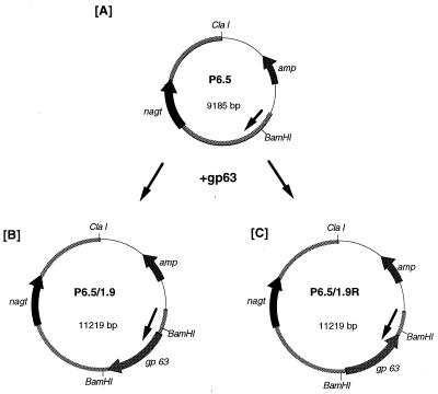
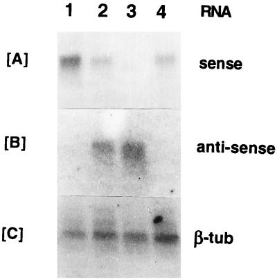
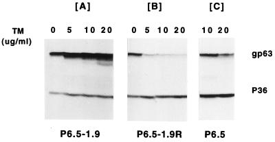
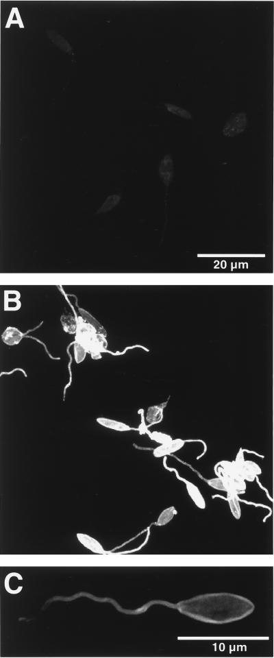
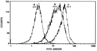
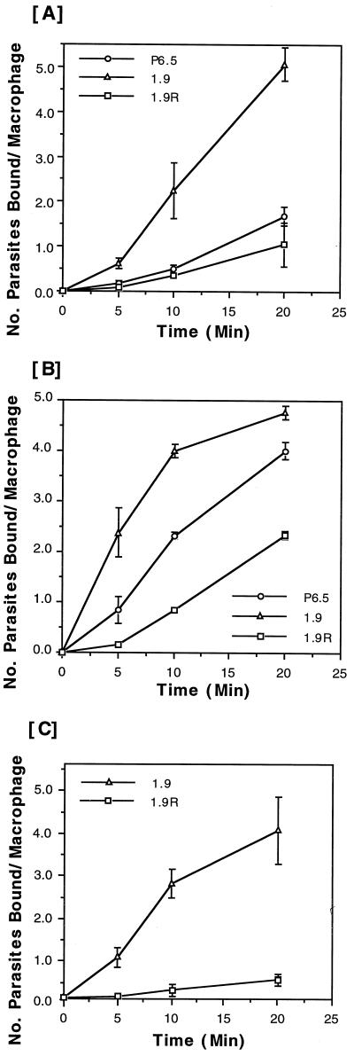
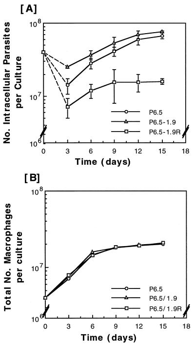
The major surface glycoprotein (gp63) of Leishmania amazonensis is a metalloprotease implicated in the infection of mammalian macrophages. The expression of gp63 and its participation in this infection were further examined by modulating the level of this molecule in a virulent gp63-abundant wild-type clone. Promastigotes were transfected with gp63 genes cloned into a Leishmania-specific vector in two different orientations, leading to the expression of gp63 sense and antisense RNAs. With increasing selective pressure, cell surface gp63 was increasingly augmented in the transfectants with sense transcripts and suppressed to a very low level in those with antisense transcripts. Thus, the expression of gp63 from chromosomal, repetitive genes is not stringently regulated at the protein level and can be substantially reduced by episomal antisense transcription of a single copy. The transfectants differed significantly only in the level of gp63, thereby allowing specific evaluation of this molecule in leishmanial infection of macrophages in vitro. Kinetic studies of infection in vitro indicate that gp63 plays a role not only in the binding of this parasite to these macrophages but also in its intramacrophage survival and replication.


Figures

FIG. 1
FIG. 1
Expression of sense and antisense gp63 transcripts with a Leishmania-specific vector in L. amazonensis. (A) P6.5, a Leishmania-specific vector (see text for its origin). (B) P6.5/1.9, a plasmid carrying the gp63 gene cloned at a BamHI site in P6.5 in the correct orientation. (C) P6.5/1.9R, a plasmid carrying the gp63 gene cloned in P6.5 in the reverse orientation. Arrowed bars in circles represent functional genes and the direction of their transcription: amp, ampicillin resistance gene of pBluescript; nagt, gene of ∼1.4 kb encoding N-acetylglucosamine-1-phosphate transferase; gp63, gene of ∼1.9 kb encoding gp63 of L. amazonensis. Thick lines of the circles including nagt represent the L. amazonensis DNA portion of P6.5 (∼6.2 kb). Thin lines of the circles represent pBluescript. The arrow inside each circle indicates the direction of Leishmania DNA transcription.
FIG. 2
FIG. 2
Northern blot analysis of transfectants for gp63 sense and antisense transcripts. Total RNA samples from different transfectants are shown. Lane 1, P6.5/1.9; lanes 2 and 3, P6.5/1.9R; lane 4, P6.5 alone. All transfectants were grown in 20 μg of TM/ml, except the sample from P6.5/1.9R in lane 2, which was grown in drug-free medium. Probes used are oligonucleotides specific to sense gp63 transcripts (A), oligonucleotides for antisense gp63 transcripts (B), and the beta-tubulin (β-tub) gene of L. amazonensis (C).
FIG. 3
FIG. 3
Western blot analysis of gp63 sense and antisense transfectants. Transfectants were grown in 0, 5, 10, and 20 μg of TM/ml. The blots were probed with antiserum (1:10,000 dilution) against gp63. p36 revealed by anti-p36 antiserum (1:10,000 dilution) was used for the loading control.
FIG. 4
FIG. 4
Confocal fluorescence microscopy of transfectants for cell surface gp63. Glutaraldehyde-fixed cells were treated first with anti-gp63 antiserum and then with fluorescein isothiocyanate-conjugated secondary antibodies; those treated with normal serum and/or with the secondary antibodies alone served as controls (see Materials and Methods for details). (A) P6.5/1.9R cells viewed at settings to reduce the images to the background level; (B) P6.5/1.9 cells with intensive signals for gp63 viewed under the same settings; (C) surface localization of gp63 in a P6.5/1.9 cell revealed under appropriate settings.
FIG. 5
FIG. 5
Flow cytometry of transfectants showing different levels of surface gp63. Cells used were those fixed with glutaraldehyde and treated as described in the legend to Fig. 4. Fluorescence-activated cell sorter analysis of 25,000 cells for each population was done for green fluorescence with a Coulter XL flow cytometer with the accompanying software programs (See Materials and Methods for details). Peaks A to C, samples of P6.5/1.9R, P6.5, and P6.5/1.9 cells, respectively. The numbers in brackets are MCFI. FITC, fluorescein isothiocyanate.
FIG. 6
FIG. 6
Binding of different transfectants expressing variable amounts of gp63 to macrophages. The binding of transfectants to macrophages was assayed at 5, 10, and 20 min, as described in Materials and Methods. Radioactive counts were normalized to the number of parasites bound per macrophage based on a standard linear curve of cell numbers versus values of radioactivity in counts per minute. (A and B) J774G8 macrophages. Results are based on two separate sets of representative data from six different experiments. (C) C57BL/6 peritoneal macrophages. Results are based on one representative experiment of two separate experiments. Samples at each time point are in triplicate.
FIG. 7
FIG. 7
Intramacrophage survival and replication of various transfectants with different levels of gp63 in J774 lines. Infection was carried out in triplicate. Each began with 4 × 10⁷ promastigotes and 4 × 10⁶ macrophages in a 25-cm2 flask. The infectivity of these parasites was assayed by quantitating the total number of intracellular parasites in each flask every 3 to 4 days after incubation (see Materials and Methods). The value for each time point was obtained from triplicate samples. Total numbers of intracellular parasites (A) and growth of J774 macrophages (B) are shown. The square on the vertical axis of panel A indicates the size of the inoculum (4 × 10⁷ promastigotes) with which the infection in each culture began. The dashed lines trace the net decrease in number of the parasites from the inoculum added on day 0 to day 3, when the intracellular parasites were first enumerated. This loss was taken to indicate extracellular and intracellular degeneration or killing of the parasites in this closed in vitro system.

References

    1. Beverley S M, Turco S J. Lipophosphoglycan (LPG) and the identification of virulence genes in the protozoan parasite Leishmania. Trends Microbiol. 1998;6:35–40. - PubMed
    1. Blackwell J M, Ezekowitz R A, Roberts M B, Channon J Y, Sim R B, Gordon S. Macrophage complement and lectin-like receptors bind Leishmania in the absence of serum. J Exp Med. 1985;162:324–331. - PMC - PubMed
    1. Bouvier J, Schneider P, Etges R. Leishmanolysin: surface metalloproteinase of Leishmania. Methods Enzymol. 1995;248:614–633. - PubMed
    1. Brittingham A, Morrison C J, McMaster W R, McGwire B S, Chang K-P, Mosser D M. Role of the Leishmania surface protease gp63 in complement fixation, cell adhesion, and resistance to complement-mediated lysis. J Immunol. 1995;155:3102–3111. - PubMed
    1. Brittingham A, Chen G, McGwire B S, Chang K-P, Mosser D. Interaction of leishmania gp63 with cellular receptors for fibronectin. Infect Immun. 1999;679:4477–4484. - PMC - PubMed

Publication types

LinkOut - more resources