

Comparative evaluation of low-molecular-mass proteins from Mycobacterium tuberculosis identifies members of the ESAT-6 family as immunodominant T-cell antigens
- PMID: 10603390
- PMCID: PMC97123
- DOI: 10.1128/IAI.68.1.214-220.2000
Comparative evaluation of low-molecular-mass proteins from Mycobacterium tuberculosis identifies members of the ESAT-6 family as immunodominant T-cell antigens
Abstract
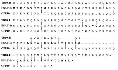
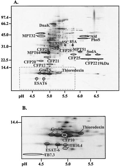
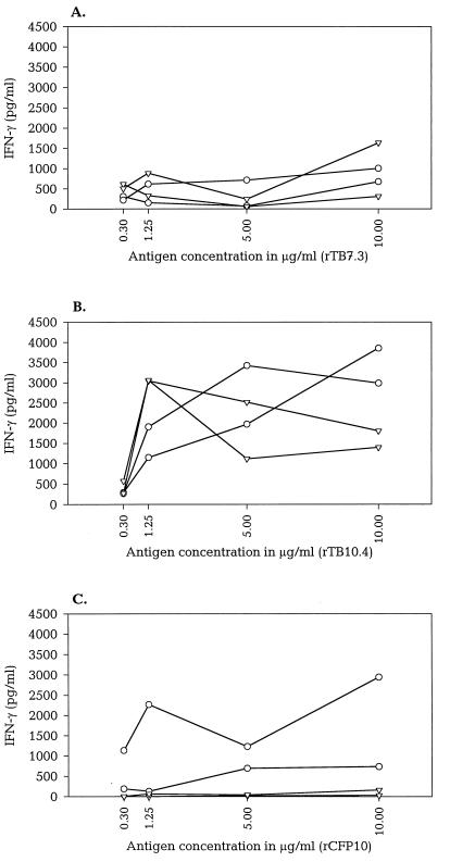
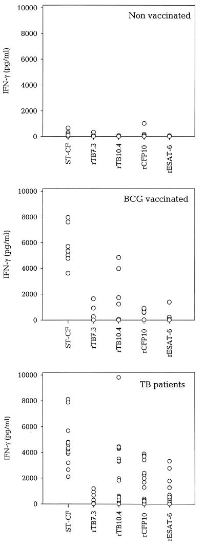
Culture filtrate from Mycobacterium tuberculosis contains protective antigens of relevance for the generation of a new antituberculosis vaccine. We have identified two previously uncharacterized M. tuberculosis proteins (TB7.3 and TB10.4) from the highly active low-mass fraction of culture filtrate. The molecules were characterized, mapped in a two-dimensional electrophoresis reference map of short-term culture filtrate, and compared with another recently identified low-mass protein, CFP10 (F. X. Berthet, P. B. Rasmussen, I. Rosenkrands, P. Andersen, and B. Gicquel. Microbiology 144:3195-3203, 1998), and the well-described ESAT-6 antigen. Genetic analyses demonstrated that TB10.4 as well as CFP10 belongs to the ESAT-6 family of low-mass proteins, whereas TB7.3 is a low-molecular-mass protein outside this family. The proteins were expressed in Escherichia coli, and their immunogenicity was tested in cultures of peripheral blood mononuclear cells from human tuberculosis (TB) patients, Mycobacterium bovis BCG-vaccinated donors, and nonvaccinated donors. The two ESAT-6 family members, TB10.4 and CFP10, were very strongly recognized and induced gamma interferon release at the same level (CFP10) as or at an even higher level (TB10.4) than ESAT-6. The non-ESAT-6 family member, TB7.3, for comparison, was recognized at a much lower level. CFP10 was found to distinguish TB patients from BCG-vaccinated donors and is, together with ESAT-6, an interesting candidate for the diagnosis of TB. The striking immunodominance of antigens within the ESAT-6 family is discussed, and hypotheses are presented to explain this targeting of the immune response during TB infection.
Figures
References
-
- Andersen P, Andersen A B, Sorensen A L, Nagai S. Recall of long-lived immunity to Mycobacterium tuberculosis infection in mice. J Immunol. 1995;154:3359–3372. - PubMed
-
- Andersen P, Askgaard D, Gottschau A, Bennedsen J, Nagai S, Heron I. Identification of immunodominant antigens during infection with Mycobacterium tuberculosis. Scand J Immunol. 1992;36:823–831. - PubMed
-
- Andersen P, Heron I. Simultaneous electroelution of whole SDS-polyacrylamide gels for the direct cellular analysis of complex protein mixtures. J Immunol Methods. 1993;161:29–39. - PubMed
Publication types
MeSH terms
Substances
LinkOut - more resources
Full Text Sources
Other Literature Sources
Medical
Molecular Biology Databases
