

Comparative analysis of the mucosal adjuvanticity of the type II heat-labile enterotoxins LT-IIa and LT-IIb
- PMID: 10603399
- PMCID: PMC97132
- DOI: 10.1128/IAI.68.1.281-287.2000
Comparative analysis of the mucosal adjuvanticity of the type II heat-labile enterotoxins LT-IIa and LT-IIb
Abstract
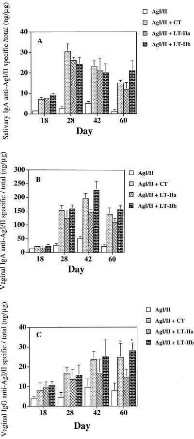
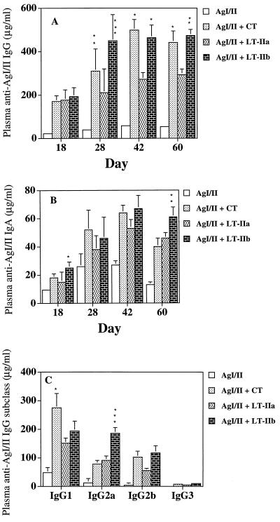
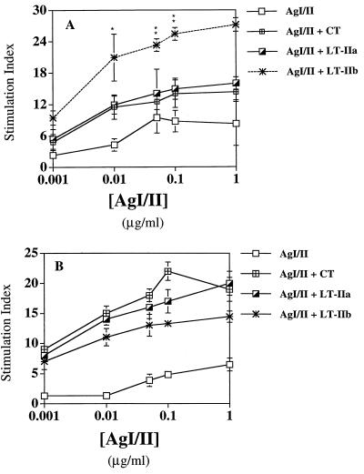
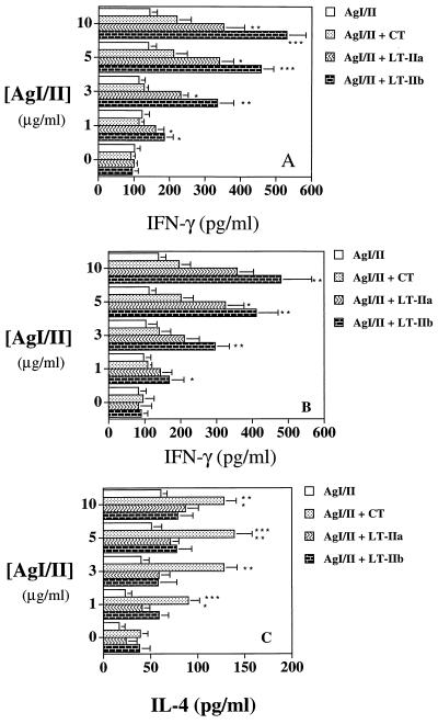
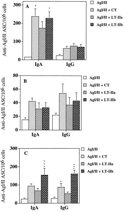
Cholera toxin (CT) and the heat-labile enterotoxin of Escherichia coli (LT-I) are members of the serogroup I heat-labile enterotoxins (HLT) and can serve as systemic and mucosal adjuvants. However, information is lacking with respect to the structurally related but antigenically distinct serogroup II HLT, LT-IIa and LT-IIb, which have different binding specificities for ganglioside receptors. The purpose of this study was to assess the effectiveness of LT-IIa and LT-IIb as mucosal adjuvants in comparison to the prototypical type I HLT, CT. BALB/c mice were immunized by the intranasal (i.n.) route with the surface protein adhesin AgI/II of Streptococcus mutans alone or supplemented with an adjuvant amount of CT, LT-IIa, or LT-IIb. Antigen-specific antibody responses in saliva, vaginal wash, and plasma were assayed by enzyme-linked immunosorbent assay. Mice given AgI/II with LT-IIa or LT-IIb by the i.n. route had significantly higher mucosal and systemic antibody responses than mice immunized with AgI/II alone. Anti-AgI/II immunoglobulin A (IgA) antibody activity in saliva and vaginal secretions of mice given AgI/II with LT-IIa or LT-IIb was statistically similar in magnitude to that seen in mice given AgI/II and CT. LT-IIb significantly enhanced the number of AgI/II-specific antibody-secreting cells in the draining superficial cervical lymph nodes compared to LT-IIa and CT. LT-IIb and CT induced significantly higher plasma anti-AgI/II IgG titers compared to LT-IIa. When LT-IIb was used as adjuvant, the proportion of plasma IgG2a relative to IgG1 anti-AgI/II antibody was elevated in contrast to the predominance of IgG1 antibodies promoted by AgI/II alone or when CT or LT-IIa was used. In vitro stimulation of AgI/II-specific cells from the superficial lymph nodes and spleen revealed that LT-IIa and LT-IIb induced secretion of interleukin-4 and significantly higher levels of gamma interferon compared to CT. These results demonstrate that the type II HLT LT-IIa and LT-IIb exhibit potent and distinct adjuvant properties for stimulating immune responses to a noncoupled protein immunogen after mucosal immunization.
Figures
Similar articles
-
Mucosal adjuvant properties of mutant LT-IIa and LT-IIb enterotoxins that exhibit altered ganglioside-binding activities.Infect Immun. 2005 Mar;73(3):1330-42. doi: 10.1128/IAI.73.3.1330-1342.2005. Infect Immun. 2005. PMID: 15731030 Free PMC article.
-
Mutants of type II heat-labile enterotoxin LT-IIa with altered ganglioside-binding activities and diminished toxicity are potent mucosal adjuvants.Infect Immun. 2007 Feb;75(2):621-33. doi: 10.1128/IAI.01009-06. Epub 2006 Nov 21. Infect Immun. 2007. PMID: 17118982 Free PMC article.
-
LT-IIc, a new member of the type II heat-labile enterotoxin family, exhibits potent immunomodulatory properties that are different from those induced by LT-IIa or LT-IIb.Vaccine. 2011 Jan 17;29(4):721-7. doi: 10.1016/j.vaccine.2010.11.020. Epub 2010 Nov 21. Vaccine. 2011. PMID: 21095251 Free PMC article.
-
The role of ADP-ribosylation and G(M1)-binding activity in the mucosal immunogenicity and adjuvanticity of the Escherichia coli heat-labile enterotoxin and Vibrio cholerae cholera toxin.Immunol Cell Biol. 1998 Jun;76(3):270-9. doi: 10.1046/j.1440-1711.1998.00745.x. Immunol Cell Biol. 1998. PMID: 9682971 Review.
-
Cholera toxin, LT-I, LT-IIa and LT-IIb: the critical role of ganglioside binding in immunomodulation by type I and type II heat-labile enterotoxins.Expert Rev Vaccines. 2007 Oct;6(5):821-34. doi: 10.1586/14760584.6.5.821. Expert Rev Vaccines. 2007. PMID: 17931161 Free PMC article. Review.
Cited by
-
Recombinant antigen-enterotoxin A2/B chimeric mucosal immunogens differentially enhance antibody responses and B7-dependent costimulation of CD4(+) T cells.Infect Immun. 2001 Jan;69(1):252-61. doi: 10.1128/IAI.69.1.252-261.2001. Infect Immun. 2001. PMID: 11119513 Free PMC article.
-
Novel Strategy To Protect against Influenza Virus-Induced Pneumococcal Disease without Interfering with Commensal Colonization.Infect Immun. 2016 May 24;84(6):1693-1703. doi: 10.1128/IAI.01478-15. Print 2016 Jun. Infect Immun. 2016. PMID: 27001538 Free PMC article.
-
Enhanced antigen uptake by dendritic cells induced by the B pentamer of the type II heat-labile enterotoxin LT-IIa requires engagement of TLR2.Vaccine. 2010 May 7;28(21):3696-705. doi: 10.1016/j.vaccine.2010.03.016. Epub 2010 Mar 21. Vaccine. 2010. PMID: 20332049 Free PMC article.
-
Heat-labile enterotoxins as adjuvants or anti-inflammatory agents.Immunol Invest. 2010;39(4-5):449-67. doi: 10.3109/08820130903563998. Immunol Invest. 2010. PMID: 20461887 Free PMC article. Review.
-
Binding to gangliosides containing N-acetylneuraminic acid is sufficient to mediate the immunomodulatory properties of the nontoxic mucosal adjuvant LT-IIb(T13I).Clin Vaccine Immunol. 2010 Jun;17(6):969-78. doi: 10.1128/CVI.00076-10. Epub 2010 Apr 14. Clin Vaccine Immunol. 2010. PMID: 20392887 Free PMC article.
References
-
- Biewenga J, Hameleers D M H, Koornstra P J, Kuper C F. Immuno- and enzyme-histochemical and functional studies on nasal associated lymphoid tissue (NALT) in the rat. In: Tsuchiya M, Nagura H, Hibi T, Moro I, editors. Frontiers of mucosal immunology. Vol. 2. Amsterdam, The Netherlands: Excerpta Medica; 1991. pp. 603–604.
-
- Elson C O, Ealding W. Genetic control of the murine immune response to cholera toxin. J Immunol. 1985;135:930–932. - PubMed
-
- Finkelman F D, Holmes J, Katona I M, Urban J F, Jr, Beckmann M P. Lymphokine control of in vivo immunoglobulin isotype selection. Annu Rev Immunol. 1990;8:303–333. - PubMed
Publication types
MeSH terms
Substances
Grants and funding
LinkOut - more resources
Full Text Sources
Other Literature Sources
Miscellaneous
