. 2000 Jan;68(1):342-51.
doi: 10.1128/IAI.68.1.342-351.2000.

In vitro Brucella suis infection prevents the programmed cell death of human monocytic cells

Affiliations

In vitro Brucella suis infection prevents the programmed cell death of human monocytic cells

A Gross et al. Infect Immun. 2000 Jan.

Abstract

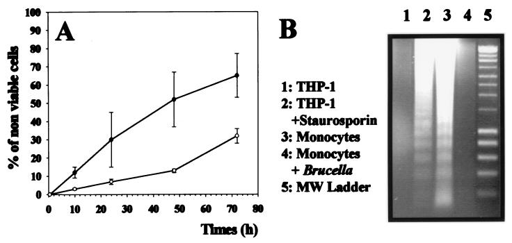
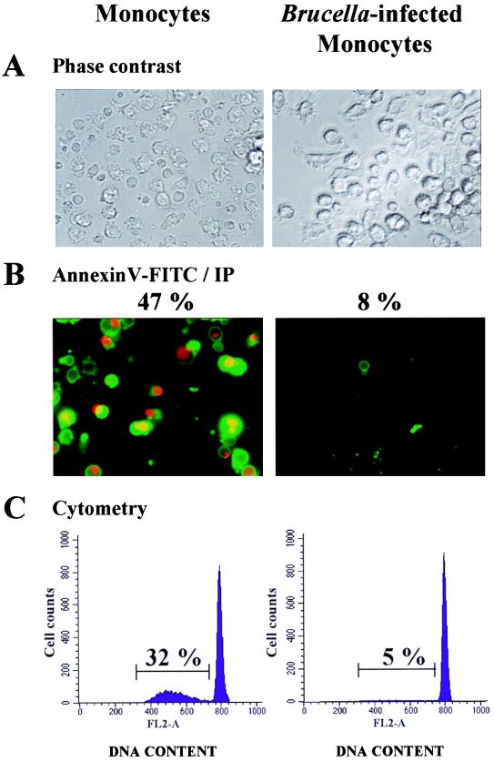
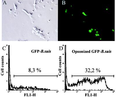
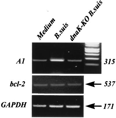
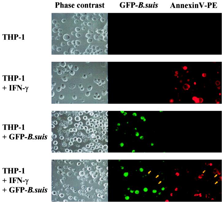
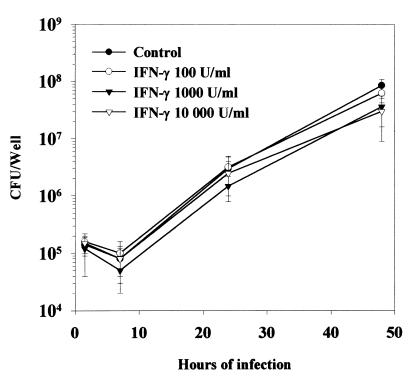
During the complex interaction between an infectious agent and a host organism, the pathogen can interfere with the host cell's programmed death to its own benefit. Induction or prevention of host cell apoptosis appears to be a critical step for determining the infection outcome. Members of the gram-negative bacterial genus Brucella are intracellular pathogens which preferentially invade monocytic cells and develop within these cells. We investigated the effect of Brucella suis infection on apoptosis of human monocytic phagocytes. The present study provides evidence that Brucella infection inhibited spontaneously occurring apoptosis in human monocytes. Prevention of monocyte apoptosis was not mediated by Brucella lipopolysaccharide and required bacterial survival within infected cells. Both invaded and noninvaded cells were protected, indicating that soluble mediators released during infection were involved in the phenomenon. Analysis of Brucella-infected monocytes revealed specific overexpression of the A1 gene, a member of the bcl-2 family implicated in the survival of hematopoietic cells. Brucella infection also rendered macrophage-like cells resistant to Fas ligand- or gamma interferon-induced apoptosis, suggesting that Brucella infection protected host cells from several cytotoxic processes occurring at different steps of the immune response. The present data clearly show that Brucella suis modulated the monocyte/macrophage's apoptotic response to the advantage of the pathogen, thus preventing host cell elimination. This might represent a strategy for Brucella development in infected hosts.


Figures

FIG. 1
FIG. 1
(A) B. suis infection prevents monocyte death. Human monocytes were infected with B. suis (○) or not (●) in 96-well plates or stimulated with E. coli LPS or 1,000 U of TNF-α per ml and cultured in complete medium. At different times postinfection, the percentages of nonviable cells in the different cultures were evaluated as described in Materials and Methods. (Values for LPS or TNF-α activation are indicated in the text.) Data are means and standard deviations from quadruplicate determinations. (B) DNA fragmentation assays on agarose gels. Cytosolic low-molecular-weight DNA was isolated from different monocytic phagocytes and electrophoresed on 1.2% agarose gels for 3 h at 100 V. Lanes 1 and 2 (control experiments), DNA isolated from THP-1 cells (lane 1) or from THP-1 cells treated with staurosporine for 7 h (lane 2). Lane 3, DNA isolated from human monocytes cultured for 48 h in complete medium. Lane 4, DNA from a 48-h culture of B. suis-infected monocytes. Lane 5, 200-bp DNA ladder molecular size (MW) markers (Smart ladder; Eurogentec, Seraing, Belgium).
FIG. 2
FIG. 2
B. suis infection prevents spontaneously occurring apoptosis in human monocytes. Monocytes infected (or not) with B. suis (MOI = 20) were cultured for 2 days in complete medium. Cells infected in eight-chamber culture slides were directly stained with PI (IP) and fluorescein (FITC)-conjugated annexin V. They were then concomitantly analyzed by phase-contrast microscopy (A) or fluorescence microscopy (B). Magnification, ×400. For each culture condition, the percentage of apoptotic cells was determined by counting at least 800 cells. Among them, those which displayed green fluorescence and those which simultaneously displayed red fluorescence and condensed chromatin were considered apoptotic cells. (C) In parallel experiments, cells from the same donors were infected in six-well plates, cultured for 2 days, harvested, permeabilized, and stained with PI. Single cells were then gated by classical procedures which exclude doublets and aggregates (FL2-W versus FL2-A) and analyzed with a flow cytometer. The percentages of resting or B. suis-infected monocytes with hypodiploid DNA were calculated and are indicated. Both types of experiment were repeated with at least 10 different donors.
FIG. 3
FIG. 3
Determination of percentage of B. suis-infected monocytes. Human monocytes were infected with GFP-B. suis or opsonized GFP-B. suis (MOI = 20). Twenty-four hours later, the percentage of infected cells was determined. (A and B) Determination by counting the numbers of total and fluorescent cells. A culture of monocytes infected with opsonized GFP-B. suis visualized by phase-contrast microscopy (A) and by fluorescence microscopy (B) is shown. For each determination, 800 cells were analyzed on four different slides. Magnification, ×400. (C and D) Determination by cytometry. Percentages of infected cells were determined by gating monocytes under forward- and side-scatter parameters and measuring the number of cells expressing green fluorescence (GFP protein); 10,000 cells were analyzed. Cytofluorographs of 24-h monocyte cultures infected with GFP-B. suis (C) or opsonized GFP-B. suis (D) are shown. The dark surface is resting monocytes.
FIG. 4
FIG. 4
Reverse transcription-PCR detection of A1, bcl-2, and GAPDH mRNAs in B. suis- and dnaK-KO B. suis-infected monocytes. Monocytes were infected (or not) with B. suis or dnaK-KO B. suis (MOI = 20). Four hours later, total mRNAs were isolated, and after reverse transcription, PCR was performed with 2 ng of cDNA for each sample. PCR products obtained after 25 cycles (A1), 19 cycles (GAPDH), or 35 cycles (bcl-2) were then analyzed on 1.2% agarose gels supplemented with ethidium bromide. Lane 1, uninfected cells; lane 2, B. suis-infected cells; lane 3, dnaK-KO B. suis-infected monocytes. Numbers on the right indicate amplicon length (in base pairs).
FIG. 5
FIG. 5
B. suis infection of VD3-THP-1 cell cultures renders both invaded and noninvaded cells resistant to IFN-γ-induced apoptosis. VD3-THP-1 cells were infected (or not) with opsonized GFP-B. suis in eight-chamber culture slides and after gentamicin addition were cultured with (or without) IFN-γ (10,000 U/ml). Two days later, cells were stained with phycoerythrin (PE)-labeled annexin V (red fluorescence). The red and green fluorescences of the cells visualized by phase-contrast microscopy were analyzed by fluorescence microscopy to determine the number of GFP-B. suis-infected cells (green fluorescence), the number of apoptotic cells (red fluorescence), and the number of infected cells in apoptosis (green plus red fluorescence; yellow arrow). This figure is representative of seven identical experiments reported in Table 4.
FIG. 6
FIG. 6
Intracellular behavior of B. suis within IFN-γ-treated VD3-THP-1 cells. VD3-THP-1 cells (106 cells/well) were infected with opsonized B. suis (MOI = 20) in 24-well plates, as described in Materials and Methods, in the presence of IFN-γ at the indicated concentrations or in RPMI 1640 alone (control). The cells were then cultured in the presence of corresponding concentrations of IFN-γ. At different time periods, they were lysed and the number of viable intracellular bacteria was determined and expressed in CFU per well as described previously (2, 3). All results are means and standard deviations from four separate experiments performed in duplicate.

Similar articles

Cited by

References

    1. Adachi S, Cross A R, Babior B M, Gottlieb R A. Bcl-2 and the outer mitochondrial membrane in the inactivation of cytochrome c during Fas-mediated apoptosis. J Biol Chem. 1997;272:21878–21882. - PubMed
    1. Ashany D, Song X, Lacy E, Nikolic-Zugic J, Friedman S, Elkon K. Th1 CD4+ lymphocytes delete activated macrophages through Fas/Apo-1 antigen pathway. Proc Natl Acad Sci USA. 1995;92:11255–11261. - PMC - PubMed
    1. Behr-Perst S I, Munk M E, Schaberg T, Ulrichs T, Schulz R J, Kaufmann S H E. Phenotypically activated γδ T lymphocytes in the peripheral blood of patients with tuberculosis. J Infect Dis. 1999;180:141–149. - PubMed
    1. Campbell G A, Adams L G, Sowa B A. Mechanisms of binding of Brucella abortus to mononuclear phagocytes from cows naturally resistant or susceptible to brucellosis. Vet Immunol Immunopathol. 1994;41:295–306. - PubMed
    1. Caron E. Thesis. Montpellier, France: Université de Montpellier II; 1994.

Publication types

MeSH terms