A transgenic rabbit model for human hypertrophic cardiomyopathy
- PMID: 10606622
- PMCID: PMC409884
- DOI: 10.1172/JCI7956
A transgenic rabbit model for human hypertrophic cardiomyopathy
Abstract
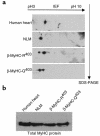
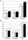
Certain mutations in genes for sarcomeric proteins cause hypertrophic cardiomyopathy (HCM). We have developed a transgenic rabbit model for HCM caused by a common point mutation in the beta-myosin heavy chain (MyHC) gene, R400Q. Wild-type and mutant human beta-MyHC cDNAs were cloned 3' to a 7-kb murine beta-MyHC promoter. We injected purified transgenes into fertilized zygotes to generate two lines each of the wild-type and mutant transgenic rabbits. Expression of transgene mRNA and protein were confirmed by Northern blotting and 2-dimensional gel electrophoresis followed by immunoblotting, respectively. Animals carrying the mutant transgene showed substantial myocyte disarray and a 3-fold increase in interstitial collagen expression in their myocardia. Mean septal thicknesses were comparable between rabbits carrying the wild type transgene and their nontransgenic littermates (NLMs) but were significantly increased in the mutant transgenic animals. Posterior wall thickness and left ventricular mass were also increased, but dimensions and systolic function were normal. Premature death was more common in mutant than in wild-type transgenic rabbits or in NLMs. Thus, cardiac expression of beta-MyHC-Q(403) in transgenic rabbits induced hypertrophy, myocyte and myofibrillar disarray, interstitial fibrosis, and premature death, phenotypes observed in humans patients with HCM due to beta-MyHC-Q(403).
Figures


Similar articles
-
Decreased left ventricular ejection fraction in transgenic mice expressing mutant cardiac troponin T-Q(92), responsible for human hypertrophic cardiomyopathy.J Mol Cell Cardiol. 2000 Mar;32(3):365-74. doi: 10.1006/jmcc.1999.1081. J Mol Cell Cardiol. 2000. PMID: 10731436
-
Evolution of expression of cardiac phenotypes over a 4-year period in the beta-myosin heavy chain-Q403 transgenic rabbit model of human hypertrophic cardiomyopathy.J Mol Cell Cardiol. 2004 May;36(5):663-73. doi: 10.1016/j.yjmcc.2004.02.010. J Mol Cell Cardiol. 2004. PMID: 15135661 Free PMC article.
-
Tissue Doppler imaging consistently detects myocardial contraction and relaxation abnormalities, irrespective of cardiac hypertrophy, in a transgenic rabbit model of human hypertrophic cardiomyopathy.Circulation. 2000 Sep 19;102(12):1346-50. doi: 10.1161/01.cir.102.12.1346. Circulation. 2000. PMID: 10993850 Free PMC article.
-
Molecular genetics and pathogenesis of hypertrophic cardiomyopathy.Minerva Med. 2001 Dec;92(6):435-51. Minerva Med. 2001. PMID: 11740432 Free PMC article. Review.
-
Familial hypertrophic cardiomyopathy: a paradigm of the cardiac hypertrophic response to injury.Ann Med. 1998 Aug;30 Suppl 1:24-32. Ann Med. 1998. PMID: 9800880 Review.
Cited by
-
Animal Models to Study Cardiac Arrhythmias.Circ Res. 2022 Jun 10;130(12):1926-1964. doi: 10.1161/CIRCRESAHA.122.320258. Epub 2022 Jun 9. Circ Res. 2022. PMID: 35679367 Free PMC article. Review.
-
Induced Pluripotent Stem Cell-Derived Cardiomyocytes from a Patient with MYL2-R58Q-Mediated Apical Hypertrophic Cardiomyopathy Show Hypertrophy, Myofibrillar Disarray, and Calcium Perturbations.J Cardiovasc Transl Res. 2019 Oct;12(5):394-403. doi: 10.1007/s12265-019-09873-6. Epub 2019 Feb 22. J Cardiovasc Transl Res. 2019. PMID: 30796699
-
Rabbit models of heart disease.Drug Discov Today Dis Models. 2008 Fall;5(3):185-193. doi: 10.1016/j.ddmod.2009.02.001. Epub 2009 Mar 17. Drug Discov Today Dis Models. 2008. PMID: 32288771 Free PMC article. Review.
-
Transgenic rabbit model for human troponin I-based hypertrophic cardiomyopathy.Circulation. 2005 May 10;111(18):2330-8. doi: 10.1161/01.CIR.0000164234.24957.75. Epub 2005 May 2. Circulation. 2005. PMID: 15867176 Free PMC article.
-
The R403Q myosin mutation implicated in familial hypertrophic cardiomyopathy causes disorder at the actomyosin interface.PLoS One. 2007 Nov 7;2(11):e1123. doi: 10.1371/journal.pone.0001123. PLoS One. 2007. PMID: 17987111 Free PMC article.
References
-
- Maron BJ, et al. Prevalence of hypertrophic cardiomyopathy in a general population of young adults. Echocardiographic analysis of 4111 subjects in the CARDIA Study. Coronary Artery Risk Development in (Young) Adults. Circulation. 1995;92:785–789. - PubMed
-
- Wigle ED, Rakowski H, Kimball BP, Williams WG. Hypertrophic cardiomyopathy. Clinical spectrum and treatment. Circulation. 1995;92:1680–1692. - PubMed
-
- Ferrans VJ, Rodriguez ER. Specificity of light and electron microscopic features of hypertrophic obstructive and nonobstructive cardiomyopathy. Qualitative, quantitative and etiologic aspects. Eur Heart J. 1983;4(Suppl. F):9–22. - PubMed
-
- Maron BJ, et al. Sudden death in young competitive athletes. Clinical, demographic, and pathological profiles. JAMA. 1996;276:199–204. - PubMed
-
- Marian AJ, Roberts R. Molecular genetic basis of hypertrophic cardiomyopathy: genetic markers for sudden cardiac death. J Cardiovasc Electrophysiol. 1998;9:88–99. - PubMed
Publication types
MeSH terms
Substances
Grants and funding
LinkOut - more resources
Full Text Sources
Other Literature Sources

