. 1999 Dec;104(12):1739-49.
doi: 10.1172/JCI7779.

Bone marrow CD34(+) cells and megakaryoblasts secrete beta-chemokines that block infection of hematopoietic cells by M-tropic R5 HIV

Affiliations

Bone marrow CD34(+) cells and megakaryoblasts secrete beta-chemokines that block infection of hematopoietic cells by M-tropic R5 HIV

M Majka et al. J Clin Invest. 1999 Dec.

Abstract

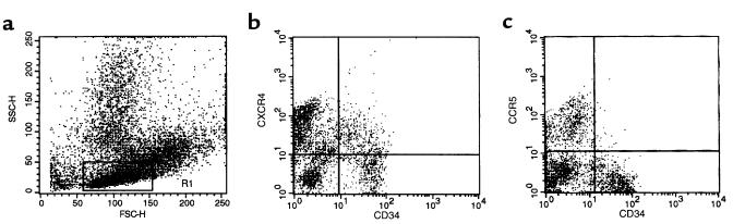
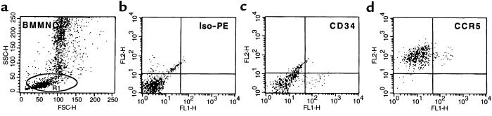
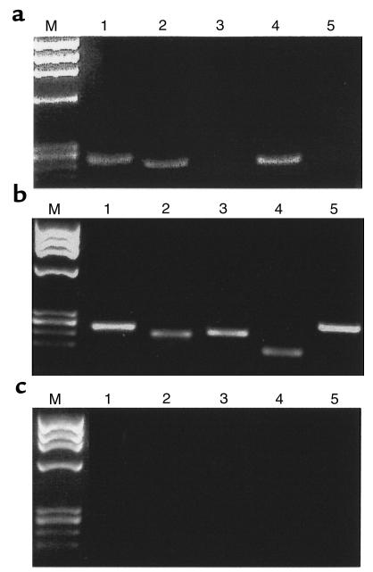
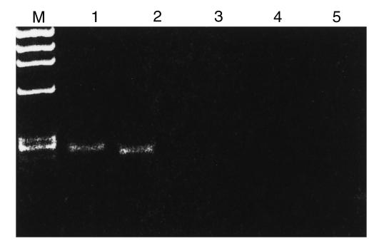
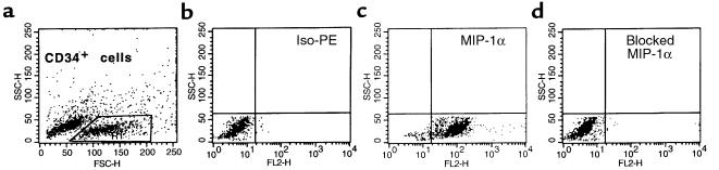
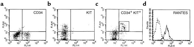
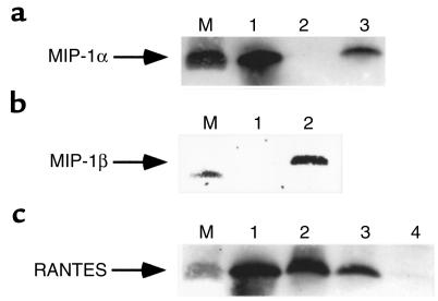
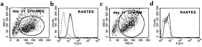
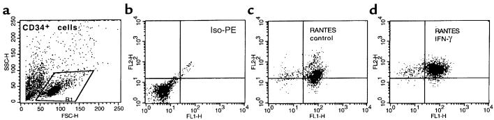
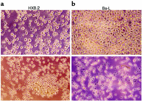
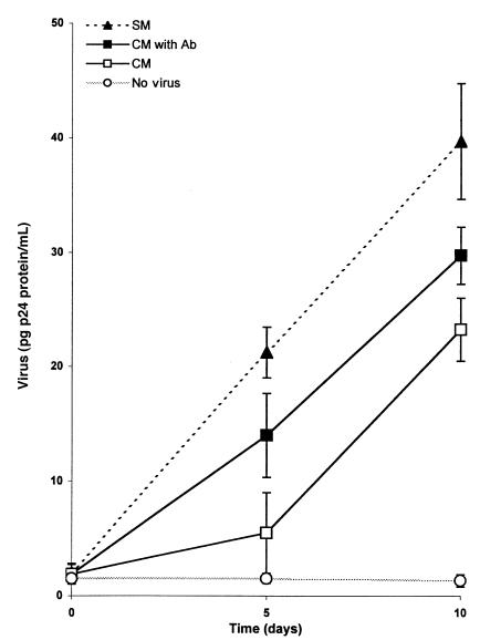
CD34(+) cells are nonpermissive to infection by HIV strains X4 and R5, despite the fact that many CD34(+) cells express high levels of the viral receptor protein CD4 and the coreceptor CXCR4 on their surface. In these cells, the co-receptor CCR5 protein, which, like CXCR4, is a chemokine receptor, is detected mainly intracellularly. We hypothesized that CD34(+) cells secrete CCR5-binding chemokines and that these factors interfere with HIV R5 interactions with these cells, possibly by binding CCR5 or by inducing its internalization. We found that human CD34(+) cells and CD34(+)KIT(+) cells, which are enriched in myeloid progenitor cells, expressed and secreted the CCR5 ligands RANTES, MIP-1alpha, and MIP-1beta and that IFN-gamma stimulated expression of these chemokines. In contrast, SDF-1, a CXCR4 ligand, was not detectable in the CD34(+)KIT(+) cells, even by RT-PCR. Conditioned media from CD34(+) cell culture significantly protected the T lymphocyte cell line PB-1 from infection by R5 but not X4 strains of HIV. Interestingly, the secretion of endogenous chemokines decreased with the maturation of CD34(+) cells, although ex vivo, expanded megakaryoblasts still secreted a significant amount of RANTES. Synthesis of CCR5-binding chemokines by human CD34(+) cells and megakaryoblasts therefore largely determines the susceptibility of these cells to infection by R5 HIV strains. We postulate that therapeutic agents that induce the endogenous synthesis of chemokines in human hematopoietic cells may protect these cells from HIV infection.


Figures

Figure 1
Figure 1
Expression of chemokine receptors on bone marrow CD34+ BMMNC. BMMNC were isolated from bone marrow aspirates of healthy donors by Ficoll-gradient centrifugation and then stained with mAbs against CD34 antigen (FITC) and chemokine receptors (PE). (a) Forward (FSC) and side scatter (SSC) analysis of BMMNC. Lymphocyte region is defined by R1. (b) Analysis of cells dual-labeled with FITC anti-CD34 mAb and PE anti-CXCR4 mAb. (c) Analysis of cells dual-labeled with FITC anti-CD34 mAb and PE anti-CCR5 mAb. Data from at least 5 different donors were analyzed with similar results. Data from a representative donor is presented.
Figure 2
Figure 2
Intracellular expression of CCR5 on bone marrow CD34+ BMMNC. BMMNC were isolated from bone marrow aspirates of healthy donors by Ficoll-gradient centrifugation. Cells were permeabilized before staining with mAbs against CD34 antigen (FITC) and CCR5 receptor (PE). (a) Forward (FSC) and side scatter (SSC) analysis of permeabilized BMMNC. Lymphocyte region is defined by R1. (b) Iso-PE staining. (c) FITC anti-CD34. (d) Analysis of cells dual-labeled with FITC anti-CD34 mAb and PE anti-CCR5 mAb. Data from at least 3 different donors were analyzed with similar results. Data from a representative donor is presented. We obtained similar data by using 3 other mAbs (clones 2D7 and 549 from R&D Systems Inc., and CTC5 from Protein Design Laboratories Inc.) for CCR5 detection.
Figure 3
Figure 3
RT-PCR analysis of chemokine expression in human CD34+ cells. (a) MIP-1α (lane 1), RANTES (lane 2), SDF-1 (lane 3), MIP-1β (lane 4), and MCP-1 (lane 5). (b) Positive RT-PCR control reactions for MIP-1α (lane 1), MIP-1β (lane 2), RANTES (lane 3), MCP-1 (lane 4), and SDF-1 (lane 5). Expression of MIP-1α, MIP-1β, and RANTES were detected in mRNA isolated from BMMNC, whereas expression of MCP-1 and SDF-1 were detected in mRNA isolated from bone marrow–derived stroma fibroblasts. (c) Negative RT-PCR control reactions (using H2O instead of mRNA) for MIP-1α (lane 1), RANTES (lane 2), SDF-1 (lane 3), MIP-1β (lane 4), and MCP-1 (lane 5). Lane M, molecular weight marker (ΦX174 DNA/HaeIII).
Figure 4
Figure 4
RT-PCR analysis of chemokine expression in human CD34+KIT+ cells sorted by FACS®. Lanes 1–3 show MIP-1α, RANTES, and SDF-1, respectively. Lanes 4–6 show negative RT-PCR control reactions for MIP-1α, RANTES, and SDF-1, respectively. Data from 3 different donors was analyzed with similar results. Data from a representative donor are presented. Lane M, molecular weight marker (ΦX174 DNA/HaeIII).
Figure 5
Figure 5
Intracellular staining for MIP-1α in CD34+ cells isolated using MiniMACS beads and subsequently permeabilized. (a) Forward and side scatter analysis of CD34+ cells isolated by MiniMACS beads. (b) Iso-PE staining. (c) PE anti–MIP-1α mAb. (d) MIP-1α detection has been specifically blocked by addition of MIP-1α. Data from 3 different donors were analyzed with similar results. Data from a representative donor are presented.
Figure 6
Figure 6
Intracellular staining for RANTES in CD34+KIT+ MNC. (a) FITC anti-CD34 staining. (b) Cy5 anti-KIT staining. (c) R2 cells dual-labeled with FITC anti-CD34 mAb and Cy5 anti-KIT mAb. (d) Expression of intracellular RANTES in CD34+KIT+ cells from R2. Data from 3 different donors were analyzed with similar results. Data from a representative donor are presented. Cells in area R2 are dual-labeled CD34+KIT+ cells.
Figure 7
Figure 7
Western blots showing expression of chemokines in cell lysates from human hematopoietic cells. (a) Expression of MIP-1α in CD34+ cells (lane 3) in CFU-Meg–derived αIIb3+ cells (lane 2). Lane 1: recombinant MIP-1α (positive control). Lane M: protein size marker (∼7 kDa). (b) Expression of MIP-1β in CD34+ cells (lane 2) and in CFU-Meg–derived αIIb3+ cells (lane 1). Lane M: protein size marker (∼7 kDa). (c) Expression of RANTES in human platelets (lane 1), CFU-Meg–derived cells (lane 2), and CD34+ cells (lane 3). RANTES is not expressed in CFU-GM–derived cells (lane 4). Lane M: protein size marker (∼7 kDa).
Figure 8
Figure 8
Western blots showing expression of SDF-1 in lysates from human bone marrow stroma fibroblasts (lane 1), human CFU-Meg–derived αIIb3+ cells (lane 2), and human CD34+ cells (lane 3).
Figure 9
Figure 9
Intracellular staining for RANTES in megakaryoblasts and myeloblasts that were expanded ex vivo for 11 days under serum-free conditions. (a) Forward and side scatter analysis of day 11 CFU-Meg–derived cells. (b) Expression of RANTES in day 11 megakaryoblasts. (c) Forward and side scatter analysis of day 11 CFU-GM–derived cells. (d) Expression of RANTES in day 11 myeloblasts. Data from 4 different donors were analyzed with similar results. Data from a representative donor are presented.
Figure 10
Figure 10
Intracellular staining for RANTES in CD34+ cells that were isolated using MiniMACS beads and then permeabilized. (a) Forward and side scatter analysis of CD34+ cells isolated using MiniMACS beads. (b) Iso-PE staining. (c) PE anti-RANTES mAb. (d) PE anti-RANTES mAb in CD34+ cells stimulated for 24 hours with IFN-γ (1,000 U/mL). Data from 3 different donors were analyzed with similar results. Data from a representative donor are presented.
Figure 11
Figure 11
PB-1 cells infected with different strains of HIV in the presence of conditioned medium collected from CD34+ cell cultures, which was added fresh (top) or heat inactivated before addition (bottom). (a) Cells infected with HXB.2 (X4 HIV). (b) Cells infected with Ba-L (R5 HIV). Pictures were taken 5 days after infection.
Figure 12
Figure 12
PB-1 cells infected with R5 HIV (Ba-L) in the presence of conditioned medium (CM) collected from CD34+ cell cultures, conditioned medium that contained a cocktail of neutralizing Abs (50 μg/mL α–MIP-1α, 50 μg/mL α–MIP-1β, and 10 μg/mL α-RANTES) (CM with Ab), and control culture medium (SM) that was mock cleared with an equal amount of nonreactive goat Ig (110 μg/mL). Results are representative of 3 separate experiments and are presented as mean p24 (ng/mL) of triplicate measurements. The multiplicity of infection for both viruses was 0.02. There was a significant decrease in Ba-L p24 production at day 5 and day 10 in the cultures when PB-1 cells were infected in the presence of supernatant from CD34+ cell cultures (P < 0.001). The decrease in Ba-L p24 production was only partially abrogated by preclearing conditioned medium from CD34+ cells with neutralizing mAbs.

References

    1. Fauci AS. Host factors and the pathogenesis of HIV-induced disease. Nature. 1996;384:529–534. - PubMed
    1. Hoxie, J.A. 1995. Hematologic manifestations of AIDS. In Hematology: basic principles and practice. R. Hoffman et al., editors. Churchill Livingstone, New York. 2171–2200.
    1. Moses A, Nelson J, Bagby GC. The influence of human immunodeficiency virus-1 on hematopoiesis. Blood. 1998;91:1479–1495. - PubMed
    1. Lee B, Doranz BJ, Ratajczak MZ, Doms RW. An intricate web: chemokine receptors, HIV-1 and hematopoiesis. Stem Cells. 1998;16:79–88. - PubMed
    1. Cairns JS, D’Souza MP. Chemokines and HIV-1 second receptors: the therapeutic connection. Nat Med. 1998;4:563–568. - PubMed

Publication types