. 2000 Jan 15;28(2):416-23.
doi: 10.1093/nar/28.2.416.

Better conditions for mammalian in vitro splicing provided by acetate and glutamate as potassium counterions

Affiliations

Better conditions for mammalian in vitro splicing provided by acetate and glutamate as potassium counterions

V Reichert et al. Nucleic Acids Res. .

Abstract

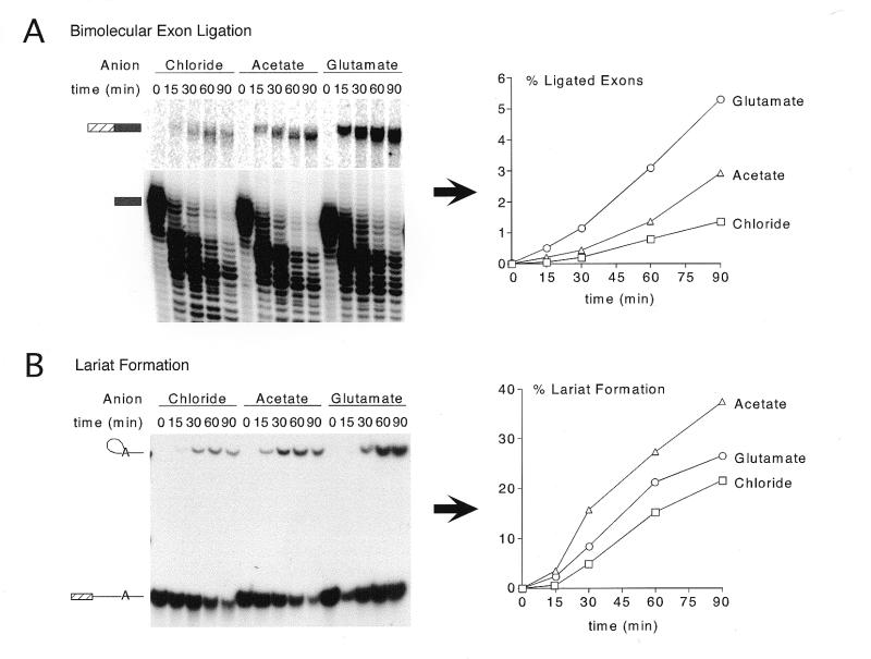
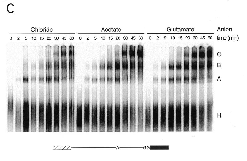
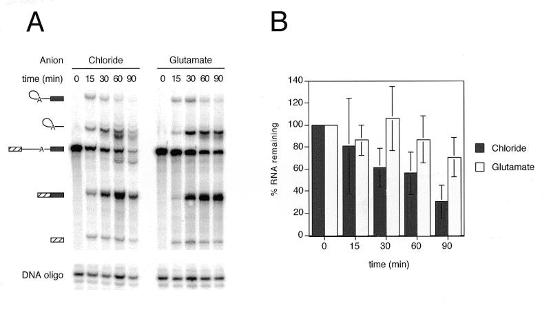
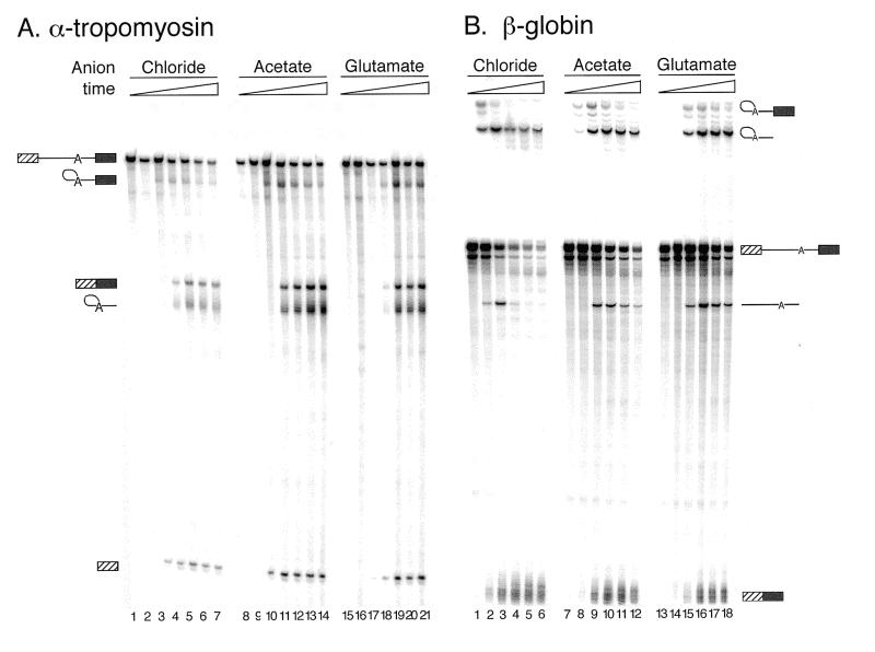
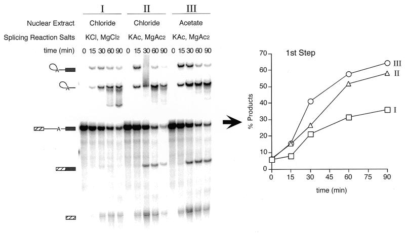
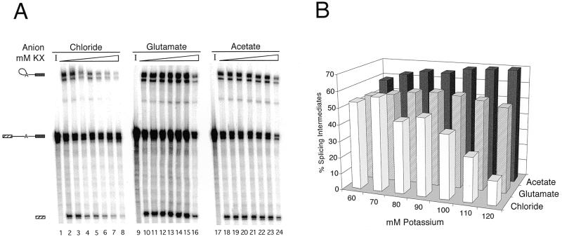
We demonstrate here that replacing potassium chloride (KCl) with potassium acetate (KAc) or potassium glutamate (KGlu) routinely enhances the yield of RNA intermediates and products obtained from in vitro splicing reactions performed in HeLa cell nuclear extract. This effect was reproducibly observed with multiple splicing substrates. The enhanced yields are at least partially due to stabilization of splicing precursors and products in the KAc and KGlu reactions. This stabilization relative to KCl reactions was greatest with KGlu and was observed over an extended potassium concentration range. The RNA stability differences could not be attributed to heavy metal contamination of the KCl, since ultrapure preparations of this salt yielded similar results. After testing various methods for altering the salts, we found that substitution of KAc or KGlu for KCl and MgAc(2)for MgCl(2)in splicing reactions is the simplest and most effective. Since the conditions defined here more closely mimic in vivo ionic concentrations, they may permit the study of more weakly spliced substrates, as well as facilitate more detailed analyses of spliceosome structure and function.


Figures

Figure 1
Figure 1
Comparison of first and second step product yields and splicing complexes in chloride, acetate and glutamate. (A) Splicing timecourses using the bimolecular exon ligation assay (19) to monitor the second step of splicing (exon ligation) independent of lariat formation (see text). The 5′ substrate (35 nM) was pre-incubated at 30°C for 30 min, and then bimolecular exon ligation initiated by addition of 3′ substrate (175 nM) and incubation continued for the times indicated. Splicing reactions followed standard conditions (80 mM potassium) using either an undialyzed, chloride extract or extracts dialyzed against KAc or KGlu Buffer E. KAc or KGlu and MgAc2 were substituted for KCl and MgCl2 where appropriate. 3′ substrate (lower; light exposure) and ligated exon product (upper; dark exposure) are indicated to the left of the gel. Percent ligated exon product versus time is shown graphically to the right. Calculations were performed as described in Materials and Methods. (B) Splicing timecourses using a truncated derivative of pAdMLΔAG that can only perform lariat formation (see text) (19). Splicing conditions were identical to those described for (A). Lariat product and 5′ substrate are indicated to the left of the gel. Percent lariat formation is shown graphically to the right. (C) Native gel showing splicing complex formation in reactions containing chloride, acetate or glutamate. Use of a modified full-length AdML substrate with a mutated 3′ splice site (bottom) allowed accumulation of intermediate-containing C complex spliceosomes. All splicing reactions were incubated under standard splicing conditions except that KAc or KGlu were substituted for KCl and MgAc2 for MgCl2 where indicated using the partial substitution conditions (see Results and Fig. 5).
Figure 1
Figure 1
Comparison of first and second step product yields and splicing complexes in chloride, acetate and glutamate. (A) Splicing timecourses using the bimolecular exon ligation assay (19) to monitor the second step of splicing (exon ligation) independent of lariat formation (see text). The 5′ substrate (35 nM) was pre-incubated at 30°C for 30 min, and then bimolecular exon ligation initiated by addition of 3′ substrate (175 nM) and incubation continued for the times indicated. Splicing reactions followed standard conditions (80 mM potassium) using either an undialyzed, chloride extract or extracts dialyzed against KAc or KGlu Buffer E. KAc or KGlu and MgAc2 were substituted for KCl and MgCl2 where appropriate. 3′ substrate (lower; light exposure) and ligated exon product (upper; dark exposure) are indicated to the left of the gel. Percent ligated exon product versus time is shown graphically to the right. Calculations were performed as described in Materials and Methods. (B) Splicing timecourses using a truncated derivative of pAdMLΔAG that can only perform lariat formation (see text) (19). Splicing conditions were identical to those described for (A). Lariat product and 5′ substrate are indicated to the left of the gel. Percent lariat formation is shown graphically to the right. (C) Native gel showing splicing complex formation in reactions containing chloride, acetate or glutamate. Use of a modified full-length AdML substrate with a mutated 3′ splice site (bottom) allowed accumulation of intermediate-containing C complex spliceosomes. All splicing reactions were incubated under standard splicing conditions except that KAc or KGlu were substituted for KCl and MgAc2 for MgCl2 where indicated using the partial substitution conditions (see Results and Fig. 5).
Figure 2
Figure 2
RNA degradation is reduced by substitution of KGlu for KCl. (A) A representative denaturing polyacrylamide gel used for determining RNA stability in chloride versus glutamate. Full-length AdML pre-mRNA was incubated according to standard splicing conditions, and KGlu and MgAc2 were substituted for KCl and MgCl2 using partial substitution. Reactions were incubated for the times shown and splicing products and intermediates are indicated to the left of the gel. The kinased DNA oligo indicated at the bottom of the gel was added to the splicing stop buffer to allow normalization for pipetting differences. (B) Data were averaged from three splicing timecourses of full-length AdML [as described in (A)]. The percent of input RNA (I) remaining at each time point was calculated as described in the text. Error bars represent standard deviations.
Figure 3
Figure 3
Splicing timecourses comparing ultrapure KCl (Aldrich and AlfaAesar) to standard KCl (Fisher), KAc and KGlu. Full-length AdML substrate was incubated under standard splicing conditions (80 mM potassium) using partial substitution to add the various potassium counterions. Splicing time points were 0, 15, 30, 60 and 90 min. RNA species are indicated to the left of the gel. Observed amounts of ligated exon product versus time are shown to the right. Values for ligated exon product represent raw counts taken from the PhosphorImager volume report. The asterisk (*) indicates that this point was discounted because lane 4 was clearly overloaded.
Figure 4
Figure 4
Substitution of KAc or KGlu for KCl improves spliced product yield for substrates other than AdML. Splicing timecourses with substrates containing derivatives of the (A) human α-tropomyosin intron 2 (27) or (B) rabbit β-globin intron 2 (26). For these experiments, the partial substitution conditions were used and standard splicing conditions were otherwise followed. Splicing time points for α-tropomyosin pre-mRNA (A) were 0, 15, 30, 60, 90, 120 and 180 min. Splicing time points for β-globin (B) were 0, 15, 30, 60, 90 and 150 min. RNA species were separated on 8% (29:1) polyacrylamide gels. Splicing intermediates and products are as indicated to the sides of the gels.
Figure 5
Figure 5
Partial substitution of chloride with acetate or glutamate yields levels of spliced product comparable to microdialysis. Using a full-length AdML substrate, splicing was compared under the following conditions: (I) chloride nuclear extract with additional KCl and MgCl2 in the splicing buffer; (II) chloride nuclear extract with KAc and MgAc2 in the splicing buffer; and (III) acetate nuclear extract with KAc and MgAc2 in the splicing buffer. The percentage of first step products shown to the right was calculated as described in Materials and Methods.
Figure 6
Figure 6
Relative first step product yields over a range of potassium concentrations with chloride, acetate or glutamate counterions. (A) AdMLΔAG was incubated under standard splicing conditions for 60 min, except that KAc or KGlu and MgAc2 were substituted for KCl and MgCl2 according to the partial substitution conditions. Potassium concentrations were 60, 70, 80, 90, 100, 110 and 120 mM; lanes 1, 9 and 17 contained 80 mM potassium but were incubated for 0 min [used as input (I) values to calculate RNA stability]. The doublet bands present in this gel are due to 3′ end heterogeneity of the T7 transcript (41,42). (B) Indicated splicing efficiencies represent first step product species (lariat intermediate and 5′ exon) and were calculated as described in Materials and Methods.

References

    1. Hardy S.F., Grabowski,P.J., Padgett,R.A. and Sharp,P.A. (1984) Nature, 308, 375–377. - PubMed
    1. Krainer A.R., Maniatis,T., Ruskin,B. and Green,M.R. (1984) Cell, 36, 993–1005. - PubMed
    1. Hernandez N. and Keller,W. (1983) Cell, 35, 89–99. - PubMed
    1. Padgett R.A., Hardy,S.F. and Sharp,P.A. (1983) Proc. Natl Acad. Sci. USA, 80, 5230–5234. - PMC - PubMed
    1. Kramer A. and Keller,W. (1990) In Dahlberg,J.E. and Abelson,J.N. (eds), Methods in Enzymology— RNA Processing, Part B: Specific Methods, Vol. 181. Academic Press, San Diego, CA, pp. 3–11.

Publication types