. 2000 Jan 15;28(2):570-81.
doi: 10.1093/nar/28.2.570.

The dhfr oribeta-binding protein RIP60 contains 15 zinc fingers: DNA binding and looping by the central three fingers and an associated proline-rich region

Affiliations

The dhfr oribeta-binding protein RIP60 contains 15 zinc fingers: DNA binding and looping by the central three fingers and an associated proline-rich region

C R Houchens et al. Nucleic Acids Res. .

Abstract

Initiation of DNA replication occurs with high frequency within oribeta, a short region 3' to the Chinese hamster dhfr gene. Homodimers of RIP60 (replication initiation-region protein 60 kDA) purified from nuclear extract bind two ATT-rich sites in oribeta and foster the formation of a twisted 720 bp DNA loop in vitro. Using a one hybrid screen in yeast, we have cloned the cDNA for human RIP60. RIP60 contains 15 C(2)H(2)zinc finger (ZF) DNA binding motifs organized in three clusters, termed hand Z1 (ZFs 1-5), hand Z2 (ZFs 6-8) and hand Z3 (ZFs 9-15). A proline-rich region is located between hands Z2 and Z3. Gel mobility shift and DNase I footprinting experiments show hands Z1 and Z2 independently bind the oribeta RIP60 sites specifically, but with different affinities. Hand Z3 binds DNA, but displays no specificity for RIP60 sites. Ligation enhancement, DNase I footprinting, and atomic force microscopy assays show that hand Z2 and a portion of the associated proline-rich region is sufficient for protein multimerization on DNA and DNA looping in vitro. Polyomavirus origin-dependent plasmid replication assays show RIP60 has weak replication enhancer activity, suggesting that RIP60 does not harbor a transcriptional transactivation domain. Because vertebrate origins of replication have no known consensus sequence, we suggest that sequence-specific DNA binding proteins such as RIP60 may act as accessory factors in origin identification prior to the assembly of pre-initiation complexes.


Figures
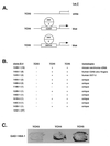
Figure 1
Figure 1
One hybrid screen in yeast for RIP60. (A) A genetic screen in S.cerevisiae was used to identify cDNA-encoded fusion proteins that bind the RIP60 target sequence (DSR) and activate expression of a linked reporter gene (LacZ or histidine) by recruiting a fused GAL4 activation domain (GAD) to the promoter. Shown are lacZ reporter strains in which lacZ expression is controlled by five copies of the DSR target sequence (YCH4) or a single copy of the DSR embedded in its native flanking sequences (YCH5). YCH3 was used as a control. (B) Summary of the one hybrid screen. From 14 million transformants, 37 clones were found that activated expression in YCH4 and/or YCH5 but not in YCH3. (C) Specificity test in yeast for RIP60 binding. In the one hybrid screen multiple in-frame fusions were identified that overlapped with clone 146A-1. Each of these fusions contained the Z2 and PRR region of RIP60 and activated lacZ expression in both YCH4 and YCH5, but not in YCH3.
Figure 2
Figure 2
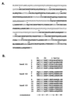
RIP60 is a polydactyl ZF protein. Two otherwise identical cDNAs with different poly(A) tails were isolated from a HeLa cell lambda phage library using a clone related to 146A as probe. (A) The open reading frame of both clones predicts a 567 amino acid protein that includes 15 ZFs (bold) and both RIP60 peptide sequences obtained by microsequencing (underlined). (B) RIP60 contains 15 C2H2 Kruppel-like ZFs organized in three hands termed Z1 (ZFs 1–5), Z2 (ZFs 6–8) and Z3 (ZFs 9–15). A proline-rich region (PRR) predicted to form three polyproline helices separates hands Z2 and Z3.
Figure 3
Figure 3
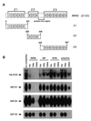
Specific DNA binding by RIP60 is located in both hands Z1 and Z2. (A) Schematic representation of RIP60 (Z123) showing the organization of hands Z1, Z2, Z3 and the proline-rich region. (B) Analysis of RIP60 DNA binding activity by gel mobility shift assays. GST fusion proteins containing hands Z1, Z2 or Z3 were expressed in E.coli, purified and analyzed for DNA binding activity. Because expression of full length RIP60 in E.coli was difficult, analysis of RIP60 binding activity was conducted with nuclear extracts from cells transfected with pCMV-HA-RIP60 (see Results). Binding specificity was assessed by competition with the DSR RIP60 binding site, an intron binding factor site (IBF), or the AT-rich regions from either the SV40 or polyomavirus origins of replication.
Figure 4
Figure 4
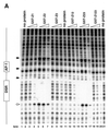
DNase I footprinting of RIP60 on the DHFR-E bent DNA fragment. The indicated RIP60 GST fusion proteins were incubated with end labeled DHFR-E probes and digested with DNase I as described in the Materials and Methods. The digestion products were resolved on sequencing gels and the footprinting patterns were visualized by autoradiography. (A) Nuclease protection patterns for the top strand of DHFR-E. Hypersensitive sites are indicated by arrows. The open arrow indicates a prominent hypersensitive site observed with RIP60 purified from cell extract and by in vivo genomic footprinting of the bent DNA motif in CHO cells (not shown). (B) Nuclease protection patterns for the bottom strand of DHFR-E. Hypersensitive sites are indicated by arrows.
Figure 4
Figure 4
DNase I footprinting of RIP60 on the DHFR-E bent DNA fragment. The indicated RIP60 GST fusion proteins were incubated with end labeled DHFR-E probes and digested with DNase I as described in the Materials and Methods. The digestion products were resolved on sequencing gels and the footprinting patterns were visualized by autoradiography. (A) Nuclease protection patterns for the top strand of DHFR-E. Hypersensitive sites are indicated by arrows. The open arrow indicates a prominent hypersensitive site observed with RIP60 purified from cell extract and by in vivo genomic footprinting of the bent DNA motif in CHO cells (not shown). (B) Nuclease protection patterns for the bottom strand of DHFR-E. Hypersensitive sites are indicated by arrows.
Figure 5
Figure 5
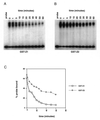
GST-Z2 binds the DSR more avidly than does GST-Z1. GST-Z1 and GST-Z2 were bound to labeled DSR probe at room temperature under standard gel shift conditions. After 20 min a 1000-fold molar excess of unlabeled probe was added, the incubation was continued and samples were withdrawn at the indicated time points and loaded directly onto gels as described in the Materials and Methods. (A) Autoradiograph of GST-Z1 binding after addition of unlabeled probe (time 0). (B) Autoradiograph of GST-Z2 binding after addition of unlabeled probe. (C) Quantification of percent probe bound as a function of time after the addition of unlabeled probe.
Figure 6
Figure 6
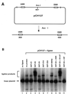
Binding of RIP60 GST-Z2 to the USR and DSR enhances ligation of linear pCH127. A ligation enhancement assay was used to test for the ability of RIP60 fusion proteins to mediate DNA looping between the USR and DSR. (A) pCH127 contains a 1.1 kb fragment of the DHFR ori-β region that includes both the USR and DSR RIP60 binding sites. (B) pCH127 was linearized with AccI and incubated with the indicated protein factors for 15 min prior to the addition of ligase and ATP for 5 min. The products of the reactions were resolved by gel electrophoresis, blotted to nylon, and visualized by hybridization with 32P-labeled pCH127. Ligation products representing open circles or linear dimers are indicated by the square bracket. Addition of DSR competitor DNA eliminated increased ligation by GST-Z2 (lane 9) whereas the AT-rich region from the SV40 origin of replication had no effect (lane 10). Dephosphorylated linear pCH127 did not support ligation with (lane 11) or without (not shown) GST-Z2.
Figure 7
Figure 7
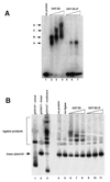
The proline-rich region stimulates DNA binding and DNA looping by hand Z2. (A) Gel mobility shift assays were used to assess the effect of the proline-rich region of DNA binding by GST-Z2. Equivalent amounts of GST-Z2 and GST-Z2ΔP were incubated with 32P-labeled DSR probe, the products were resolved by non-denaturing electrophoresis, and binding was visualized by autoradiography. GST-Z2 forms at least four gel shift complexes with the DSR (A–D) whereas GST-Z2ΔP forms a single complex. Competition assays showed binding by GST-Z2ΔP was specific for the DSR (data not shown). (B) GST-Z2 and GST-Z2ΔP were tested for their ability to mediate DNA looping using a ligation enhancement assay. In this instance ligation of the linear pCH127 control was allowed to proceed for 20 min in order to visualize a more complex array of ligation products. Lanes 6–8 and 9–11 contained 5, 50 or 500 ng of the indicated protein, respectively. (C) DNA looping between the USR and DSR by GST-Z2. The arrows indicate free ends of pCH127 that protrude from the loop complex. (D) The PRR is required for DNA looping. When incubated with GST-Z2ΔP looping between the USR and DSR in linear pCH127 was not observed by AFM.
Figure 7
Figure 7
The proline-rich region stimulates DNA binding and DNA looping by hand Z2. (A) Gel mobility shift assays were used to assess the effect of the proline-rich region of DNA binding by GST-Z2. Equivalent amounts of GST-Z2 and GST-Z2ΔP were incubated with 32P-labeled DSR probe, the products were resolved by non-denaturing electrophoresis, and binding was visualized by autoradiography. GST-Z2 forms at least four gel shift complexes with the DSR (A–D) whereas GST-Z2ΔP forms a single complex. Competition assays showed binding by GST-Z2ΔP was specific for the DSR (data not shown). (B) GST-Z2 and GST-Z2ΔP were tested for their ability to mediate DNA looping using a ligation enhancement assay. In this instance ligation of the linear pCH127 control was allowed to proceed for 20 min in order to visualize a more complex array of ligation products. Lanes 6–8 and 9–11 contained 5, 50 or 500 ng of the indicated protein, respectively. (C) DNA looping between the USR and DSR by GST-Z2. The arrows indicate free ends of pCH127 that protrude from the loop complex. (D) The PRR is required for DNA looping. When incubated with GST-Z2ΔP looping between the USR and DSR in linear pCH127 was not observed by AFM.
Figure 8
Figure 8
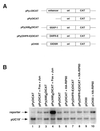
Polyomavirus origin-dependent replication assay for replication enhancer activity. (A) A series of plasmids containing the wild type polyomavirus (Py) core origin of replication were used in transient replication assays to test RIP60 for replication enhancer activity. pPyOICAT contains the Py core origin and the native α-enhancer region. The negative control plasmid pPyOICAT lacks the α-enhancer, whereas the positive control plasmid pPy(AM)6OICAT contains six AP-1 sites in place of the α-enhancer. Test plasmids pPy(DHFR-E)OICAT and pCH30 contained the DHFR-E fragment or five copies of the DSR in place of the α-enhancer, respectively. (B) Representative results of transient Py-origin dependent plasmid replication assays. The indicated reporter plasmids were cotransfected into NIH 3T3 cells with unmethylated pUC19 plasmid DNA, an expression vector for Py large T antigen, and the indicated expression vectors for c-fos, c-jun, or RIP60. After imaging by autoradiography, the signals in each band were quantified in a phosphoimager and the ratio of signal between the reporter and the unmethylated pUC19 control plasmid was calculated. Coexpression of Fos and Jun stimulated replication of pPy(AM)6OICAT ~40-fold in this experiment.

Similar articles

Cited by

References

    1. Stillman B. (1996) Science, 274, 1659–1664. - PubMed
    1. Newlon C. (1997) Cell, 91, 717–720. - PubMed
    1. Bell S.P. and Dutta,A. (1997) Annu. Rev. Cell Dev. Biol., 13, 293–332. - PubMed
    1. Diffley J.F. (1998) Curr. Biol., 8, 771–773. - PubMed
    1. Bousett K. and Diffley,J.F. (1998) Genes Dev., 12, 480–490. - PMC - PubMed

Publication types

MeSH terms

Associated data