. 2000 Jan 15;28(2):e4.
doi: 10.1093/nar/28.2.e4.

The specificity of protein-DNA crosslinking by formaldehyde: in vitro and in drosophila embryos

Affiliations

The specificity of protein-DNA crosslinking by formaldehyde: in vitro and in drosophila embryos

J Toth et al. Nucleic Acids Res. .

Abstract

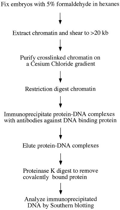
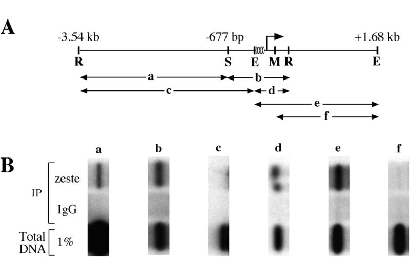
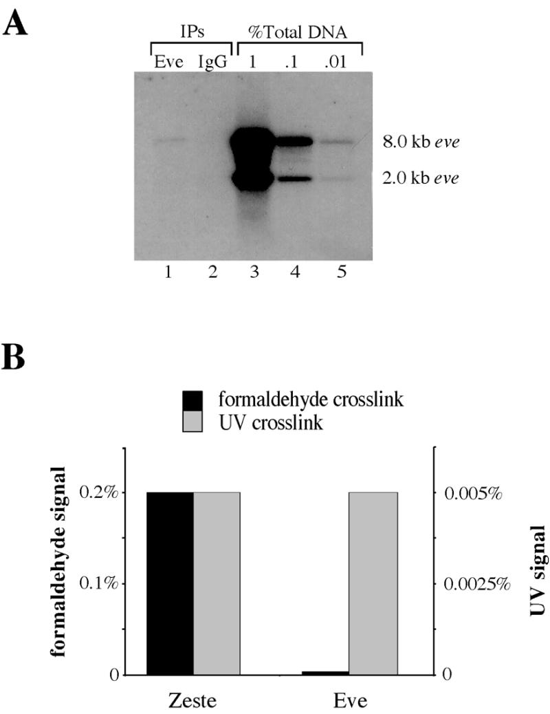
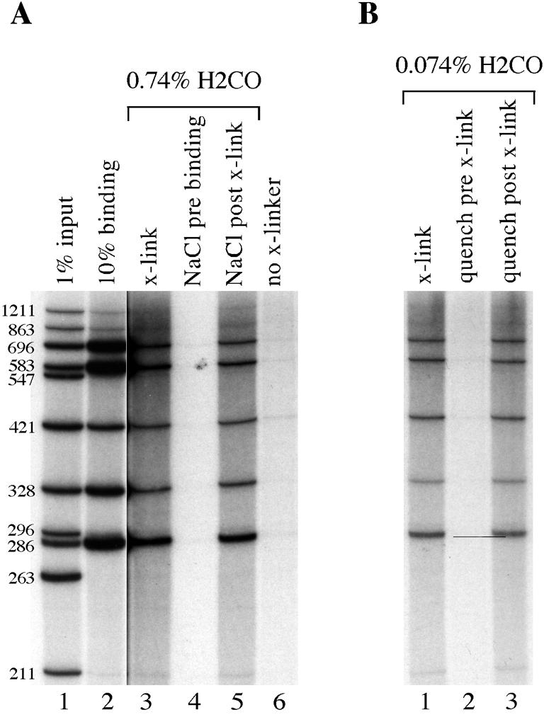
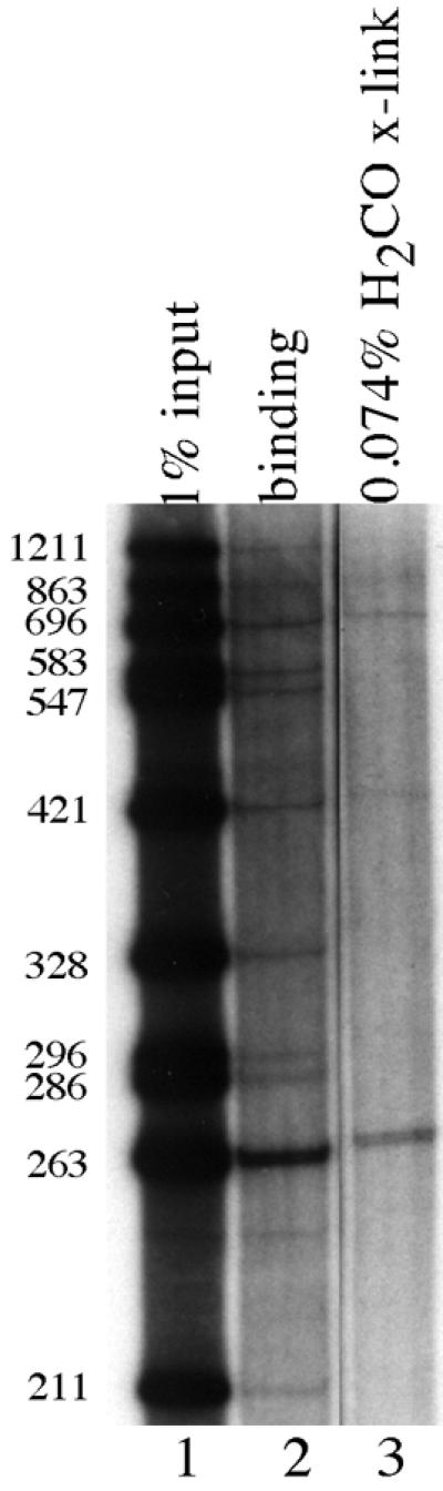
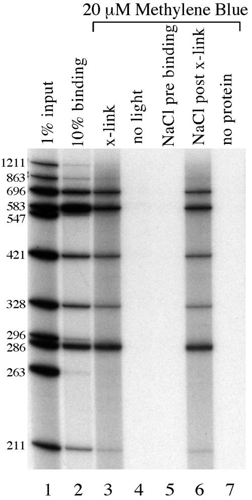
Formaldehyde crosslinking has been widely used to study binding of specific proteins to DNA elements in intact cells. However, previous studies have not determined if this crosslinker preserves the bona fide pattern of DNA binding. Here we show that formaldehyde crosslinking of Drosophila embryos maps an interaction of the transcription factor Zeste to a known target element in the Ultrabithorax promoter. This data agrees broadly with previous mapping of the same Zeste binding sites by in vivo UV crosslinking, though the formaldehyde method does give a low, possibly artifactual signal on other DNA fragments that is not detected by the UV method. We also demonstrate, using an in vitro assay, that formaldehyde crosslinking accurately reflects the DNA binding specificities of both Zeste and a second transcription factor, Eve. The crosslinking reagent methylene blue is shown to preserve DNA binding specificity in vitro as well. Our results suggest that crosslinking by formaldehyde, and possibly also by methylene blue, provide an accurate guide to the interaction of proteins with their high affinity target sites in cells.


Figures

Figure 1
Figure 1
Outline of our in vivo formaldehyde crosslinking protocol.
Figure 2
Figure 2
Zeste binds specifically to the Ubx proximal promoter. (A) Immunoprecipitation of EcoRI digested, formaldehyde crosslinked chromatin from 0–12 h embryos with either anti-Zeste antibody (lane 1) or with non-specific IgG (lane 2). A dilution series of the total amount of input DNA in the immunoprecipitation is also shown (lanes 3–5). The Southern blot was probed with a 3.5 kb fragment of the Ubx proximal promoter that spans from –3.1 to +0.4 kb: the size of the EcoRI fragment detected is indicated. (B) The same Southern blot used in (A) was stripped and reprobed with an 8.7 kb EcoRI fragment from the actin 5C gene (35). The lane designations are the same as in (A).
Figure 3
Figure 3
Zeste is crosslinked most strongly to a 297 bp region of the Ubx promoter. (A) Diagram of the restriction fragments produced by various digests of the Ubx proximal promoter. The positions of EcoRI (R), EheI (E), MluI (M) and StuI (S) restriction sites are marked, and the RNA start site at +1 is indicated by an arrow. The restriction fragments to which binding has been examined are a 2.8 kb EcoRI–StuI fragment that spans nucleotides –3.54 kb to –677 bp (a), a 1 kb EcoRI–StuI fragment from –677 to +365 bp (b), a 3.4 kb EcoRI–EheI fragment from –3.54 kb to –177 bp (c), a 540 bp EcoRI–EheI fragment from –177 to +365 bp (d), a 1.8 kb EheI–EheI fragment from –177 bp to +1.63 kb (e) and a 1.5 kb MluI–EheI fragment from +120 bp to +1.63 kb (f). Ovals denote the positions of high affinity Zeste binding sites. (B) Results of formaldehyde crosslinking experiments examining binding to the DNA fragments shown in (A). Vertical columns show blots of immunoprecipitations of crosslinked chromatin from wild-type 0–12 h embryos (a–f). The top row contains DNA fragments immunoprecipitated with anti-Zeste antibodies, the middle row contains the result obtained with non-specific antibody and the bottom row contains 1% of the input DNA. The Southern blots examining binding to fragments a–d were probed with the same 3.5 kb fragment of the Ubx proximal promoter as used in Figure 2A. The blots examining binding to fragments e and f were probed with an adjacent EcoRI DNA fragment that lies between nucleotides +365 bp and +3.68 kb.
Figure 4
Figure 4
In vivo formaldehyde crosslinking does not convincingly detect DNA binding by Eve. (A) Southern blot of EcoRI digested, formaldehyde crosslinked chromatin from 4–5 h embryos immunoprecipitated either with anti-Eve antibodies (lane 1) or with non-specific IgG (lane 2). The blot was probed with eve gene sequence spanning nucleotides –6.4 kb to –400 bp. A dilution series of input DNA is shown on the left (lanes 3–5) and the size of the EcoRI fragment detected is indicated. (B) Normalized crosslinking efficiency of Zeste and Eve detected by the in vivo UV and formaldehyde techniques. The percent formaldehyde crosslinking signals for Eve and Zeste are shown by the gray bars and indicated by the scale on the left. The percent UV crosslinking is shown by the black bars and the values are indicated on the right hand scale. The values for Zeste are for crosslinking to the 3.5 kb proximal promoter fragment of the Ubx gene and for Eve are for the eve upstream promoter fragment used in (A). The data for UV crosslinking are from Solomon et al. (7).
Figure 5
Figure 5
In vitro formaldehyde crosslinking of Eve protein reproduces the same pattern of DNA fragments as an in vitro binding reaction. Autoradiograms of 6% denaturing polyacrylamide gels. (A) Lane 1, 1% of the DNA used in all DNA binding and crosslinking reactions; lane 2, 10% of the DNA immunoprecipitated from a standard in vitro DNA binding reaction containing Eve protein; lanes 3–6, all of the DNA recovered from various formaldehyde crosslinking experiments containing Eve protein and either 0.74% (lanes 3–5) or no formaldehyde (lane 6); lanes 4 and 5, DNA fragments recovered when NaCl was added to a final concentration of 500 mM to DNA binding reactions prior to (lane 4) or after (lane 5) formaldehyde crosslinking. The DNA fragments used in this and all other in vitro crosslinking experiments are from a BsaJI + BssHII + ScaI digest of a 3.5 kb Ubx proximal promoter from plasmid p3102. The sizes of the DNA fragments in base pairs are indicated along the left. The autoradiogram of lanes 3–6 was exposed for 12 times longer than the autoradiogram of lanes 1 and 2. (B) Lanes 1–3, all of the DNA recovered from formaldehyde crosslinking experiments containing Eve protein and 0.074% formaldehyde; lanes 2 and 3, DNAs recovered when Tris quencher was added before (lane 2) or after crosslinking (lane 3). The DNAs used in this experiment are the same as those in (A). The autoradiogram of this gel was exposed for the same length of time as the autoradiogram of lanes 3–6 in (A).
Figure 6
Figure 6
In vitro formaldehyde crosslinking localizes the binding of Zeste protein to the same region of the Ubx gene to which it crosslinks in vivo. Autoradiogram of a 6% denaturing polyacrylamide gel. Lane 1, 1% of the DNA from the same digest of Ubx DNA used in Figure 5; lane 2, all of the DNA immunoprecipitated from an in vitro DNA binding assay containing purified Zeste protein; lane 3, all of the DNA recovered from an in vitro formaldehyde crosslinking reaction with Zeste protein. The sizes of the DNA fragments in base pairs are indicated on the left. These DNA fragments are the same as those used in Figure 5, and the autoradiogram of this gel was exposed for the same length of time as lanes 3–6 of Figure 5.
Figure 7
Figure 7
Methylene blue efficiently crosslinks Eve protein to DNA in vitro. Lane 1, 1% of the input DNA used in the binding and crosslinking reactions; lane 2, 10% of the DNA isolated in a standard DNA binding reaction containing Eve protein; lanes 3–7, all of the DNA recovered from a series of in vitro crosslinking reactions with Eve protein; lanes 3 and 4, DNA immunoprecipitated from reactions containing 20 µM methylene blue that had either been irradiated with white light for 30 min (lane 3) or that had been kept in the dark (lane 4); lanes 5 and 6, methylene blue crosslinking experiments in which 500 mM NaCl had been added either prior to (lane 5) or after (lane 6) crosslinking; lane 7, DNAs recovered when protein was not included in the reaction. The DNAs used in this experiment are the same as those used in Figure 5, and the autoradiogram of this gel was exposed for the same length of time as lanes 3–6 of Figure 5.

References

    1. Johnson P.F. and McKnight,S.L. (1989) Annu. Rev. Biochem., 58, 799–839. - PubMed
    1. Faisst S. and Meyer,S. (1992) Nucleic Acids Res., 20, 3–26. - PMC - PubMed
    1. Heinemeyer T., Wingender,E., Reuter,I., Hermjakob,H., Kel,A.E., Kel,O.V., Ignatieva,E.V., Ananko,E.A., Podkolodnaya,O.A., Kolpakov,F.A., Podkolodny,N.L. and Kolchanov,N.A. (1998) Nucleic Acids Res., 26, 362–367. - PMC - PubMed
    1. Pabo C.O. and Sauer,R.T. (1992) Annu. Rev. Biochem., 61, 1053–1095. - PubMed
    1. Gilmour D.S. and Lis,J.T. (1985) Mol. Cell. Biol., 5, 2009–2018. - PMC - PubMed

LinkOut - more resources