. 2000 Jan 15;28(2):e6.
doi: 10.1093/nar/28.2.e6.

A method for preparing genomic DNA that restrains branch migration of Holliday junctions

Affiliations

A method for preparing genomic DNA that restrains branch migration of Holliday junctions

T Allers et al. Nucleic Acids Res. .

Abstract

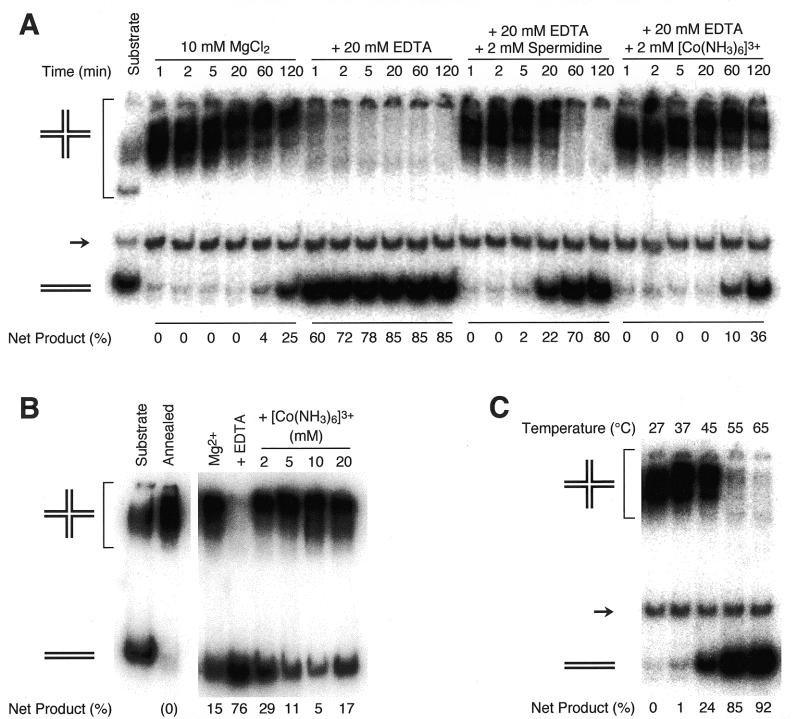
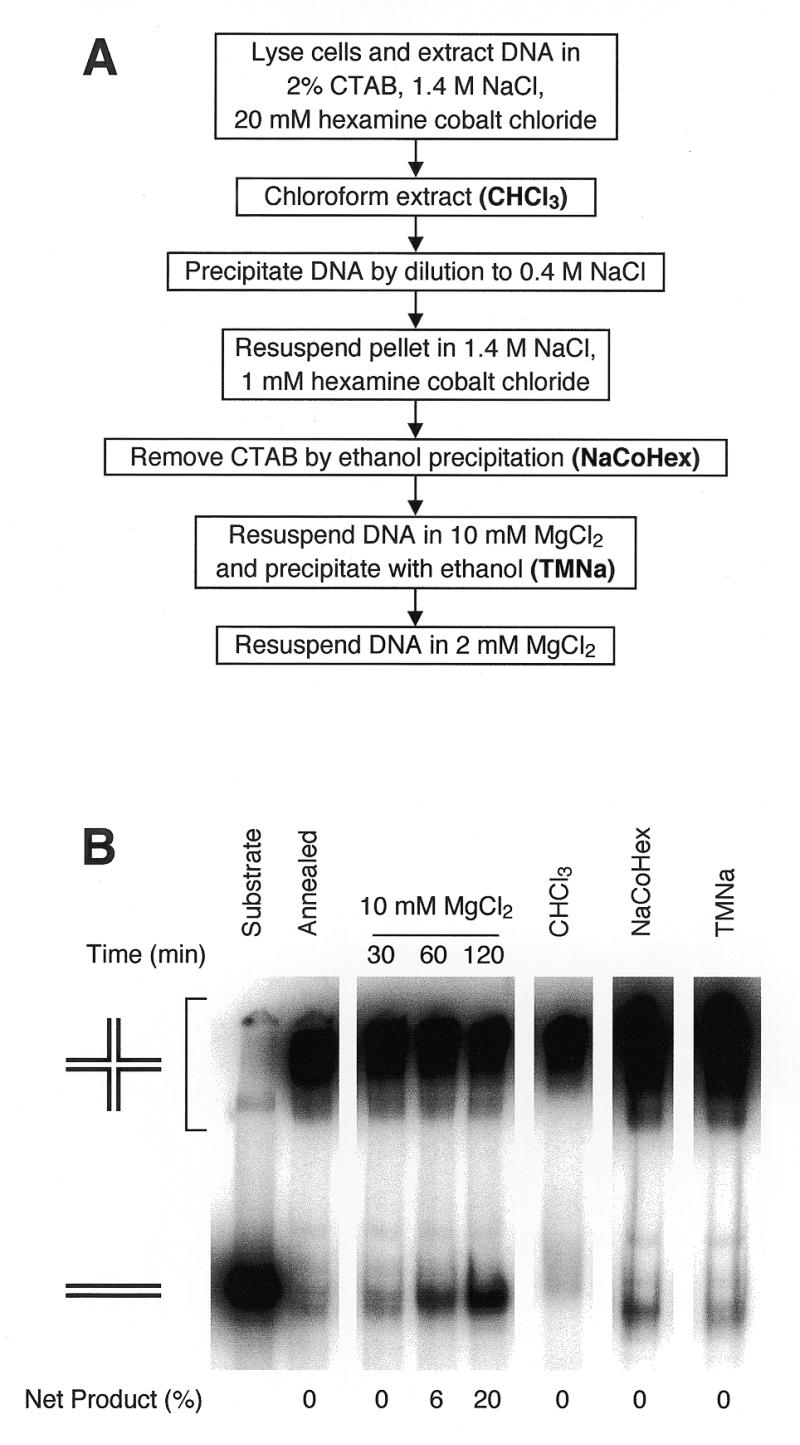
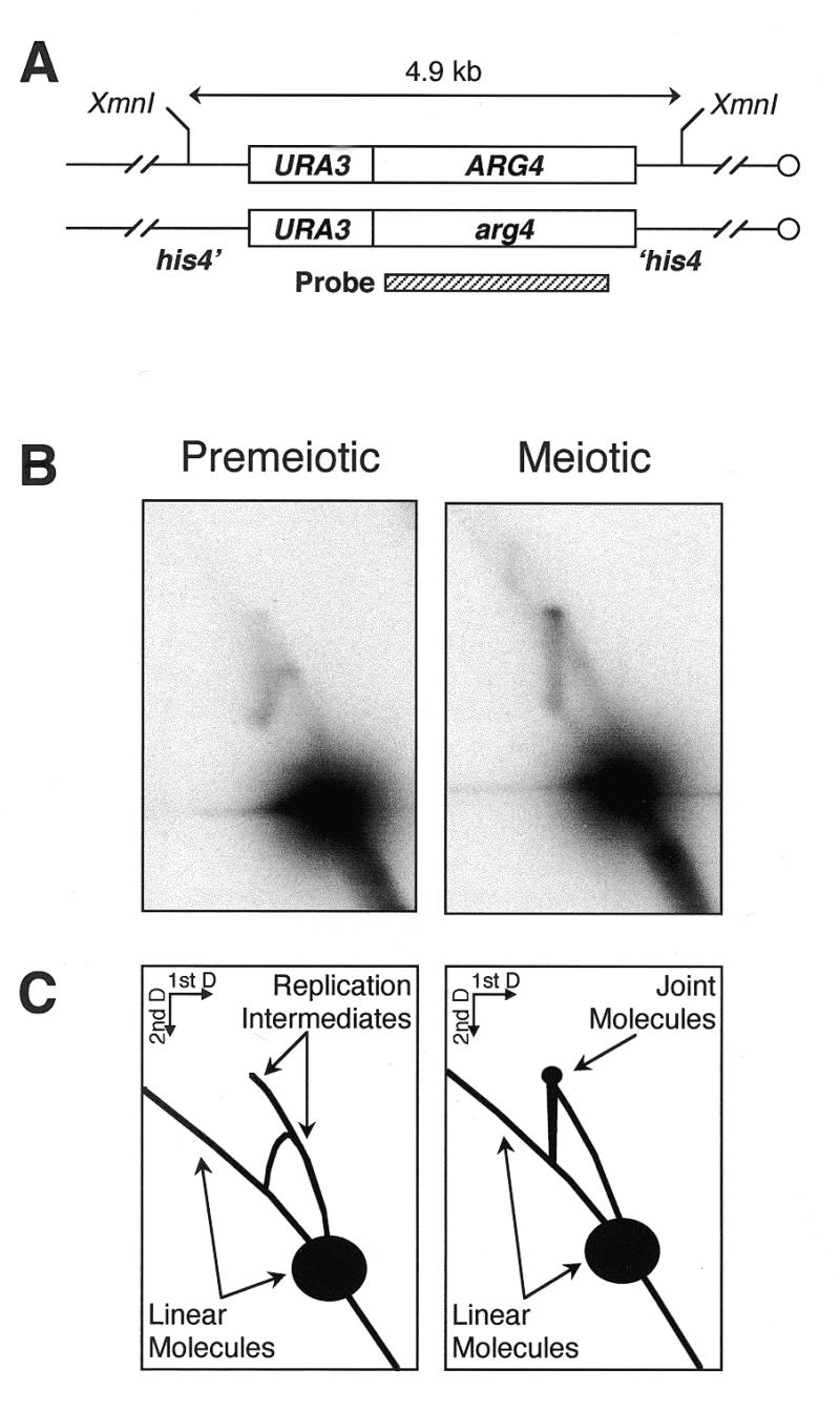
The Holliday junction is a central intermediate in genetic recombination. This four-stranded DNA structure is capable of spontaneous branch migration, and is lost during standard DNA extraction protocols. In order to isolate and characterize recombination intermediates that contain Holliday junctions, we have developed a rapid protocol that restrains branch migration of four-way DNA junctions. The cationic detergent hex-adecyltrimethylammonium bromide is used to lyse cells and precipitate DNA. Manipulations are performed in the presence of the cations hexamine cobalt(III) or magnesium, which stabilize Holliday junctions in a stacked-X configuration that branch migrates very slowly. This protocol was evaluated using a sensitive assay for spontaneous branch migration, and was shown to preserve both artificial Holliday junctions and meiotic recombination intermediates containing four-way junctions.


Figures

Figure 1
Figure 1
Branch migration assay. Two homologous duplexes (hatched) each having heterologous single-strand tails (black and white) are rapidly annealed to form a four-stranded structure with a Holliday junction. Two sets of substrates were used: S1 and S2 (300 bp homologous duplex) and R1 and R2 (273 bp homologous duplex). Spontaneous branch migration proceeds as a random walk until the junction reaches the distal end of the homologous region, leading to the irreversible dissociation of two duplex products. Asterisks indicate 5′-32P-label.
Figure 2
Figure 2
Effect of polyvalent cations and temperature on branch migration. (A) Effect of polyvalent cations. 32P-labeled S1 and excess unlabeled S2 substrates were annealed in TNM buffer (10 mM MgCl2) and aliquoted to reactions containing the indicated supplements. Reactions were incubated at 37°C for the indicated time, and dissociation of the Holliday junction (upper band) to duplex products (lower band) by branch migration was monitored by gel electrophoresis. The middle band (→) is 32P-labeled duplex DNA lacking single-stranded tails, which does not participate in the reaction. Net product refers to the fraction of total 32P label migrating as monomer duplex, after subtraction of residual unannealed substrate (in this case, duplex remaining after 1 min in 10 mM MgCl2). (B) Effect of Co3+(NH3)6Cl3 concentration. R1 and R2 were annealed in TNM buffer and aliquoted to reactions containing 10 mM MgCl2, 10 mM MgCl2 + 20 mM EDTA or 10 mM MgCl2 + 20 mM EDTA + Co3+(NH3)6Cl3 at the indicated concentrations. Reactions were incubated at 37°C for 2 h, and branch migration was determined as described in (A). (C) Effect of temperature. S1 and S2 were annealed in TNM buffer, which was then supplemented with 20 mM EDTA and 10 mM Co3+(NH3)6Cl3. Reactions were incubated for 1 h at the indicated temperatures, and branch migration was determined as described in (A).
Figure 3
Figure 3
DNA extraction with CTAB in the presence of Co3+(NH3)6Cl3. (A) Outline of the protocol. Experimental details are given in Materials and Methods. (B) Branch migration during yeast DNA extraction using the protocol. R1 and R2 were annealed in TNM buffer. As a positive control for branch migration, an aliquot was incubated at 37°C in TNM buffer (10 mM MgCl2) for the indicated times. Remaining annealed substrate was added to yeast spheroplasts, and DNA was extracted as described in Materials and Methods. A sample was taken after chloroform:isoamyl alcohol extraction (CHCl3). DNA pellets obtained after ethanol precipitation from NaCoHex solution (NaCoHex) and after ethanol precipitation from TMNa solution (TMNa) were resuspended in TMSpe buffer on ice. Branch migration was measured as described in Figure 2; net product refers to the fraction of total 32P label migrating as monomer duplex, after subtraction of residual unannealed substrate.
Figure 4
Figure 4
Joint molecules observed during yeast meiosis. (A) Structure of the locus examined. The his4::URA3-ARG4 insert in MJL2444 consists of a 1.1 kb HindIII–SmaI fragment of URA3 and a 2.3 kb Eco47III–PstI fragment of ARG4 inserted in HIS4 coding sequences. (B) DNA was extracted with CTAB, either from a premeiotic culture or 4 h after the onset of meiosis, digested with XmnI, resolved on a 2D agarose gel and hybridized with the indicated probe as described in Materials and Methods. (C) Interpretation of the results. While only DNA replication intermediates are seen in the premeiotic sample, a distinct off-arc spot is observed in the meiotic sample, at the position expected for joint molecules formed between the homologs illustrated in (A) (7).

References

    1. Holliday R. (1964) Genet. Res., 5, 282–304.
    1. Sigal N. and Alberts,B. (1972) J. Mol. Biol., 71, 789–793. - PubMed
    1. Meselson M.S. and Radding,C.M. (1975) Proc. Natl Acad. Sci. USA, 72, 358–361. - PMC - PubMed
    1. Resnick M.A. (1976) J. Theor. Biol., 59, 97–106. - PubMed
    1. Orr-Weaver T.L., Nicolas,A. and Szostak,J.W. (1988) Mol. Cell. Biol., 8, 5292–5298. - PMC - PubMed