

Evidence for antigen presentation to sensitized T cells by thyroid peroxidase (TPO)-specific B cells in mice injected with fibroblasts co-expressing TPO and MHC class II
- PMID: 10606962
- PMCID: PMC1905538
- DOI: 10.1046/j.1365-2249.2000.01087.x
Evidence for antigen presentation to sensitized T cells by thyroid peroxidase (TPO)-specific B cells in mice injected with fibroblasts co-expressing TPO and MHC class II
Abstract
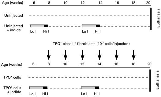
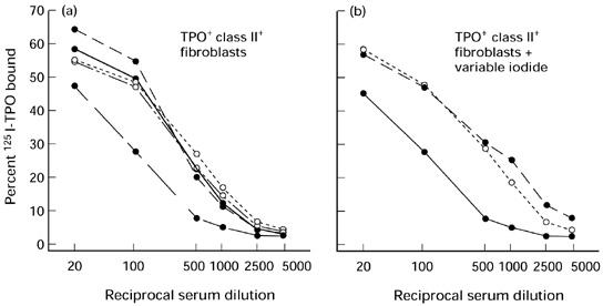
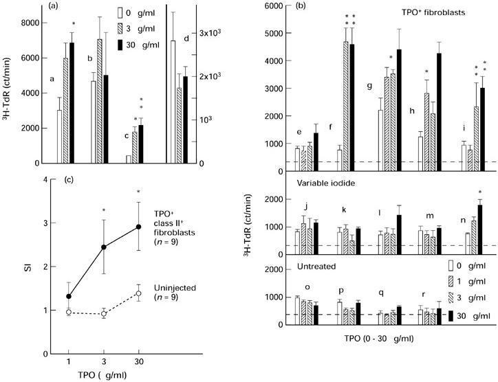
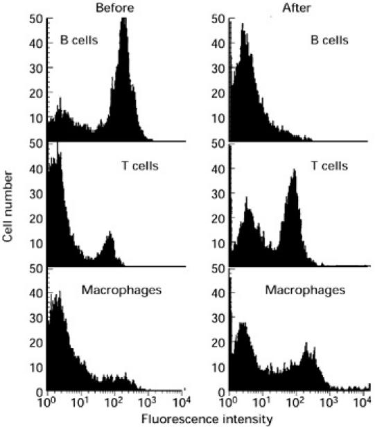
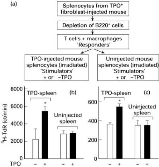
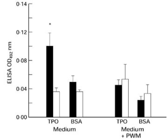
Injection of AKR/N mice with fibroblasts co-expressing MHC class II and TPO in the absence of adjuvant induces IgG-class TPO antibodies that resemble spontaneously arising human thyroid autoantibodies. We have used this model to examine the effect of iodide on TPO antibody induction as well as to analyse the interaction between T and B cells. Despite its importance as a major environmental factor in thyroid autoimmunity, variable iodide intake had no detectable effects on TPO antibody levels, lymphocytic infiltration of the thyroid or thyroid hormone levels. In terms of T cell responsiveness, splenocytes from TPO fibroblast-injected mice, but not from control mice, proliferated in response to TPO. Intriguingly, B cell-depleted splenocytes (mainly T cells without reduction of macrophages) proliferated in response to TPO only when co-cultured with irradiated autologous splenocytes from TPO fibroblast-injected mice but not from control mice. These data suggest that TPO-specific B cells are involved in antigen presentation to sensitized T cells and are supported by the ability of spleen cells from TPO cell-injected (but not control) mice to secrete TPO antibodies spontaneously in culture. In conclusion, we provide the first evidence for the presence of thyroid autoantigen-specific B cells and their ability to present their autoantigen to sensitized T cells in mice induced to develop TPO antibodies resembling autoantibodies in humans.
Figures
Similar articles
-
Cytokines, IgG subclasses and costimulation in a mouse model of thyroid autoimmunity induced by injection of fibroblasts co-expressing MHC class II and thyroid autoantigens.Clin Exp Immunol. 2000 Nov;122(2):170-9. doi: 10.1046/j.1365-2249.2000.01362.x. Clin Exp Immunol. 2000. PMID: 11091271 Free PMC article.
-
Cellular thyroid peroxidase (TPO), unlike purified TPO and adjuvant, induces antibodies in mice that resemble autoantibodies in human autoimmune thyroid disease.J Clin Endocrinol Metab. 1999 May;84(5):1651-7. doi: 10.1210/jcem.84.5.5666. J Clin Endocrinol Metab. 1999. PMID: 10323395
-
Autoimmune response to the thyroid in humans: thyroid peroxidase--the common autoantigenic denominator.Int Rev Immunol. 2000;19(6):587-618. doi: 10.3109/08830180009088514. Int Rev Immunol. 2000. PMID: 11129117 Review.
-
Relationship between thyroid peroxidase T cell epitope restriction and antibody recognition of the autoantibody immunodominant region in human leukocyte antigen DR3 transgenic mice.Endocrinology. 2005 Nov;146(11):4961-7. doi: 10.1210/en.2005-0760. Epub 2005 Aug 4. Endocrinology. 2005. PMID: 16081633
-
Thyroid peroxidase as an autoantigen.Thyroid. 2007 Oct;17(10):939-48. doi: 10.1089/thy.2007.0169. Thyroid. 2007. PMID: 17822378 Review.
Cited by
-
Localization of the immunodominant region on human thyroid peroxidase in autoimmune thyroid diseases: an update.J Autoimmune Dis. 2005 Mar 15;2(1):2. doi: 10.1186/1740-2557-2-2. J Autoimmune Dis. 2005. PMID: 15769293 Free PMC article.
-
Thyroid peroxidase (TPO) expressed in thyroid and breast tissues shows similar antigenic properties.PLoS One. 2017 Jun 2;12(6):e0179066. doi: 10.1371/journal.pone.0179066. eCollection 2017. PLoS One. 2017. PMID: 28575127 Free PMC article.
-
Evaluation of conformational epitopes on thyroid peroxidase by antipeptide antibody binding and mutagenesis.Clin Exp Immunol. 2004 Apr;136(1):137-44. doi: 10.1111/j.1365-2249.2004.02422.x. Clin Exp Immunol. 2004. PMID: 15030525 Free PMC article.
-
Targeted biological therapies for Graves' disease and thyroid-associated ophthalmopathy. Focus on B-cell depletion with Rituximab.Clin Endocrinol (Oxf). 2011 Jan;74(1):1-8. doi: 10.1111/j.1365-2265.2010.03806.x. Clin Endocrinol (Oxf). 2011. PMID: 20455896 Free PMC article. Review.
-
Cytokines, IgG subclasses and costimulation in a mouse model of thyroid autoimmunity induced by injection of fibroblasts co-expressing MHC class II and thyroid autoantigens.Clin Exp Immunol. 2000 Nov;122(2):170-9. doi: 10.1046/j.1365-2249.2000.01362.x. Clin Exp Immunol. 2000. PMID: 11091271 Free PMC article.
References
-
- McLachlan SM, Rapoport B. The molecular biology of thyroid peroxidase: cloning, expression and role as autoantigen in autoimmune thyroid disease. Endocr Rev. 1992;13:192–206. - PubMed
-
- Bigazzi PE, Rose NR. Spontaneous autoimmune thyroiditis in animals as a model of human disease. Prog Allergy. 1975;19:245–74. - PubMed
-
- Wick G, Most J, Schauenstein K, et al. Spontaneous autoimmune thyroiditis—a bird's eye view. Immunol Today. 1985;6:359–65. - PubMed
-
- Rasooly L, Burek CL, Rose NR. Iodine-induced autoimmune thyroiditis in NOD-H2h4 mice. Clin Immunol Immunopathol. 1996;81:287–92. - PubMed
Publication types
MeSH terms
Substances
Grants and funding
LinkOut - more resources
Full Text Sources
Research Materials
