. 2000 Jan;119(1):47-56.
doi: 10.1046/j.1365-2249.2000.01116.x.

Induction of microthrombotic thrombocytopenia in normal mice by transferring a platelet-reactive, monoclonal anti-gp70 autoantibody established from MRL/lpr mice: an autoimmune model of thrombotic thrombocytopenic purpura

Affiliations

Induction of microthrombotic thrombocytopenia in normal mice by transferring a platelet-reactive, monoclonal anti-gp70 autoantibody established from MRL/lpr mice: an autoimmune model of thrombotic thrombocytopenic purpura

K Hashimoto et al. Clin Exp Immunol. 2000 Jan.

Abstract

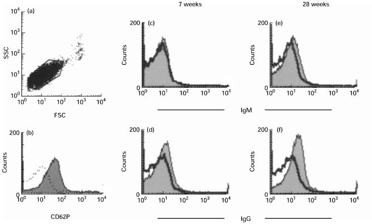
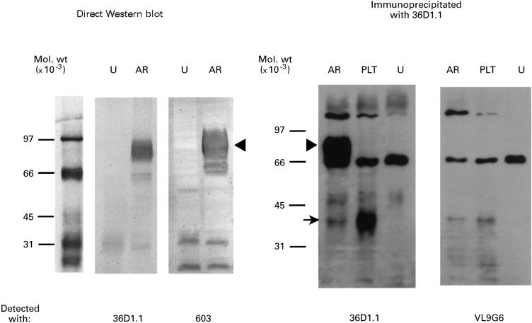
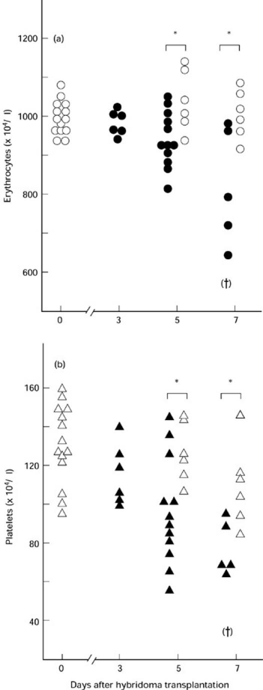
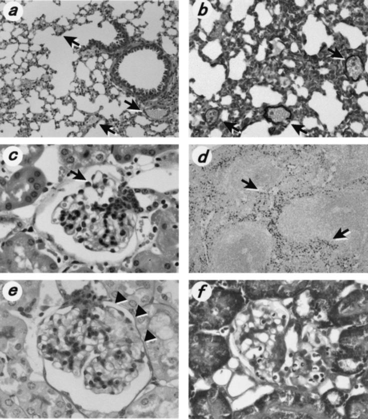
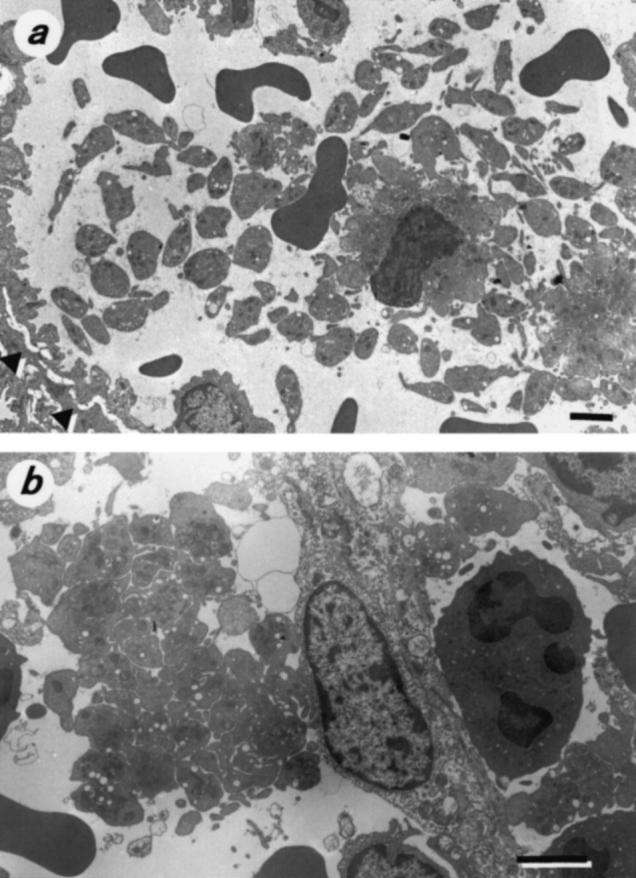
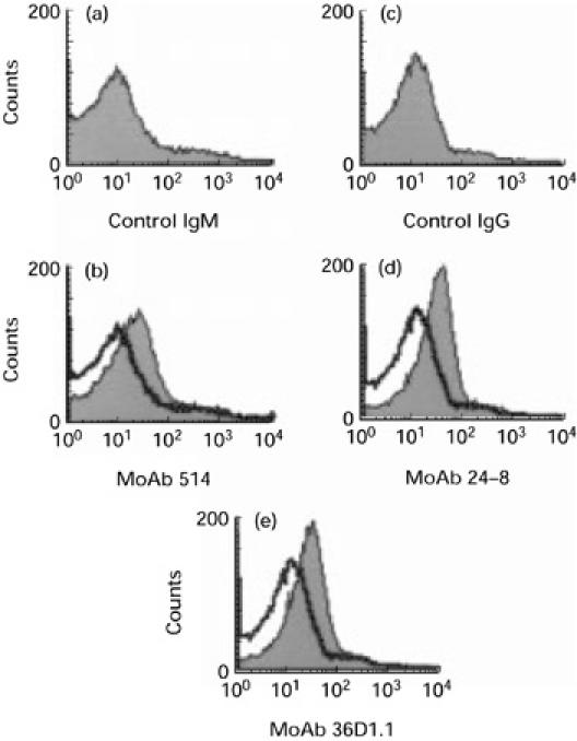
MRL/MpJ-lpr/lpr (MRL/lpr) mice spontaneously develop immune complex-mediated glomerulonephritis and thrombocytopenia. Although the presence of cross-reactive anti-phospholipid antibodies in sera of MRL/lpr mice has been demonstrated, possible relationships between detected autoantibodies and the development of thrombocytopenia have not been elucidated. Recent genetic analyses in a few different strains of lupus-prone mice have pointed out a close correlation between autoantibodies reactive with endogenous retroviral env gene product, gp70, and the development and severity of glomerulonephritis. In the process of establishing possibly nephritogenic anti-gp70 autoantibody-producing hybridoma cells from MRL/lpr mice, we identified an IgG2a-producing anti-gp70 hybridoma clone that induced microvascular intraluminal platelet aggregation, thrombocytopenia, and amenia upon transplantation into syngeneic non-autoimmune mice. This and two other anti-gp70 antibodies bound onto the surface of mouse platelets, and purified IgG2a of the anti-gp70 autoantibody induced glomerular lesions with characteristics of thrombotic thrombocytopenic purpura when injected into non-autoimmune mice. The pathogenic anti-gp70 autoantibody specifically precipitated a platelet protein with an approximate relative molecular mass of 40 000.


Figures

Fig. 1
Fig. 1
Demonstration by flow cytometry of the presence of platelet-associated autoantibodies in MRL/lpr mice. (a,b) A typical gating condition adopted in the present study to analyse mouse platelets. FSC, Forward scatter; SSC, side scatter. When stained with anti-mouse CD62P MoAb after stimulation with thrombin, cells within the polygonal gate (a) showed a single peak (shadowed area in (b)) in flow cytometry analysis, confirming their homogeneity. (b) Dotted line shows the pattern of staining obtained when the same preparation of mouse platelets was incubated with the isotype-matched control antibody. (c–f) Detection of surface-bound IgM and IgG on mouse platelets. Platelets were isolated from MRL/+ and MRL/lpr mice at 7 and 28 weeks old. In each chart a thick solid line shows the staining pattern of MRL/+ mouse platelets, while a shadowed area indicates that of MRL/lpr mouse platelets.
Fig. 2
Fig. 2
Direct Western blotting and immunoprecipitation followed by Western blotting to detect the antigen reactive with the anti-gp70 MoAb. Extracts from uninfected Mv1Lu mink cells (U), NZB-AR cells chronically infected with an NZB xenotropic virus (AR), or (BALB/c × MRL/+)F1 mouse platelets (PLT) were either directly subjected to SDS–PAGE (left) or immunoprecipitated with MoAb 36D1.1 before being separated by electrophoresis (right). Lanes mol.wt show positions of molecular weight markers with numbers on the left indicating relative molecular mass × 10−3 of each marker protein. Direct Western blotting confirmed the reactivity of MoAb 36D1.1 to retroviral gp85 (gp70 + p15E) that was also detected with the reference anti-gp70 MoAb, 603 [31]. The same protein was also precipitated from the extract of NZB-AR cells, but not from the extract of uninfected Mv1Lu cells, with MoAb 36D1.1 and detected with the same anti-gp70 MoAb (arrowhead). However, the precipitated gp85 was not detectable with the isotype-matched control antibody, VL9G6. A protein of approximately 40 000 in relative molecular mass was precipitated from mouse platelets and detected with MoAb 36D1.1 (arrow). Note that a faint band of apparently the same molecular mass was also detectable in the immunoprecipitate prepared from NZB-AR cells, but this band was absent from the blots of the extract prepared from uninfected Mv1Lu cells.
Fig. 3
Fig. 3
Changes in the number of peripheral blood erythrocytes (a) and platelets (b) in (BALB/c × MRL/+)F1 mice after transplantation with the anti-gp70 or control hybridoma cells. Each open symbol represents a blood cell number of an individual control mouse transplanted with hybridoma VL9G6, while closed symbols show those of the mice transplanted with anti-gp70 hybridoma 36D1.1. One mouse transplanted with 36D1.1 cells died before day 7 (†). *Significant difference between the indicated groups (P < 0.05).
Fig. 4
Fig. 4
Representative histopathology of mice transplanted with anti-gp70 hybridoma 36D1.1. (a) The lung with several sections of small arterioles (arrows) and venules that are filled with fine-granular platelet thrombi. (Haematoxylin and eosin, × 100.) (b) A representative section of the affected lung stained with phosphotungstenic acid-haematoxylin reagent to examine the presence of fibrin (× 200). Fine-granular thrombi filling the arterioles (arrows) do not contain fibrin. (c) A representative section of the kidney. Note extreme dilatation of capillary lumina in the upper half (arrow), and segmental hyaline-like changes with disappearance of mesangial cells in the lower half. (Haematoxylin and eosin, × 260.) (d) Berlin blue staining of the spleen (× 20) showing accumulation of haemosiderin in the red pulp (arrows) at 5 days after transplantation of hybridoma 36D1.1. (e,f) Representative glomerular pathology of mice injected with purified MoAb 36D1.1. Note the segmental hyaline-like change with disappearance of mesangial cells in (e) (arrowheads, periodic acid-Schiff stain, × 260). (f) Capillaries are abnormally dilated (Masson's trichrome stain, × 260).
Fig. 5
Fig. 5
Electron micrographs showing in vivo platelet aggregation in mice transplanted with hybridoma 36D1.1. (a) Aggregates of platelets within the lumen of a small arteriole in the lung. Arrowheads indicate elastic laminae. Note an erythrocyte is embroiled in an aggregate of platelets near the centre of the picture. Bar = 2 μm. (b) Aggregates of platelets in the splenic sinus. Bar = 2 μm.
Fig. 6
Fig. 6
The results of flow cytometric analyses demonstrating the expression of gp70 antigenicity on the surface of mouse platelets. (a,c) Staining patterns obtained with the control MoAb. These patterns are duplicated in (b,d,e) with a solid line for comparison. (b,d,e) Staining patterns obtained with anti-gp70 MoAb (shaded). Not only the pathogenic anti-gp70 MoAb 36D1.1 but two other anti-gp70 MoAbs of independent origins, 514 [29] and 24-8 [30], reacted to the surface of mouse platelets.

Similar articles

Cited by

References

    1. Moake JL. Studies on the pathophysiology of thrombotic thrombocytopenic purpura. Semin Hematol. 1997;34:83–9. - PubMed
    1. Ruggenenti P, Luts J, Remuzzi G. Pathogenesis and treatment of thrombotic microangiopathy. Kidney Int. 1997;51(Suppl. 58):S97–S101. - PubMed
    1. Neild GH. Hemolytic uremic syndrome/thrombotic thrombocytopenic purpura. pathophysiology and treatment. Kidney Int. 1998;53(Suppl. 64):S45–S49. - PubMed
    1. Tandon NN, Rock G, Jamieson GA. Anti-CD36 antibodies in thrombotic thrombocytopenia purpura. Br J Haematol. 1994;88:816–25. - PubMed
    1. Schultz DR, Arnold PI, Jy W, et al. Anti-CD36 autoantibodies in thrombotic thrombocytopenic purpura and other thrombotic disorders: identification of an 85kD form of CD36 as a target antigen. Br J Haematol. 1998;103:849–57. - PubMed

Publication types

MeSH terms